The Varus Thrust Gait: A career ender.


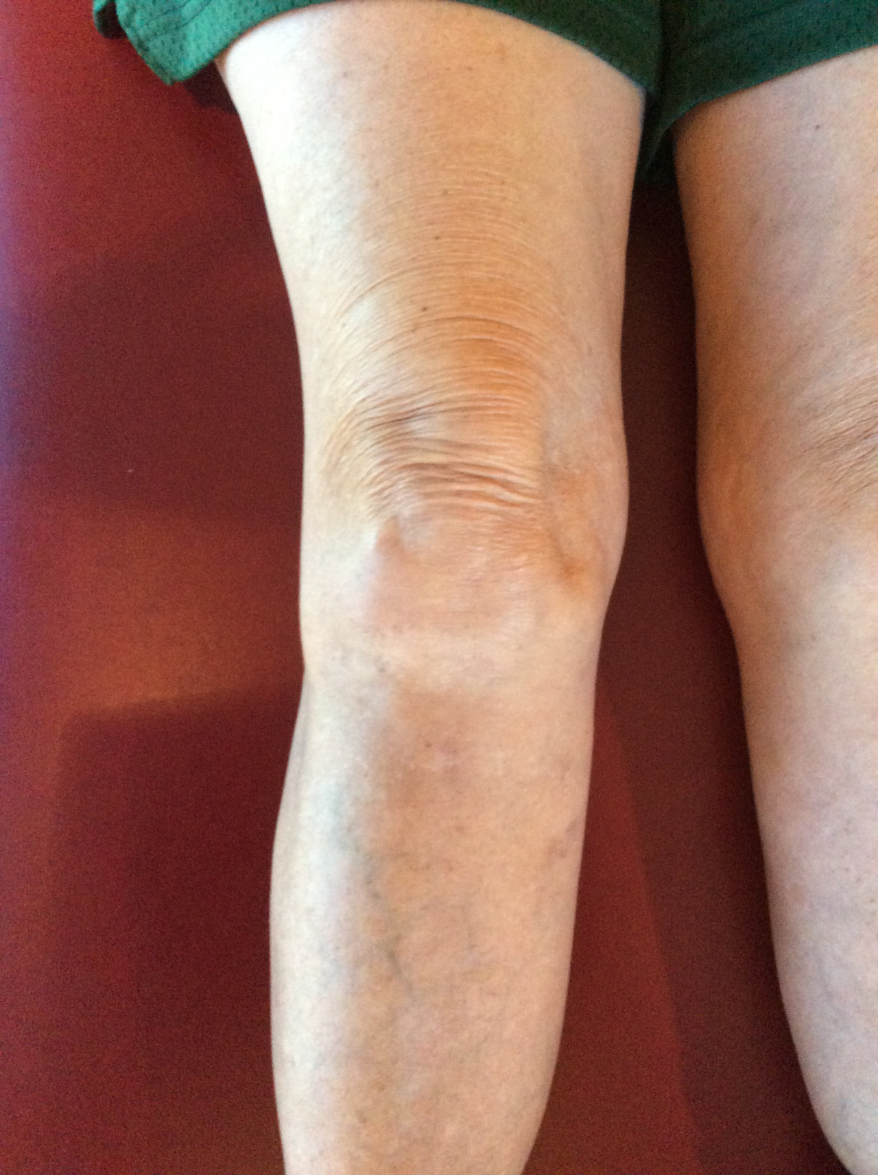
As the viewer should note in the video, the right knee is undergoing a sudden abrupt varus (lateral) shift during the gait loading response.  The tib-femoral joint is a sagittal hinge, not a frontal-lateral plane hinge, so this is clearly pathomechanical movement. This knee will likely undergo premature knee cartilage and meniscal degeneration.
However, there are other thoughts and considerations here.  The big question is, likely, how did this happen and what is wrong ? The cause of this issue is likely more simple than complicated however there may also be multiple factors coming together in a perfect storm. However, make no mistake, in order to understand a varus thrust gait, one has to understand the why and how of the gait presentation. Additionally, one must have a clinical knowledge of the restraining systems of the knee, both active and passive, and have a high degree of clinical suspicion and working knowledge of how to assess for these types of problems.

Things to consider:  
- old ACL/PCL and posterolateral corner damage (read this post here, link)
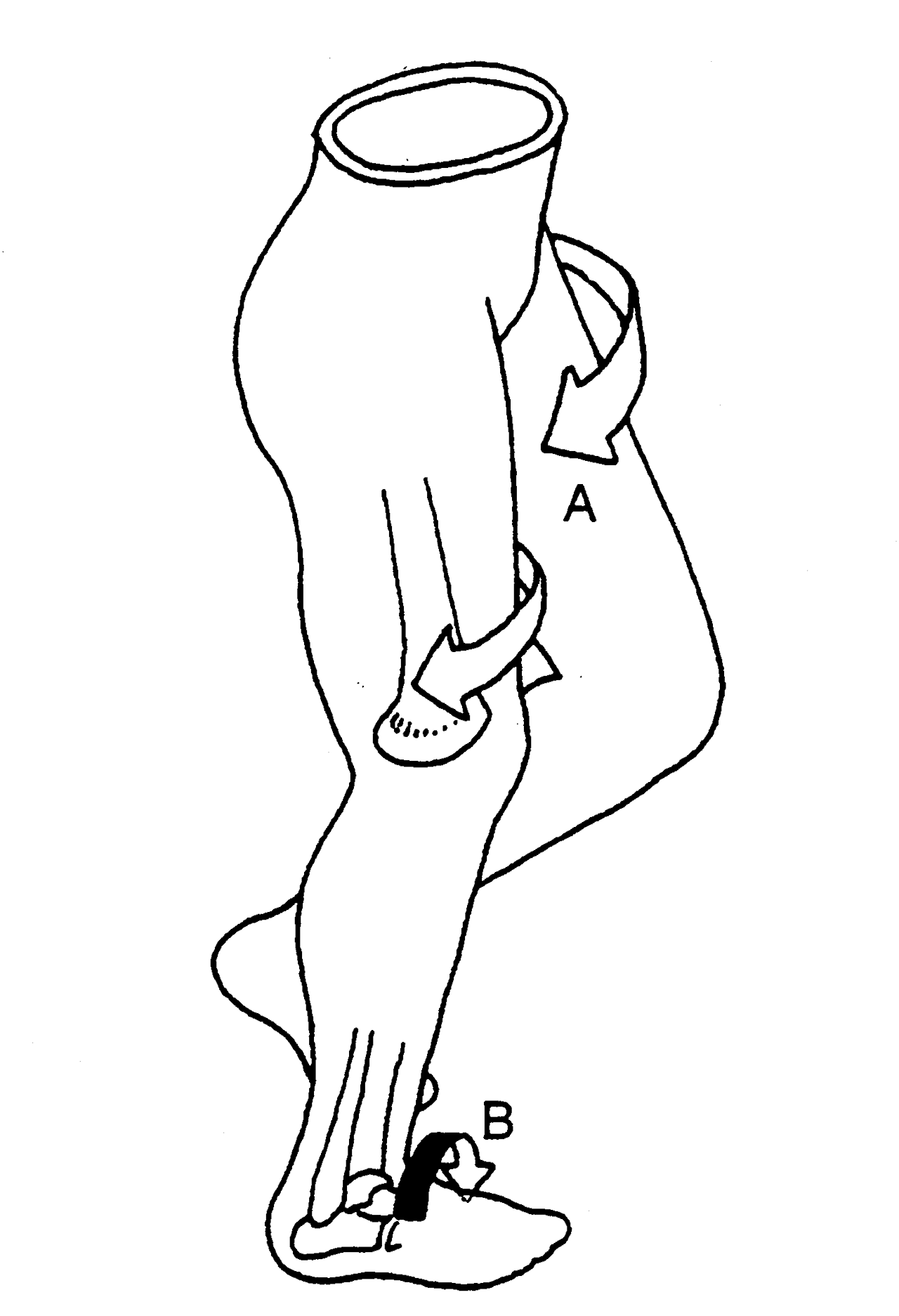
When the posterolateral corner complex of the knee is torn up from a blow to the knee or a torsional loading failure, the 3 components of the posterolateral corner (the lateral collateral ligament (LCL), the popliteal tendon, and the popliteo-fibular ligament complex). This complex attaches just in front of the origin of the lateral gastrocnemius tendon off the lateral femoral epicondyle. This complex can be blown out from either a PCL or ACL injury mechanism, these big player ligaments are rarely torn in isolation.
- is there a Pivot Shift phenomenon, likely.  A positive Pivot Shift test will be present. One must know how to perform this test to confirm its presence, it can be a tricky test if one does not know the load vectors to apply and what the shift feels like and where it occurs during the test. This can be a very subtle positive test, again, first hand experience is everything. 
- one must find this before surgery occurs for the ACL or PCL. Failure to find and address this damaged complex will likely result in rotational stability problems once return to play occurs. IT will not likely be noted in the initial post-operative months as the aggressive loading response will not be performed early on. Failure to address this problem will likely put ACL-PCL reconstruction success at a high risk.


Other critical factors to consider in the Varus Thrust Gait:
- is there medial knee osteoarthritis ?
- what is the foot type and what are the mechanics ?  ie. Forefoot varus, Forefoot supinatus, rearfoot variances
- does the patient have excessive pronation challenges that create massive internal spin into the tibia ?
- is the hip frontal and rotation plane stable?  Can the patient adequately control rotation at the hip level ?
- is there a Cross Over gait phenomenon with narrow based step width ? (search our blog and youtube for  "gait guys crossover gait").  A narrow step width will create an "unstacked" limb and promote more rotational risk into the limb, often playing out at the least tolerable joint to rotation . . . the knee.
- Does the client have Tibial Varum ? Genu Varum, Genu Valgum ? These can promote and complicate the Varus Thrust gait.
- Does the client have Tibial torsion or Femoral Torsion variants ? These can promote and complicate the Varus Thrust gait.

- is there weakness of the lateral gastrocnemius or biceps femoris (to name just two the directly cross over this posterolateral interval and can offer joint compression/stability ? What about weaknesses in the medial leg ? Not that these are anywhere sufficient to offset a PLRI (posterolateral rotatory instability), but, they are secondary helpers/restraints.

One should clearly see now that the Varus Thrust gait is potentially complicated and multifactorial. One MUST understand:
1. many components of normal gait and normal anatomy from foot to pelvis, at least.
2. be able to assess for aberrant mechanics and pathologies within all joints of the lower limb
3. be able to assess for post operative rotational stability and laxity (*even a healed, yet partially attenuated, Posterolateral corner complex that was not noted or addressed in the ACL-PCL reconstruction can come back to haunt even the best reconstruction. Those little rotational instabiliites will build over the years and render attenuation of the other secondary posterior restraints in the knee. Like a Lisfranc injury, sometimes things take a few years to brew and blossom before the "career ender" instability shows up. Trust us, we have seen it enough times.  

Rule: if one does not know it exists, one will miss it. If one does not know how to assess it, one will miss it. If one does not know normal anatomy, torsional variants, foot types and gait types, one is likely to be lost and left fumbling.  Our clients deserve more. 

Dr. Shawn Allen

Varus Thrust and Knee Frontal Plane Dynamic Motion in Persons with Knee Osteoarthritis. Osteoarthritis Cartilage. 2013 Nov; 21(11): 1668–1673. Published online 2013 Aug 12.
Alison H. Chang, PT, DPT, MS, Joan S. Chmiel, PhD, Kirsten C. Moisio, PT, PhD, Orit Almagor, MS, Yunhui Zhang, MS, September Cahue, MPH, and Leena Sharma, MD

Whoa # 2

And what about # 260?

Things are certainly different for this chap compared to # 172. His take off from his right foot looks pretty good. His left foot looks like it is going to cross over with some medial knee fall as it descends to hit the ground. Just before it hits the ground, it should be supinated in some degree of dorsiflexion (he is sprinting, so we expect some plantar flexion because he is sprinting), inversion and adduction. His pelvis is nice and level. His arm swing looks pretty good with only a slight cross over on the right as it is coming through. With form this good, no wonder he is out front!


and what have we been saying?Gait problems leave clues. Asymmetry is a BIG clue“Asymmetrical lower extremity neuromuscular control is predictive of repetitive stress injury in recreational runners, according to findings presented at the Combin…

and what have we been saying?

Gait problems leave clues. Asymmetry is a BIG clue

“Asymmetrical lower extremity neuromuscular control is predictive of repetitive stress injury in recreational runners, according to findings presented at the Combined Sections Meeting of the American Physical Therapy Association in February in Anaheim.”

http://lermagazine.com/issues/march/years-after-achilles-tear-injured-limb-demonstrates-elevated-knee-loading

tumblr_o1y939Qxjn1qhko2so3_1280.jpg
tumblr_o1y939Qxjn1qhko2so1_1280.jpg
tumblr_o1y939Qxjn1qhko2so2_1280.jpg
tumblr_o1y939Qxjn1qhko2so4_1280.jpg

The Banana Toe: The Force has to go somewhere. It’s a Jedi Gait Rule.

* note: there are 4 photos to today’s blog post. Be sure you click through all 4.

When you toe off, you have to toe off from somewhere in the foot unless you like an apropulsive hip flexion gait, where  you just lift you foot off the ground from foot flat, kind of like a true neurologic “foot drop” gait client would.  But, if you are lucky enough not to have a true foot drop, you are going to push off somewhere in the foot.  You can do it off the lateral foot (low gear toe off) and lesser toes, or you can do it off the big toe (high gear) the way we were built to do it. 

The above pictures show a nasty dorsal crown of osteophytes that is limiting hallux (big toe) extension/dorsiflexion. This is true hallus rigidus and hallux limitus. When this client attempts to toe off, the joint cannot normally partake in the activity, there is no Windlass effect, no posturing up over the sesamoids for mechanical advantage etc.   

In this scenario, there are two places you can put it, up into the next proximal joint(s) meaning the met-cuneiform joint or further down into the interphalangeal joint. In other words. the loads go proximal or distal to the limited joint, and they eventually play out there, over and over and over gain. The former option would basically mean you are pronating/dorsiflexing through the midfoot which is never good (can you say Saddle exostosis ! ouch !) or the latter option is to dorsiflex through the interphalangeal joint  and over time that toe begins to attenuate plantar ligamentous structures and extend beyond its normal limits resulting in the “Chiquita” toe (a upward bent toe resembling a banana shape). This will disable the long flexor of the great toe (FHL: flexor hallucis longus).and inhibit mechanical advantage of the extensor digitorum brevis.  If you struggle with the “how and why” behind this sentence in terms of restoration attempts, you need to watch my video here. It will offer you deeper insights.

Will this toe become painful ? yes, in time it is quite possible.  Is there much you can do? Sure, a rocker bottomed shoe will help take the load at toe off instead of forcing it into this toe or the midfoot.  Will an orthotic  help ? Well, this is a loaded question. If you are putting the forces into the midfoot choice as described above, the orthotic will block that motion and you will likely default option into the toe presentation above. So you are merely just moving loading forces around. It can be helpful, but you are quite possibly “robbing Peter to pay Paul” as they say.  The video I asked you to watch can be helpful but it will force that metatarsophalangeal joint into extension, a range it does not have, so it is not a remedy and not recommended.  Perhaps some awareness and slight increase in FHL(long toe flexor) use can be attained however.  These are tricky cases, simple in theory, but execution can be fussy and requires patient awareness and education. We like the rocker bottomed shoe as a nice easy solution and some increased FHL use awareness.  Help them find a little more FHL use by putting a pencil under the crease of the toe and help them to drive the tip of the toe down just a little out of that banana extension posture. It can help them control the overloading of the dorsal aspect of the interphalangeal joint.

As always, lets carry this forward into gait thoughts.  How is  hip extension going to be in this client ? How is glute strength ? Hip joint range ? Hip extension motor patterning ? Will the client go into anterior pelvic tilt to borrow the last range of hip extension ? Will the hamstrings have to accommodate ? Lots of yummy biomechanical and neurological mental gymnastics here. Bottom line answer to all the above ?  “ it depends, they will have to accommodate and compensate”.  And as the Jedi Gait Rule goes, “the Force as to go somewhere”.

Shawn Allen, one of the gait guys

tumblr_o0pa2hQiOL1qhko2so1_1280.jpg
tumblr_o0pa2hQiOL1qhko2so2_1280.jpg
tumblr_o0pa2hQiOL1qhko2so3_1280.jpg
tumblr_o0pa2hQiOL1qhko2so4_1280.jpg
tumblr_o0pa2hQiOL1qhko2so5_1280.jpg
tumblr_o0pa2hQiOL1qhko2so6_1280.jpg
tumblr_o0pa2hQiOL1qhko2so7_r1_250.png
tumblr_o0pa2hQiOL1qhko2so8_r1_250.jpg

What were they thinking? Oh, they weren’t thinking…

Here is a simple case of knowing your anatomy. 

make sure to use the toggle bar to the right and left of the picture to see all the pictures : )

This woman came in with right sided lateral knee pain with hiking and skiing; worse with fatigue, better with rest. The pain was localized at the lateral joint line and at the tibial fibular joint. 

She had been previously been diagnosed with tibial fibular hypermobility and subsequently had an arthrodesis (fusion) performed at that joint for knee pain. The surgery helped for a short time and a newer, slightly different pain developed. 

Yes, she has a moderate genu valgus, R > L. Yes, she has a left, anatomically short (tibial) Left leg. Yes, she has has NO MOBILITY at the tib/fib articulation and the focus of pain is just above at the joint line and at the lateral aspect of the patello femoral joint. 

The tibial fibular joint is a syndesmosis (not a true synovial or diarthrodial joint) that is supposed to have a a superio/inferior gliding motion (see diagram) with ankle dorsiflexion, due to the wedge shape of the talar dome and talo crural articulation. It also is supposed to have an anterior/posterior gliding movement at the superior aspect of the joint and a reciprocal movement in the opposite direction at the ankle (see diagram).

Whenever we take away movement in one area, it needs to occur somewhere else; in this case, at the femoral tibial joint and patello femoral joints.

Does it make sense that her left sided leg would cause hypermobility on the right side with a supinatory moment of the foot on the left to attempt to lengthen the leg and a pronatory movement of the foot on the right, in addition to valgus angulation of the joint on the right to attempt to “shorten” that extremity? Would this increased valgus angluation of the knee, in turn, cause abnormal, lateral, tracking of the patella? Wouldn’t the increased pronatory moment cause a more supple foot on that side with increased requirements for “push off” on that side with increased calf recruitment? Do you think that may impair proprioception on that side?

What if you put a sole lift in the left shoe (like we did) to help to alleviate some of the discrepancy and gave her some anterior compartment exercises (toes up walking, lift/spread/reach exercises, heel walking, simple balance on 1 leg exercises? Her world becomes a much better place to live in and she can return to the activities she loves to do with her 65 year old friends, like hiking 14′ers, skiing and mountain biking,

What we do to one joint affects all the others. You cannot make one change without expecting others. Be on the lookout and know your anatomy! This case was relatively straight forward. Many are not. Do a thorough exam and expect the unexpected. 

tumblr_nm5tkl3rsl1qhko2so1_1280.gif
tumblr_nm5tkl3rsl1qhko2so2_1280.gif

How well do you understand stance phase mechanics?

Here is a recent question we fielded and thought it would make a great post. 

Question/Comment: I’m slightly confused about closed chain hip motion in the stance leg.


Maybe if I explain what my thought process is you can correct me.  Lets use
left stance phase with the right leg swinging through.

After right mid-swing, the pelvis will be rotating towards the left.  The
motion of the pelvis on the left femur would be relative femur internal
rotation.  I understand that the right leg is externally rotating
(supinating) and that normal open chain kinematics of hip extension is
coupled with external rotation.  But if the pelvis is moving towards the
left AND the left femur externally rotates, wouldn’t that create too much
rotation?  So what I’m saying is that a pelvis that is oriented to the left
with a left femur that externally rotates creates an odd motion in my head
(which may be where the problem lies).  If you’ve ever seen a western where
the gun slingers do that weird walk to a shoot out…that’s what an
externally rotating femur during terminal stance looks like to me.

I’ve discussed this with other clinicians.  Some are in agreement with me,
some think it’s externally rotating, and some don’t know what I’m talking
about.  In my patients I also see a loss of hip IR more than hip ER.  These
patients that lose hip IR seem to have more difficulty in terminal
stance/toe-off phase more than the ones that lose hip ER.

If you could help me understand these kinematics and clear this up for me I
would greatly appreciate it.

Thank you, A

our reply: 

Taking your example with the L leg in stance:
When the L heel contacts the ground, the friction of the ground (hopefully) slow the calcaneus and the talus slide anteriorly on the calcaneus. 

Because of the shape of the calcaneal facets, the talus plantar flexes, adducts and everts. This sets the stage for pronation to occur: the calcaneus everts and the lower leg internally rotates, with the thigh following. The right side of the pelvis is moving to the L (counter clockwise rotation). This should occur (ideally) until midstance. At midstance, the opposite ® foot begins to enter swing phase; this should initiate supination of the stance phase leg (L). At this point, the L foot should be beginning to supinate the the leg and thigh beginning external rotation. It (thigh and leg) should reach maximal external rotation at toe off (maximal counter clockwise rotation of the pelvis) and remain in external rotation until heel strike/initial contact on the L side again. At this point, the pelvis begins clockwise rotation.

It is necessary for the thigh and leg to externally rotate while the pelvis is rotating counter clockwise, because of the constraints of the iliofemoral, pubeofemoral and ishiofemoal ligaments.

We too often see a loss of internal rotation of the hip in symptomatic populations more often than external rotation.

We hope this clarifies things for you.

Thank you again for the question and taking the time to write.

The Gait Guys

Lets make a resolution…Or not…
Cool guy, cool picture, cool scenery. Motivational? He is the fitness guru the developed the “Insanity” workout series, amongst others.
But looks may be deceiving.  
For reference, draw a line…

Lets make a resolution…Or not…

Cool guy, cool picture, cool scenery. Motivational? He is the fitness guru the developed the “Insanity” workout series, amongst others.

But looks may be deceiving.  

For reference, draw a line from the philitrium, interpec interval, symphisis pubis to area bisected between feet. 

Did you notice the following?

  • crossing arms across midline; look how far that left arm is abducted.
  • look at pelvis list to left. If you bring that arm in, you need to compensate somewhere
  • did you notice the hip hike on the left? That may have something to do with the excessive internal rotation of the thigh on the left. Is that because of the pelvis shift to the left (to compensate) or is he making up for limited internal rotation of the right hip?
  • what about the subtle head tilt to the right? is that driving the compensation or is it another compensation?

Questions, questions, questions…

We are choosing to make a resolution without him for the time being : ) More on this photo another day.

tumblr_nc9gscGC521qhko2so2_1280.jpg
tumblr_nc9gscGC521qhko2so1_1280.jpg
tumblr_nc9gscGC521qhko2so3_1280.jpg
tumblr_nc9gscGC521qhko2so4_1280.jpg
tumblr_nc9gscGC521qhko2so5_1280.jpg
tumblr_nc9gscGC521qhko2so6_1280.jpg

Whoa!  It is amazing what the human frame can withstand…

This 300 pound individual is retired from working with tow trucks from a towing company as well as a service station.   He believes working with the tow trucks, particularly jumping out of them contributed to the O.A. of the ankles.

He has osteoarthritic ankles, a rear foot varus of 15 degrees left side, 5 degrees right.  He is currently in the New Balance 1040 shoe.  He would like some new orthotics built. He Fowler tests positive on his current orthotic set up (with the foot on the ground, dorsiflex the foot at the 1st metatarsal phalangeal joint (ie big toe joint), simulating terminal stance; the orthotic should hug the arch through the range of motion; ie about 45-60 degress of great toe dorsiflexion, which he incredibly has). He is unable to one leg stand because of the O.A. on the ankles and pain.

He has bi-lat. internal tibial torsion, Left > Right and moderate tibial varum, L > R. He has very little internal rotation of the hips bi-lat. Ankle dorsiflexion is about 5 degrees bilaterally.

He is currently in an older New Balance motion control shoe. You can see how he has worn the shoes into varus. More neutral shoes hurt his feet; attempts to put his rear foot into valgus causes increased ankle pain. Exercise compliance is minimal.

WHAT WOULD YOU DO?

The Gait Guys. Teaching and educating with each post.

Does this guy have a short leg or what? How good are your eyes?

One again, we had the gait cam, investigating gait on the east coast. What do we see in this gent?

  • heel strike on out side of left foot with increased progression angle

he appears to be stabilizing the left side during stance phase. notice the upper torso shift to the left during left stance phase

  • abbreviated arm swing on right

note that ankle rocker is adequate on the left

  • body lean to right on right stance phase

gluteus medius weakness on right? short leg on right?

Good.

  • Did you also notice the loss of ankle rocker on the right, compared to the left? This results in less hip extension on that side as well.
  • He flexes his right thigh less than his right during pre swing and swing

external obliques should be firing to initiate hip flexion, perpetuated by the psoas, iliacus and rectus femoris. This does not appear to be happening.

All of this is great BUT nothing like being able to actually examine your patients is there? You can see how gait analysis can tell us many things, but they need to be confirmed by a physical exam.

The Gait Guys. Educating (and hopefully enlightening) with each post. Keep your eyes open and your thinking from the ground up : )


Got Arm Swing?

We have written many times about arm swing. Click here for some of our posts here on Tumblr.

Here we are again at the beach. Look at the beautiful difference in arm swing from side to side in the guy carrying the bag. Makes you want to tell him to use a backpack, eh?

Never mind what it does to his gait

  • decreased arm swing on the carrying side
  • increased step length on the left side
  • increased thigh flexion of the left side
  • increased body lean and head tilt to right side (Take a look at this paper)

think about the increased metabolic cost. Think about what this  type of input (increased amplitude of movement unilaterally) is doing to your cortex!

keep your movements symmetrical, folks!

The Gait Guys

A profound loss of hip extension…

While sitting on the beach, our mind never rests. Even when on vacation we continue to watch how people move.

Luckily today, I had the gait cam (Dr Allen is holding down the Gait Guys Fort), so live from Sunset Beach, it’s Sunday night. See of you can see what I saw.

Sitting with my wife and watching the kids dig in the sand, this gal with the flexed posture caught my eye.

Why is she so flexed forward? The profound loss of hip extension made it impossible for her to stand up straight! It was difficult to say if she has bilateral hip osteoarthritis, or possible bilateral THR’s (total hip replacements), maybe just really tight hip flexors, painful bunions that do not like toe off, or even all of the above. She may have a leg length discrepancy, as she leans to the left on left stance phase; of course she could have weak hip abductors on the left. It does not appear she has good control of her core.

What do we see?

  • flexion at the waist
  • loss of hip extension
  • body lean to left at left midstance
  • shortened step length
  • loss of ankle rocker
  • premature heel rise
  • decreased arm swing (she is carrying something in her left hand)

No one is safe from the gait cam! Stay tuned for more beach footage this week!

We remain, The Gait Guys, even on vacation.

This is part 2 of a 2 part post; with the video from the case previously discussed

please note the following in the video:

  • body lean to left during left stance phase (to clear right longer leg)
  • circumduction of right lower extremity  (to clear right longer leg)
  • lack of arm swing bilaterally (cortical involvement)
  • patient looking down while walking (decomposition of gait)
  • shortened step length (decomposition of gait)
  • increased tibial varum bilaterally

ASSESSMENT:  This patient’s short leg and internal tibial torsion impediments to her full recovery. She has increased tibial varum noted which is complicating the picture. This is causing pathomechanics and an abductory moment not only at the knee but also in the lumbar, thoracic and cervical spines.

WHAT DID WE DO?:                    

  • We attempted to do the one leg standing exercise. She needed to hold on and did not feel stable on the left hip while performing this.  This is probably more of confidence rather than ability issue. 
  • We gave her the stand/sit exercise to try to improve gluteal recruitment.
  • We also gave her the lift/spread/reach exercise to attempt to strengthen her feet.
  • A full-length 5 mm lift was cut for the left shoe  She felt more stable when walking on this.
  • She was treated with IC, PIR and manipulative therapy and neuromuscular stim of the knee as well as left hip area above, below and at the joint line of the knee as well as gluteus medius and minimus.   
  • We may need to consider building a more aggressive orthotic with a forefoot varus post depending upon her progress and response to care  

 The Gait Guys. Making it real, each and every post here on the blog.

special thanks to SZ for allowing us to publish her case, so others can learn

tumblr_nc9grebaIQ1qhko2so1_1280.jpg
tumblr_nc9grebaIQ1qhko2so2_1280.jpg
tumblr_nc9grebaIQ1qhko2so3_1280.jpg

Wow! What would you do?

This is part 1 of a 2 part post. Look for the other one a few minutes after this one with a video up top for the conclusion

PRESENTING PROBLEM: This 54 YO female patient presents with with left sided knee pain.  She had a total knee replacement (TKR) done in 2011.  She’s had a significant amount of discomfort on the medial aspect of the knee since then. She had an MRI of the hip done thinking the problem was there, and found nothing.   She is walking with a bad limp, left leg is half inch shorter than the right.  Pain is worse at night, changes with weather. 

She has knee pain on the lateral aspect (points to tibial plateau and joint line) with swelling that goes down to the ankle left side.  She has been wearing a “Good Feet” OTC orthotic on the left side which she states helps quite a bit.

Generally speaking, stretching and analgesics make the discomfort better.    Ibuprofen 400 mg. b.i.d. can take the edge off  Soft sided brace (neoprene sleeve) makes a difference as well. The hard sided brace gives her difficulty.

WORK HISTORY: She works for a preschool.  Her job involves standing and getting up and down a lot.  

FAMILY HISTORY:  She has left sided lid ptosis, this evidently is familial.  

PHYSICAL EXAM:  She stood 5’ 1” and weighed approx. 150 pounds.

Viewing the knees bi-lat., the left knee is markedly externally rotated.

She does have a left short leg; tibial and femoral.  She has bilateral tibial torsion (look at the tibial tuberosities and drop a line straight down; it should pass through the 2nd metatarsal head) and marked internal tibial torsion on the left side (>60 degrees) with femoral retrotorsion (less than 8 degree angle of femoral head with the shaft) on this side.  There is no rotation of the thigh or leg past zero degrees midline. .  She had 10 degrees of tibial varum on the left hand side.  Her Q-angle is 10 degrees on that side.  There is plantar flexion inversion of the foot.  Left lower extremity has less sensation secondary to the her TKR  surgery.

Gait evaluation reveals a fair amount of midfoot pronation noted on the left hand side in addition to an intoed gait.  She has to lean her body over to the left to get the right leg to clear.

Some mild weakness noted of hip abduction musculature left hand side gluteus medius, middle and anterior fibers. Knee stability tests were negative.

Neurologically, otherwise, she had full integrity with respect to sensation, motor strength and deep tendon reflexes in the upper and lower extremities.

Please see part 2 of this post for additional info including our assessment and what WE did.

 The Gait Guys. Making it real, each and every post here on the blog.

special thanks to SZ for allowing us to publish her case, so others can learn

tumblr_n92mssK8su1qhko2so2_r1_1280.gif
tumblr_n92mssK8su1qhko2so3_r1_1280.gif

So you want to do a Gait Analysis: Part 4

This is the 4th in a multi part series. If you missed part 1, click here. For part 2, click here, part 3, click here

These are the basics, folks. We hope this is a review for many.

A quick review of the walking gait cycle components:

There are two phases of gait: stance and swing

Stance consists of:

  • Initial contact
  • Loading response
  • Midstance
  • Terminal stance
  • Pre-swing

Swing consists of:

  • initial (early) swing
  • mid swing
  • terminal (late) swing

today, lets explore Terminal stance

Terminal stance is one of the last stages of stance phase. Following midstance, where maximal pronation should be occurring, the stance phase foot should now begin supinating, initiated by the the opposite foot in swing phase moving forward of the center of gravity. 

Lets look at what is happening here at the major anatomical areas:

Foot

  • Supination begins from the opposite, swing phase leg (see above)
  •  the calcaneus inverts to neutral
  •  the center of gravity of the foot raises from its lowest point at midstance
  • The lower leg should begin externally rotating (as it follows the talus)
  • The thigh should follow the lower leg and should also be externally rotating; sometimes to a greater extent due to the shape and size of the medial condyle of the femur (which is larger than the lateral)
  • these actions are perpetuated by the gluteus maximus and posterior fibers of the gluteus medius, as well as posterior compartment of the lower leg including the flexor digitorum longus, flexor hallucis longus, peroneus longus and tibialis posterior
Ankle
  • The ankle should be 5 degrees dorsiflexed and in ankle rocker
  • the calcaneocuboid locking mechanism should be engaging to assist the peroneus longus in getting the head of the 1st metatarsal to the ground

Knee

  • near or at full extension. This is perpetuated by the quadriceps and biceps femoris, contracting concentrically and attenuated by the semi membranosis and tendonosis. The popliteus contracts eccentrically as soon as the knee passes midstance to keep the rates of external rotation of the tibia and femur in congruence.

Hip

  • The hip should be extending to 10 degrees.

Can you picture what is happening? Try and visualize these motions in your mind. Can you understand why you need to know what is going on at each phase to be able to identify problems? If you don’t know what normal looks like, you will have a tougher time figuring out what is abnormal.

Ivo and Shawn. Gait and foot geeks extraordinaire. Helping you to build a better foundation to put all this stuff you are learning on.

pictured used with permission from Foot Orthoses and Other Conservative Forms of Foot Care

tumblr_n7yjctusWm1qhko2so1_1280.jpg
tumblr_n7yjctusWm1qhko2so2_1280.jpg
tumblr_n7yjctusWm1qhko2so3_1280.jpg
tumblr_n7yjctusWm1qhko2so4_1280.jpg
tumblr_n7yjctusWm1qhko2so7_1280.jpg
tumblr_n7yjctusWm1qhko2so9_1280.jpg

You can only “borrow” so much before you need to “pay it back”

How can feet relate to golf swing?

This 52 year old right handed gentleman presented with pain at the thoracolumbar junction after playing golf. He noticed he had a limited amount of “back swing” and pain at the end of his “follow through”.

Take a look a these pix and think about why.

Hopefully, in addition to he having hairy and scarred legs (he is a contractor by trade), you noted the following

  • Top left: note the normal internal rotation of the right hip; You need 4 degrees to walk normally and most folks have close to 40 degrees. He also has internal tibial torsion.
  • Top right: loss of external rotation of the right hip. Again, you need 4 degrees (from neutral) of external rotation of the hip to supinate and walk normally.
  • Top center:normal internal rotation of the left hip; internal tibial torsion
  • 3rd photo down: limited external rotation of the left hip, especially with respect ti the amount of internal rotation present; this is to a greater degree than the right
  • 4th and 5th photos down: note the amount of tibial varum and tibial torsion. Yes, with this much varum, he has a forefoot varus.

The brain is wired so that it will (generally) not allow you to walk with your toes pointing in (pigeon toed), so you rotate them out to somewhat of a normal progression angle (for more on progression angles, click here). If you have internal tibial torsion, this places the knees outside the saggital plane. (For more on tibial torsion, click here.) If you rotate your extremity outward, and already have a limited amount of range of motion available, you will take up some of that range of motion, making less available for normal physiological function. If the motion cannot occur at the knee or hip, it will usually occur at the next available joint cephalad, in this case the spine.

The lumbar spine has a limited amount of rotation available, ranging from 1.2-1.7 degrees per segment in a normal spine (1). This is generally less in degenerative conditions (2).

Place your feet on the ground with your feet pointing straight ahead. Now simulate a right handed golf swing, bending slightly at the waist and  rotating your body backward to the right. Now slowly swing and follow through from right to left. Note what happens to your hips: as you wind back to the right, the left hip is externally rotating and the right hip is internally rotating. As you follow through to the left, your right, your hip must externally rotate and your left hip must externally rotate. Can you see how his left hip is inhibiting his back swing and his right hip is limiting  his follow through? Can you see that because of his internal tibial torsion, he has already “used up” some of his external rotation range of motion?

If he does not have enough range of motion in the hip, where will it come from?

he will “borrow it” from a joint more north of the hip, in this case, his spine. More motion will occur at the thoracolumbar junction, since most likely (because of degenerative change) the most is available there; but you can only “borrow” so much before you need to “Pay it back”. In this case, he over rotated and injured the joint.

What did we do?

  • we treated the injured joint locally, with manipulation of the pathomechanical segments
  • we reduced inflammation and muscle spasm with acupuncture
  • we gave him some lumbar and throacolumbar stabilization exercises: founders exercise, extension holds, non tripod, cross crawl, pull ups
  • we gave him foot exercises to reduce his forefoot varus: tripod standing, EHB, lift-spread-reach
  • we had him externally rotate both feet (duck) when playing golf

The Gait Guys. Helping you to store up lots “in your bank” of foot and gait literacy, so you can help people when they need to “pay it back”, one case at a time.

(1) http://www.ncbi.nlm.nih.gov/pmc/articles/PMC2223353/

(2) http://www.ncbi.nlm.nih.gov/pmc/articles/PMC3705911/

tumblr_n7yjq09EgU1qhko2so1_1280.jpg
tumblr_n7yjq09EgU1qhko2so2_1280.jpg
tumblr_n7yjq09EgU1qhko2so3_1280.jpg
tumblr_n7yjq09EgU1qhko2so4_1280.jpg
tumblr_n7yjq09EgU1qhko2so5_1280.jpg
tumblr_n7yjq09EgU1qhko2so7_1280.jpg
tumblr_n7yjq09EgU1qhko2so9_1280.jpg
tumblr_n7yjq09EgU1qhko2so6_r1_500.jpg

So a patient presents to your office with a recent history of a L total knee replacement 8 weeks ago AND a recent history of a resurgence of low back pain, supra iliac area on the L side. Hmmmm. Hope the flags went up for you too!

His global lumbar ROM’s were 70/90 flexion with low back discomfort at the lumbo sacral junction, 20/30 extension with lumbosacral discomfort, left lateral bending 10 degrees with increased pain (reproduction); right lateral bending 20 degrees with a pulling sensation on the right. Extension and axial compression of the lumbar spine in left lateral bending reproduced his pain.

Neurologically he had an absent patellar reflex on the left, with diminished sensation over the knee medially and laterally. Muscle strength 5/5 in LE; sl impaired balance in Left single leg standing. There was incomplete extension of the left knee, being at 5 degrees flexion (right side was zero).

He has a right sided leg length deficiency (or a left sided excess!) of 5 mm. Take a look at the tibial lengths in the 1st 3 pictures. See how the left is longer? In the next shot, do you see how the knee cannot completely extend? Can you imagine that the discrepancy would probably be larger if it did?

Now look at the x rays. We drew a line across from the non surgical leg to make things clearer.

Now, think about the mechanics of a longer leg. That leg will usually pronate more in an attempt to shorten the leg, and the opposite side will supinate to attempt to lengthen. Can you see how this would cause clockwise pelvic rotation (in addition to anterior pelvic rotation)? Can you see this patients in the view of the knees from the top? Do you understand that the lumbar spine has very limited rotation (about 5-10 degrees, with more movement superiorly (1)  ). Does it make sense that the increased range of motion could effect the disc and facet joints and increase the patients low back pain?

So, how do we fix it? Have you seen the movie “Gattica”? Hmmm….A bit extreme. How about a full length 3mm sole lift to start, along with specific joint manipulation to restore normal motion and some acupuncture to reduce inflammation? We say that is a good start.

The Gait Guys. Increasing your gait literacy with each and every post. If you liked this post, please send it to someone else for them to enjoy and learn. 

(1) Three-Dimensional In Vivo Measurement of Lumbar Spine Segmental Motion Ruth S. Ochia, PhD, Nozomu Inoue, MD, PhD, Susan M. Renner, MS, Eric P. Lorenz, MS, Tae-Hong Lim, PhD, Gunnar B. Andersson, J. MD, PhD, Howard S. An, MD Spine. 2006;31(15):2073-2078.

Can you see it?
Here we are again. We have looked at this picture before; once about head tilt, and another about flip flops and form.
Take a good look at this picture and what is different about the child in blue all the way to the right and all th…

Can you see it?

Here we are again. We have looked at this picture before; once about head tilt, and another about flip flops and form.

Take a good look at this picture and what is different about the child in blue all the way to the right and all the others with the exception of the boy in pink, that we really cannot see?

Can you see it? No, we don’t mean the flip flops (but if you caught that all the boys were in sneakers and all the girls are in flip flops, you are good!)

How about looking at arm swing? Remember this post on arm swing and crossover gait, with the simple cue for correction? All of the children EXCEPT the boy in blue, are drawing their arms ACROSS their body (ie: flexion, internal rotation and adduction). Take a look at their legs. Yep, crossover gait (flexion, internal rotation and adduction). Little boy blues arms are going relatively straight and going in the saggital plane, where the others are going in the coronal plane.

We are not saying that blue does not have some gait challenges, like his torso shift to the left (or pelvic drift to the right), most likely do to gluteus medius weakness or inappropriate firing of the gluteus medius on the left stance phase leg; or his head tilt to the right, which most likely represents a compensation for the right pelvic drift and left body lean.

Arm swing. A very important clue to the puzzle we call gait and compensation. It is more prevalent than you think, and, in some cases, easily corrected with a simple cue.

The Gait Guys. Making it real and pertinent, in each and every post.

Correcting a cross over gait with arm swing? Is it really THAT easy? Sometimes, yes!

We noticed this patient had a cross over gait while running (1st few seconds of video. need to know more about crossover gait? click here). We noted she was crossing her arms over her body as well. We than had her run her hands and arms straight out. See the crossover disappear? Need to know more about arm swing? click here

We the had her do the same while walking. Easier to see, eh? That’s because it is often easier to “fudge” things when you are moving faster (ie: the basal ganglia of nervous system can interpolate where the body part is supposed to be, and because of momentum, there is less need for precision). When we do things slowly (like the 3 second Test), more precision is needed. Watch this short video clip a few more times.

The arms are essentially adducting when the arms cross over. The arms are reciprocally paired with the contralateral lower extremity. When you make a change in one, you often will make a change in the other.

Subtle. Yes. Easier to see when the task becomes more difficult. Yes. Pay attention, the answer is often right there if you look closely enough.

Providing the clues to help you be smarter, better, faster, stronger; we are The Gait Guys

special thanks to “Q” for allowing us to publish this video : )

What’s up, Doc?
Nothing like a little Monday morning brain stretching and a little Pedograph action.
This person had 2nd metatarsal head pain on the left. Can you figure out why?
Let’s start at the rear foot:
limited calcaneal eversion (…

What’s up, Doc?

Nothing like a little Monday morning brain stretching and a little Pedograph action.

This person had 2nd metatarsal head pain on the left. Can you figure out why?

Let’s start at the rear foot:

  • limited calcaneal eversion (pronation) L > R. The teardrop shape is more rounded on the left. This indicates some rigidity here.
  • note the increased pressure at the  medial calcaneal facets on each side with the increased printing
  • very little fat pad displacement overall

Now let’s look at the mid foot:

  • decreased mid foot pronation on the L. See how thin the line is going from the rear foot to the forefoot along the lateral column? This indicates a high lateral longitudinal arch

Now how about the fore foot?

  • increased printing under the met heads bilaterally; L >> R
  • increased printing of 1st met head L >> R
  • increased printing at medial proximal phalynx of hallux  L >> R
  • increased printing of distal phalanges of all toes L >> R

 Figure it out?

What would cause increased supination on the L?

  • short leg on L
  • more rigid foot on L
  • increased pronation on the R

Did you notice the elongated 2nd metatarsals (ie: Morton’s toe) on each foot?

Here is what is going on:

  • there is no appreciable leg length deformity, functional or anatomical
  • The Left foot is more rigid than the Right, thus less rear, mid and fore foot pronation, thus it is in relative supination compared to the right foot

do this: stand and make your L foot more rigid than the right; take a step forward with your right foot, what do you notice?

  • Can you feel how when your foot is supinated
  • can you see how difficult it is to have ankle rocker at this point? remember: supination is plantar flexion, inversion and adduction
  • Can you feel the weight of the body shift to the outside of the foot and your toes curl to make the foot more stable, so you do not tip to the left?
  • now, how are you going to get your center of mass forward from here? You need to press off from your big toe (hallux)

Wow, does that make sense now?

What’s the fix?

  • create a more supple foot with manipulation, massage, muscle work
  • increase ankle rocker by training the anterior compartment (shuffle walks, lift/spread/reach exercise, heel walking, Texas walk exercise, etc)
  • have them walk with their toes slightly elevated
  • we are sure you can think of more ways as well!

The Gait Guys. Increasing your gait literacy with each and every post. If you liked this post, tell others and spread the word. If you didn’t like this post, tell us! We value your constructive feedback.

More Power Leaks: Part 3


Good Morning peoples! A few weeks ago, we introduced posts about potential areas for power leaks. click here for #1, click here for #2

The common areas for leaks are:
great toe dorsiflexion
mm strength test
loss of ankle rocker
loss of knee flexion/extension
loss of hip extension
loss of balance/ proprioception

let’s take a look at a video of the next 2, with Dr Ivo and his partner in SCR, Dr John Asthalter:

Power leak 2: Muscle strength test

you need adequate strength in both the short and long extensors of the toes, for arch integrity, the windlass mechanism as well as appropriate ankle rocker

Common compensations include:

externally rotating the foot and coming off the inside of the great toe. this often causes a callus at the medial aspect of the toe. This places the foot in more pronation (plantar flexion, eversion and abduction) so it is a poorer lever.

lifting the foot (and bending the knee) excessively (knee flexion > 60 degrees) to create clearance of the toes for swing phase. This is sometimes referred to as a steppage gait.

hiking of the hip, again to create clearance for the foot

Power leak 3: ankle rocker

ankle rocker is needed to move the body mass forward in the gravitational plane. It is one of the 3 rockers (for a rocker review, click here).

Compensations for loss of ankle rocker can include:

premature heel rise

shortened step length

excessive pronation through the mid foot

external rotation of the lower extremity and “rolling off” the inside of the great toe

forefoot strike gait

Ivo and Shawn. Giving you the information you need to make informed clinical decisions and build better runners, wherever you go! Spread the word of gait literacy!