What did you notice? The Devil is in the details...

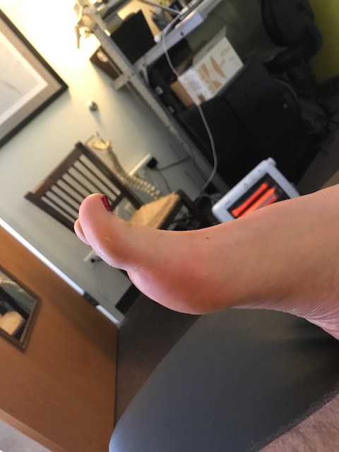
 Cavus foot? Loss of the transverse arch? Prominence of extensor tendons?

The question is: Why?

It’s about reciprocal inhibition. The concept, though observed in the 19th century, was not fully understood and accepted until it earned a Nobel prize for its creditor, Sir Charles Sherrington, in 1932. Simply put, when a muscle contracts, its antagonist is neurologically inhibited, So when your bicep contracts, your tricep is inhibited. This holds true whether you actively contract the muscle or if the muscle is irritated (causing contraction).

So how does this apply to this foot?

We see prominence of the extensor tendons (particularly the extensor digitorum brevis EDB; the longus would have caused extension at the distal interphalangeal joint). The belly of the muscle is visible, telling us that it is active. It is neurologically linked to the flexor digitorum brevis (FDB). This muscle, in turn, has slips which attach it to the abductor hallucis brevis (AHB) medially and the abductor digiti minimi (ADM) laterally. These muscles together form 2 triangles (to be discussed in another post) on the bottom of the foot, which lend to the stability of the foot and the arches, especially the transverse.

When the EDB fires, it inhibits the FDB, (which, in addition to flexing the MTP’s, assists in maintaining the arch). The EDB has an effect which drops the distal heads of the metatarsals as well (Hmm, think about all the people with met head pain) Now, look at the course of the tendons of the EDB. In a cavus foot, there is also a mild abductory moment, which flattens the arch. Conversely, the FDB in a cavus foot would serve to actually increase the arch, and would have a ,mild adductory moment. Net result? A flattened transverse arch.

Now look at the Flexor digitorum longus, overactive in tbis foot (as evidenced by the flexion of the distal interphalangeal joints, mild adduction of the toes (due to the change of direction of pull in a cavus foot) and lowering of the met heads due to hyperextesnion at the MTP joints ). This mm is reciprocally linked with the extensor digitorum longus. The prominence of the extensor tendons is do to increased activity of the EDB (go ahead, extend all your fingers and look at the tendons in your hand. Now flex the  DIP and IP joints and extend the MTP; see how they become more prominent?).

Reciprocal inhibition. It’s not just for dinner anymore…

We are and remain; The Gait Guys

Toes Spacers, anyone?

Less pain through better mechanics?

cc3.jpg

We have been using toe separators for various foot problems like hallux valgus, hammer toes and flexor dominance/extensor weakness. Our reasoning is that through changing the angle of attachment of the muscle, you alter the mechanical advantage of that muscle and help it to work more efficiently. This seems implied in the literature with respect to foot orthoses (1-3) but we could not find any data regarding toe separators. Toe separators DO seem to reduce pain and increase function (4-6). Perhaps this is through better biomechanics, mechanical deformation, proprioceptive changes, or most likely a combination of all these factors and more.  We think clinical results speak volumes. It is nice to see more data coming out on these easy to implement clinical tools. 

What is you clinical reasoning or rationale for using these devices? We would love to hear and if you have an article for reference you could share, that would be great. 


1. Scherer PR, Sanders J, Eldredge DE, Duffy SJ, Lee RY. Effect of functional foot orthoses on first metatarsophalangeal joint dorsiflexion in stance and gait. J Am Podiatr Med Assoc. 2006 Nov-Dec;96(6):474-81.

2. Halstead J, Chapman GJ, Gray JC, Grainger AJ, Brown S, Wilkins RA, Roddy E, Helliwell PS, Keenan AM, Redmond ACFoot orthoses in the treatment of symptomatic midfoot osteoarthritis using clinical and biomechanical outcomes: a randomised feasibility study. Clin Rheumatol. 2016 Apr;35(4):987-96. doi: 10.1007/s10067-015-2946-6. Epub 2015 Apr 28.

3. Bishop C, Arnold JB, May T. Effects of Taping and Orthoses on Foot Biomechanics in Adults with Flat-Arched Feet. Med Sci Sports Exerc. 2016 Apr;48(4):689-96. doi: 10.1249/MSS.0000000000000807.

4. Chadchavalpanichaya N, Prakotmongkol V, Polhan N, Rayothee P, Seng-Iad S. Effectiveness of the custom-mold room temperature vulcanizing silicone toe separator on hallux valgus: A prospective, randomized single-blinded controlled trial. Prosthet Orthot Int. 2017 Mar 1:309364617698518. doi: 10.1177/0309364617698518. [Epub ahead of print]

5. Tehraninasr A, Saeedi H, Forogh B, Bahramizadeh M, Keyhani MR. Effects of insole with toe-separator and night splint on patients with painful hallux valgus: a comparative study. Prosthet Orthot Int. 2008 Mar;32(1):79-83. doi: 10.1080/03093640701669074.

6. Tang SF, Chen CP, Pan JL, Chen JL, Leong CP, Chu NK. The effects of a new foot-toe orthosis in treating painful hallux valgus. Arch Phys Med Rehabil. 2002 Dec;83(12):1792-5. 


The season to pathologize our feet is upon us. Toe extension matters.

Screen Shot 2018-04-06 at 8.05.18 AM.png

I blew out my flip flop,
Stepped on a pop top;
Cut my heel, had to cruise on back home.
But there's booze in the blender,
And soon it will render
That frozen concoction that helps me hang on. - Jimmy Buffett

I continue to see more and more people with inadequate toe extension. It is complicated. I see those who do not even have the awareness of toe extension, loss of strength of toe extension, loss of endurance of toe extension, loss of global range of toe extension (dorsiflexion at the MTP joint), more failure of long toe extensor (EHL) strength and more prominence of increased short toe extensor strength (EDB) and more frightening, a lack of disassociation of toe extension (MTP dorsiflexion) and ankle dorsiflexion. Many clients when asked to life their toes, will drive into ankle mortise dorsiflexion; ask them to just purely toe dorsiflex and the mental games begin, a wrinkled brow, intense concentration. If you cannot extended the toes sitting, how are you going to find them in swing phase of gait when balance, and other things, are more important?
Stand and lift your toes. The arch should go up, you have engaged the Windlass Mechanism, that winds up the plantar fascia and raised the arch. If you do not have competent, unconsciously competent, toe extension, your arch is not all that it can, and should, be. If you cannot raise your toes, thus raise the arch, thus plantarflex the first metatarsal, then in gait, when the foot is on the ground, you cannot properly position the sesamoids, properly get safe terminal ranges of hallux dorsiflexion at toe off, properly position the foot for loading and unloading, adequately achieve ankle dorsiflexion, adequately offer the hip a chance for ample hip extension, offer the glutes optimal chance to work in all phases to help control spin of the limb during loading and unloading, and the list goes on and on. I am sure I left much out there, this was written in a few minutes and unedited, just a short rant for the weekend. But if you have not championed toe extension, both in an unloaded and loaded foot (on the ground), achieved control of both long and short extensor muscles to the toes (and paired them well with the long and short toe flexors), disassociated toe extension from ankle dorsiflexion, and then figured out how to properly, timely, engage all these processes into your gait unconsciously, you are working on less of an optimal system than you should be. So, if your feet hurt, hips hurt, or a plethora of other problems that you are trying to fix with orthotics or other toys, maybe start with, "can you lift your toes?". It is a piece of the puzzle, trust me.
Or, you can just stay in your flip flops and perpetuate your toe flexion and wait for bad things to take root After all, tis the season soon !
Yes, toe extension in flip flops (we must flex our toes to keep them on) is as rare as a good multi-tasking man.

Shawn Allen, one of the gait guys.

Too much extensor tone: The banana toe.

IMG_7618.JPG

Too much extensor tone.
We are often talking about the subtle balanced relationship of the long and short toe flexors and extensors. We often discuss that hammer toes are too much long flexor and short extensor tone (with too little in the short flexor and long extensor).
Here we see the opposite. We see too much long extensor tone (note the upward banana-shaped orientation of the big toe). When this foot is on the ground, the pad and distal 1/2 of the big toe does not even touch the ground, standing or in gait. IF you look closely at the blown up pic, you can sort of see (sorry, should have taken more pics) the increased callus development in the contact area of the short flexor attachment (FHB, flexor hallucis brevis).

IMG_7620.PNG

This relationship is the opposite of the above with hammer toes. Too much long extensor, too much short flexor, and not enough long flexor and short extensor. These clients need more homework for long flexor and short extensor. This is one of the reasons why we developed the exercise below in the youtube link.

Threshold Foot Drop

Threshold foot drop.  
Do you see it in this gait? No. There is a clue though, the EHL on the right (extensor hallucis longus) does not seem to be all that hearty and robust during gait, the toe is not as extended/dorsiflexes as on the left foot. A Clue ? Yes. 
This client had true blatant foot drop, but it was caught relatively immediately, and the source resolved and recovery ensued. There is still some residual weakness, as you see at the end of the video,  but making steady gains. Previously, gait showed obvious foot drop, foot slap, abrupt knee flexion (the "catch" response as we call it as the client's knee suddenly flexed forward as foot slap occurs). But, as you can see , the gait is pretty much normal now except for a little EHL strength lag. But, at the end of the video, when they heel walk, one can see the weakness, they cannot keep the ball of the foot off the ground during attempted heel walk. We like to call this "threshold weakness", it is just hovering below the surface, when taxed, it can be seen, but doesn't show up in gait. But, it does show up in longer endurance based walking events. This may be when your client's symptoms show up, as fatigue expresses limitations in the system. It just goes to show you, if you are not testing and looking for these things, you just might not find the source of your clients knee pain, foot pain, hip or low back pain. Heel and toe walking takes 10 seconds, do not forget to check them off.  It just might be the "big reveal" for you, and them !

-Shawn and Ivo, the gait guys

tumblr_o1y939Qxjn1qhko2so3_1280.jpg
tumblr_o1y939Qxjn1qhko2so1_1280.jpg
tumblr_o1y939Qxjn1qhko2so2_1280.jpg
tumblr_o1y939Qxjn1qhko2so4_1280.jpg

The Banana Toe: The Force has to go somewhere. It’s a Jedi Gait Rule.

* note: there are 4 photos to today’s blog post. Be sure you click through all 4.

When you toe off, you have to toe off from somewhere in the foot unless you like an apropulsive hip flexion gait, where  you just lift you foot off the ground from foot flat, kind of like a true neurologic “foot drop” gait client would.  But, if you are lucky enough not to have a true foot drop, you are going to push off somewhere in the foot.  You can do it off the lateral foot (low gear toe off) and lesser toes, or you can do it off the big toe (high gear) the way we were built to do it. 

The above pictures show a nasty dorsal crown of osteophytes that is limiting hallux (big toe) extension/dorsiflexion. This is true hallus rigidus and hallux limitus. When this client attempts to toe off, the joint cannot normally partake in the activity, there is no Windlass effect, no posturing up over the sesamoids for mechanical advantage etc.   

In this scenario, there are two places you can put it, up into the next proximal joint(s) meaning the met-cuneiform joint or further down into the interphalangeal joint. In other words. the loads go proximal or distal to the limited joint, and they eventually play out there, over and over and over gain. The former option would basically mean you are pronating/dorsiflexing through the midfoot which is never good (can you say Saddle exostosis ! ouch !) or the latter option is to dorsiflex through the interphalangeal joint  and over time that toe begins to attenuate plantar ligamentous structures and extend beyond its normal limits resulting in the “Chiquita” toe (a upward bent toe resembling a banana shape). This will disable the long flexor of the great toe (FHL: flexor hallucis longus).and inhibit mechanical advantage of the extensor digitorum brevis.  If you struggle with the “how and why” behind this sentence in terms of restoration attempts, you need to watch my video here. It will offer you deeper insights.

Will this toe become painful ? yes, in time it is quite possible.  Is there much you can do? Sure, a rocker bottomed shoe will help take the load at toe off instead of forcing it into this toe or the midfoot.  Will an orthotic  help ? Well, this is a loaded question. If you are putting the forces into the midfoot choice as described above, the orthotic will block that motion and you will likely default option into the toe presentation above. So you are merely just moving loading forces around. It can be helpful, but you are quite possibly “robbing Peter to pay Paul” as they say.  The video I asked you to watch can be helpful but it will force that metatarsophalangeal joint into extension, a range it does not have, so it is not a remedy and not recommended.  Perhaps some awareness and slight increase in FHL(long toe flexor) use can be attained however.  These are tricky cases, simple in theory, but execution can be fussy and requires patient awareness and education. We like the rocker bottomed shoe as a nice easy solution and some increased FHL use awareness.  Help them find a little more FHL use by putting a pencil under the crease of the toe and help them to drive the tip of the toe down just a little out of that banana extension posture. It can help them control the overloading of the dorsal aspect of the interphalangeal joint.

As always, lets carry this forward into gait thoughts.  How is  hip extension going to be in this client ? How is glute strength ? Hip joint range ? Hip extension motor patterning ? Will the client go into anterior pelvic tilt to borrow the last range of hip extension ? Will the hamstrings have to accommodate ? Lots of yummy biomechanical and neurological mental gymnastics here. Bottom line answer to all the above ?  “ it depends, they will have to accommodate and compensate”.  And as the Jedi Gait Rule goes, “the Force as to go somewhere”.

Shawn Allen, one of the gait guys

Subtle Clues to Ankle Rocker Pathology: How good are your powers of observation ?

There are clues showing you there is motor pathology to ankle dorsiflexion, if you are paying close enough attention.

When we see motor pathology in ankle dorsiflexion we immediately begin to think about impairment to hip extension range of motion, gluteal strength, motor coordination and many other issues.

Here is a simple case. Observation skills are your greatest superpower when it comes to figuring out many gait and movement problems. But, you have to know what to look for and know what they mean before you can even hope to know how to fix things.
This is a simple video. It shows active ankle dorsiflexion in supination. We asked the client (a runner with right heel and persistent sesamoid pain following a healed sesamoid fracture) to perform simple ankle dorsiflexion. This is what we saw.

It should be clear to the observer that the end of the video shows attempted right dorsiflexion pulls the 2-5 toe extensors into the pattern quite aggressively and as a dominating faction. One can see toe abduction and extension with surprisingly little help from the long hallux toe extensor (EHL).  Dorsiflexion also fatigued early on the right. There is only one reason that the lesser toe extensors (EDL & EDB) are being over recruited, it’s because the EHL and tibialis anterior are weak and/or inhibited or have been pattern corrupted for one reason or another. Depending on this smallest of anterior compartment muscles over the EHL and tib anterior will mean that ankle rocker (dorsiflexion) is impaired. It also means that abnormal forefoot valgus posturing is expected (we could make a case for valgus or varus depending on other variables present). Passive ROM assessment confirmed the impaired ankle rocker with barely greater than 90 degrees ankle dorsiflexion ROM. This impairment will possibly do many things including:

  • premature heel rise
  • premature gastrocsoleus engagement
  • accentuated rear foot eversion (Rearfoot pronation)
  • midfoot pronation
  • strain of plantar fascia
  • premature forefoot loading response (strong clue for clients sesamoid fracture and persistent pain)
  • anterior/ posterior shin splints
  • hallux VALgus /bunion formation
  • long toe flexor dominance and many other things.

This clinical find plays nicely into the clients multiple symptoms (plantar pain and sesamoid problems) and functional gait pathology.
Restoring proper motor hierarchy and synchrony to the ankle dorsiflexion team (tib anterior, peroneus tertius, EDL, EHL) will reduce the need for solitary group overuse and impart forces where they should be when they need to be present. Impair the synchrony and problems ensue.

Help your client achieve the motion at the ankle mortise and they do not have to pass the buck into the foot.  Always test for skill, endurance and strength. Endurance is the most often forgotten assessment.  If endurance is lost early, the brain will begin to block out that end range of motion because it cannot be trusted, and thus posterior compartment tightness will be detected. This is an often common source of regional achilles and para-achilles tendonopathy. If your clients symptoms take time during activity to develop looking at the endurance of motor patterns may give the clue to your solution. 

Simple case, but you have to know your normal gait parameters, know functional anatomy and know how impaired mechanics factor into injury. 

Shawn and Ivo

The gait guys

Does this foot look like your foot ? 
There are a few subtle issues here. At first glance this foot looks half-way decent but upon further observation you should note the subtle drift of all of the toes.  In the foot, the toe that delineates abducti…

Does this foot look like your foot ? 

There are a few subtle issues here. At first glance this foot looks half-way decent but upon further observation you should note the subtle drift of all of the toes.  In the foot, the toe that delineates abduction and adduction of the toes is the 2nd toe. The 2nd toe is considered the anatomic middle of the digits and forefoot. Any toe or movement that moves away from the 2nd toe is abduction and any movement towards the 2nd toe is adduction. This is obviously different than in the hand where the 3rd digit is the reference digit.  

In this foot, look at the shape of the 2nd and even the 3rd digit, they have a curve to them. Remember, form follows function and the dead give away here is that the hallux (the big toe) is drifting into adduction towards the 2nd digit. This is referred to as early hallux valgus and it is accompanied by early evidence of a bunion at the medial foot at the metatarsophalangeal joint.  When the shaft of the hallux is not in line with the shaft of the metatarsal long bone we get the angulation between the two causing the hallux valgus.  This is often from excessive pronation (either rearfoot, midfoot and/or forefoot) that collapses the tripod, splays the distal MET head via its dorsiflexion, and the development of complicated long and short hallux flexor muscle dysfunction as well as abductor hallucis (transverse and oblique head) disfunction further driving the hallux pull medially.  When the distal toes are engaged on the ground and there is still forefoot pronation occurring through the medial tripod support, the toes will be forced into a twist or spin, and in time you will get toes that appear drifted or windswept like these toes appear.  A similar phenomenon occurs at the lateral foot and a Tailor’s bunion begins to occur there as the forefoot begins to widen as the MET heads separate and the toes funnel medially (often provoked to do so by pointed footwear).  

We can also see the 4th and 5th toes curl under from the probably weak lateral head of the quadratus plantae thus encouraging unopposed oblique pull of the long flexors of the digits (FDL). See this post here for an explanation of this phenomenon.  

This is a fairly typical foot that we see in our practices.  This is not a far-gone foot but one has to catch this foot at this stage or it is rather difficult to resuscitate back to a healthy foot. Like a spinal scoliosis, once a bunion and  hallux valgus gets too far, it becomes an issue of symptom management rather than repair.  Hallux abduction must be retaught, tripod skills must be retaught, intrinsic foot muscle strength must be regained as well as strength and endurance of the tibialis anterior and toe extensors to help raise the arch again and control pronation. Sometimes a temporary orthotic can help the person to passively regain some degree of competent tripod while homework earns the changes. In some cases, an orthotic needs to be a permanent intervention if tripod stability cannot be adequately achieved.  But, we never give up and neither should you or your client, amazing things can happen over long periods of time when correction is forced.

There is plenty of life left in this foot, but you have to get to it quickly and get them in lower heeled shoes if tolerable and ones with a wider toe box.  Support the midfoot with an orthotic or built up foot bed, if necessary, but don’t leave it there. It is a crutch, and even crutches are intended to be put aside at some point. 

Shawn and Ivo, The gait guys