The Calcaneo Cuboid Locking Mechanism

Do you know what this is? You should if you walk or run!

It is the mechanism by which the tendon of the peroneus longus travels behind the lateral malleolus of the ankle, travels underfoot, around the cuboid to insert into the lateral aspect of the base of the 1st metatarsal and adjacent 1st cunieform.

When the peroneus longus contracts, in addition to plantar flexing the 1st ray, it everts the cuboid and locks the lateral column of the foot, minimizing muscular strain required to maintain the foot in supination (the locked position for propulsion). Normally, muscle strength alone is insufficient to perform this job and it requires some help from the adjacent articulations.

Screen Shot 2020-06-16 at 11.55.03 AM.PNG

In addition, the soleus maintains spuination during propulsion by plantar flexing and inverting rear foot via the subtalar joint. This is assisted by the peroneus brevis and tertius which also dorsflex and evert the lateral column, helping keep it locked. Can you see why the peroneii are so important?

Signs of a faulty calcaneo cuboid locking mechanism:

-weak peroneus longus, brevis and or tertius

-excessive rear or midfoot pronation

-low arch during ambulation-poor or low gear “push off”

-subluxated cuboid

The calcaneo cuboid locking mechanism. Essential for appropriate supination and ambulation. Insufficiency, coming to a foot you will soon examine.

Would you like to know more? Join us for our “third Wednesdays“ online webinar: Biomechanics 313. Wednesday, June 18 at 6 MST. Onlinece.com

Some Basic Tenets of Gait

Initial contact? Loading response? Mid stance? Terminal Stance? pre swing? Are these terms that are familiar to you? Hmmm. How about the wrong orthotic for someone with internal tibial torsion? Join us in this excerpt from a recent Gait and Needling seminar


https://vimeo.com/226733697

tumblr_n92uvpRnzv1qhko2so1_1280.gif
tumblr_n92uvpRnzv1qhko2so2_1280.gif
tumblr_n92uvpRnzv1qhko2so3_1280.jpg
tumblr_n92uvpRnzv1qhko2so4_1280.jpg

The Calcaneo Cuboid Locking Mechanism

Do you know what this is? You should if you walk or run!

It is the mechanism by which the tendon of the peroneus longus travels behind the lateral malleolus of the ankle, travels underfoot, around the cuboid to insert into the lateral aspect of the base of the 1st metatarsal and adjacent 1st cunieform (see above)

For more cool info on the peroneus longus, see our blog post here.

When the peroneus longus contracts, in addition to plantar flexing the 1st ray, it everts the cuboid and locks the lateral column of the foot, minimizing muscular strain required to maintain the foot in supination (the locked position for propulsion). Normally, muscle strength alone is insufficient to perform this job and it requires some help from the adjacent articulations.

In addition, the soleus maintains spuination during propulsion by plantar flexing and inverting rear foot via the subtalar joint. This is assisted by the peroneus brevis and tertius which also dorsflex and evert the lateral column, helping keep it locked. Can you see why the peroneii are so important?

signs of a faulty calcaneo cuboid locking mechanism

  • weak peroneus longus, brevis and or tertius
  • excessive rear or midfoot pronation
  • low arch during ambulation
  • poor or low gear “push off”
  • subluxated cuboid

The calcaneo cuboid locking mechanism. Essential for appropriate supination and ambulation. Insufficiency, coming to a foot you will soon examine.

The Gait Guys. Improving your GQ (Gait Quotient) each and every day with every post we write.

tumblr_n92mssK8su1qhko2so2_r1_1280.gif
tumblr_n92mssK8su1qhko2so3_r1_1280.gif

So you want to do a Gait Analysis: Part 4

This is the 4th in a multi part series. If you missed part 1, click here. For part 2, click here, part 3, click here

These are the basics, folks. We hope this is a review for many.

A quick review of the walking gait cycle components:

There are two phases of gait: stance and swing

Stance consists of:

  • Initial contact
  • Loading response
  • Midstance
  • Terminal stance
  • Pre-swing

Swing consists of:

  • initial (early) swing
  • mid swing
  • terminal (late) swing

today, lets explore Terminal stance

Terminal stance is one of the last stages of stance phase. Following midstance, where maximal pronation should be occurring, the stance phase foot should now begin supinating, initiated by the the opposite foot in swing phase moving forward of the center of gravity. 

Lets look at what is happening here at the major anatomical areas:

Foot

  • Supination begins from the opposite, swing phase leg (see above)
  •  the calcaneus inverts to neutral
  •  the center of gravity of the foot raises from its lowest point at midstance
  • The lower leg should begin externally rotating (as it follows the talus)
  • The thigh should follow the lower leg and should also be externally rotating; sometimes to a greater extent due to the shape and size of the medial condyle of the femur (which is larger than the lateral)
  • these actions are perpetuated by the gluteus maximus and posterior fibers of the gluteus medius, as well as posterior compartment of the lower leg including the flexor digitorum longus, flexor hallucis longus, peroneus longus and tibialis posterior
Ankle
  • The ankle should be 5 degrees dorsiflexed and in ankle rocker
  • the calcaneocuboid locking mechanism should be engaging to assist the peroneus longus in getting the head of the 1st metatarsal to the ground

Knee

  • near or at full extension. This is perpetuated by the quadriceps and biceps femoris, contracting concentrically and attenuated by the semi membranosis and tendonosis. The popliteus contracts eccentrically as soon as the knee passes midstance to keep the rates of external rotation of the tibia and femur in congruence.

Hip

  • The hip should be extending to 10 degrees.

Can you picture what is happening? Try and visualize these motions in your mind. Can you understand why you need to know what is going on at each phase to be able to identify problems? If you don’t know what normal looks like, you will have a tougher time figuring out what is abnormal.

Ivo and Shawn. Gait and foot geeks extraordinaire. Helping you to build a better foundation to put all this stuff you are learning on.

pictured used with permission from Foot Orthoses and Other Conservative Forms of Foot Care

tumblr_mvcjetUi171qhko2so1_540.png
tumblr_mvcjetUi171qhko2so2_540.png

Hip Abduction moment?

This was a great question we received, so we thought we would make a post of it, so everyone could benefit.

“@GregLehman: @KineticRev @TheGaitGuys do you guys have a link to your thoughts on how an ER leg allows the quads to create a hip abductor moment? Thanks”

First of all, What IS a hip abduction moment?

In posts, we often refer to a “moment”, meaning almost literally, a few seconds where a certain motion occurs. When are watching someone from behind and see their heel adduct as they get to terminal stance and pre swing (just before they toe off), you are seeing an “adductory moment” of the heel, sometimes referred to as an “adductory twist”.

Now lets think about the hip. Have you ever seen a framing square used by a carpenter? It is an “L” shaped device to make sure things are square (like hanging a door). The hip is kind of like this. It is shaped like an “L” with the neck and head forming the shorter side of the “L” and the femoral shaft forming the longer side. If you imagine the short side of the square attached to the pelvis and now hinging that away from the body, you have abduction of the hip. Normally, this task is tended to (primarily) by the middle fibers of the gluteus medius and posterior fibers of the gluteus minimus, assisted by the quadratus lumborum on the opposite side.

How can the quad be involved?

We remember that the quadriceps has four parts, the vastus lateralis, vastus intermedius and vastis medialis (collectively called “the vasti’) and the rectus femoris.

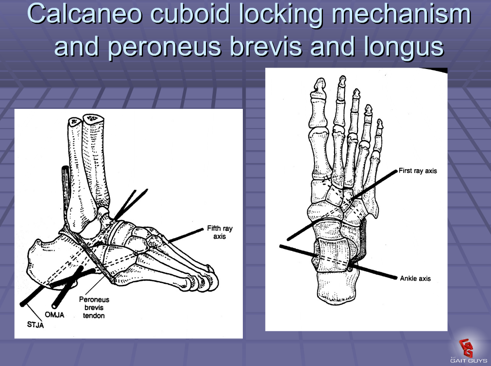
The rectus femoris proximal attachments are at the anterior inferior iliac spine (this is called the straight or anterior head) and the superior lip of the acetabulum (called the reflected or posterior head) Please see the top of the 2nd picture above, you can see the 2 heads. The distal attachment, after blending with the vasti, is into the patellar tendon and ultimately the tibial tuberosity.

The rectus is an accessory hip flexor and knee extensor, though it not normally a prime mover for either of these motions. It’s amount of action depends on the position of both the knee and hip.  When the knee is flexed, the rectus has less mechanical advantage, because it is placed in a lengthened position; same goes if the hip is extended.  It will be shortened if the hip is flexed and if the knee is extended at the same time, will have a mechanical disadvantage.

Now think about the direction of travel of each of the heads.

The “straight” head actually runs more obliquely from lateral to medial from its proximal attachment (AIIS) to the distal attachment (blending with vasti and patellar tendon); the refelected head runs a similar course, but not as oblique. If you were to externally rotate the thigh (remember, some folks may have an externally rotated foot due to external tibial torsion), it would actually give these heads more mechanical advantage (when the knee is relatively extended, such as at heel strike/ initial contact and toe off/ preswing) as abductors (remember to think from the ground up, closed chain, so the distal attachments are acting more like the origin); thus, the abductor moment we have talked about.


There you have it @Greglehman. Thanks for the great question.


The Gait Guys. Uber Gait Aficionado’s Extraordinaire. Come and learn with us. Watch us on Youtube; follow us on Facebook and Twitter, see many of our downloads on our payloadz site by clicking here.


All material copyright 2013 the Gait guys/ The Homunculus Group. All rights reserved; don’t make us call Lee.

tumblr_lnbr4hUO7T1qhko2so1_75sq.gif
tumblr_lnbr4hUO7T1qhko2so2_1280.gif

Gait Cycle Basics: Part 2

Gait Cycle components

Let’s begin today with a typical walking gait cycle.

There are 2 phases: stance and swing. It comprises approximately 62% of the gait cycle. Inman and Scranton use the following classification:


Heel strike: when the heel hits the ground

Full forefoot load: weight is transferred anteriorly to the forefoot

Heel lift: when the heel begins lifting off the ground

Toe off: the beginning of propulsion


They further divide this into a contact period (heel strike to full forefoot load), a midstance period (from full forefoot load to heel lift) and a propulsive period (from heel lift through toe off)


Jaqueline Perry, the Matriarch of gait, uses a slightly more descriptive classification which we prefer:


Initial contact: when the foot 1st touches the floor

Loading response: weight bearing on the loaded extremity from initial contact and continues until the opposite foot is lifted for swing

Midstance: the 1st ½ of single limb support, beginning when the opposite foot is lifted until weight is over the forefoot

Terminal stance: begins with heel rise and continues until the opposite foot strikes the ground

Pre swing: when initial contact of the opposite extremity begins and toes off ends

She also describes 3 tasks to be performed during a gait cycle: weight acceptance (the limb is able to bear weight), single limb support (when weight is supported by one limb with the other in swing phase), and swing limb advancement (moving the opposite limb through space to become the next stance phase leg.

The question is, how do we adequately progress over the stance phase leg? Stay tuned to our next post for a primer on the 3 rockers….

The Gait Guys…Promoting gait literacy and understanding for the clinician and the consumer.