

The Calcaneo Cuboid Locking Mechanism
/Do you know what this is? You should if you walk or run!
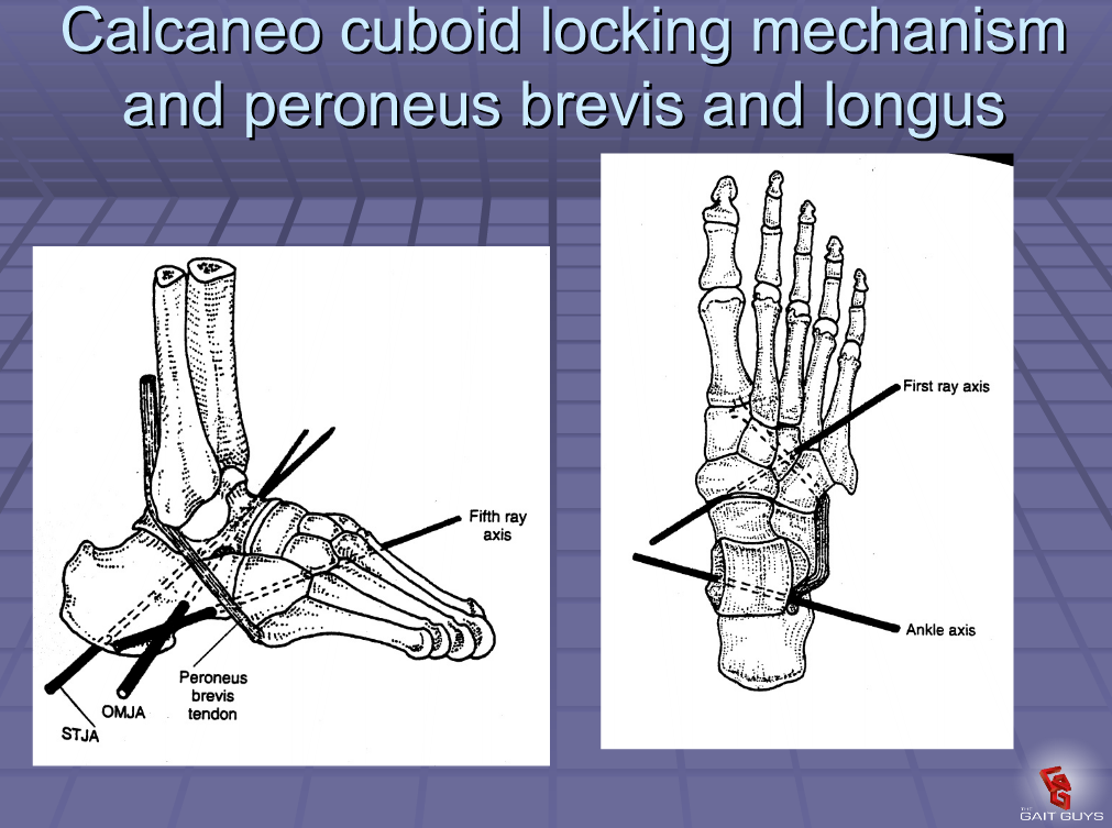
It is the mechanism by which the tendon of the peroneus longus travels behind the lateral malleolus of the ankle, travels underfoot, around the cuboid to insert into the lateral aspect of the base of the 1st metatarsal and adjacent 1st cunieform.
When the peroneus longus contracts, in addition to plantar flexing the 1st ray, it everts the cuboid and locks the lateral column of the foot, minimizing muscular strain required to maintain the foot in supination (the locked position for propulsion). Normally, muscle strength alone is insufficient to perform this job and it requires some help from the adjacent articulations.
In addition, the soleus maintains spuination during propulsion by plantar flexing and inverting rear foot via the subtalar joint. This is assisted by the peroneus brevis and tertius which also dorsflex and evert the lateral column, helping keep it locked. Can you see why the peroneii are so important?
Signs of a faulty calcaneo cuboid locking mechanism:
-weak peroneus longus, brevis and or tertius
-excessive rear or midfoot pronation
-low arch during ambulation-poor or low gear “push off”
-subluxated cuboid
The calcaneo cuboid locking mechanism. Essential for appropriate supination and ambulation. Insufficiency, coming to a foot you will soon examine.
Would you like to know more? Join us for our “third Wednesdays“ online webinar: Biomechanics 313. Wednesday, June 18 at 6 MST. Onlinece.com


