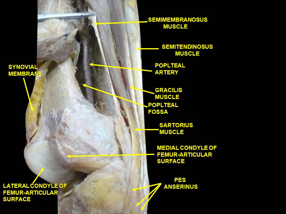
Knee pain and the the semitendinosis?

image source: https://commons.wikimedia.org/wiki/File:Slide2DADE.JPG

image source: https://commons.wikimedia.org/wiki/File:Slide2DADE.JPG

The semitendinosus hails from the posterior compartment.

During an ideal gait cycle, the semitendinosus from mid swing through nearly loading response, with a brief firing at toe off.

We remember that the abdominals should initiate thigh flexion with the iliopsoas, rectus femoris, tensor fascia lata and sartorius perpetuating the motion. Sometimes, when the abdominals are insufficient, we will substitute other thigh flexors, often the psoas and/or rectus femoris, but sometimes sartorius, especially in people with excessive midfoot pronation. Think about all of the medial rotation occurring at the knee during excessive midfoot pronation and when overpronation occurs, the extra compensatory external rotation that must occur to try and bring the knee back into the sagittal plane. The sartorius is positioned perfectly for this function, along with the semitendinosus which assists and external rotation and closed chain.

The semitendinosis is the most superficial of the hamstrings and originates between the biceps femoris, with which it shares a common tendinous attchment, which is anterior and slightly lateral and the semimembranosis which is just beneath it and slightly medial. It is fusiform and the muscle body ends about mid thigh, before becoming a long "piano string" and ultimately inserting most inferiorly of the trio, below the gracilis, on the pes anserine.

Call it pes anserinus bursitis or pes anserine tendinitis but they both add up to medial knee pain when the thigh needs help flexing. Look to this troublesome trio the next time you have recalcitrant medial knee pain.


Dr Ivo Waerlop, one of The Gait Guys

#gait, #gaitanalysis, #gaitdysfunction, #thegaitguys, #pesanserine, #semitendinosis


Imani F, Rahimzadeh P, Abolhasan Gharehdag F, Faiz SH. Sonoanatomic variation of pes anserine bursa. Korean J Pain. 2013;26(3):249-54. 

Gupta, Aman & Saraf, Abhinesh & Yadav, Chandrajeet. (2013). ISSN 2347-954X (Print) High-Resolution Ultrasonography in PesAnserinus Bursitis: Case Report and Literature Review. 1. 753-757. 

Gray H:  Anatomy of the Human Body  Lea and Febiger, Phildelphia and New York 1918

https://www.anatomy-physiotherapy.com/knee/articles/systems/musculoskeletal/lower-extremity/knee/test-your-knowledge-the-pes-anserinus

 Michaud T: in Foot Orthoses and Other Forms of Conservative Foot Care Williams & Wilkins, 1993 Pp. 50-55

 Michaud T: in Human Locomotion: The Conservative Management of Gait-Related Disorders 2011

The QP....What's the deal?

tumblr_mau352bxll1qhko2so1_540.png

Possibly heard of, rarely implicated and not often treated, this is one muscle you should consider taking a look at.

The quadratus plantae is generally considered to arise from two heads of differing and variable  fiber type composition, with the lateral head having slightly more Type 1 endurance fibers (1) The two heads are separated from each other by the long plantar ligament, though it can arise from from one (somewhat more common)  to 3 heads (very rare).  The attachments can be variable, The medial head is larger and more muscular, attached to the medial calcaneus, lateral aspect of the long plantar ligament and often from the plantar calcaneocuboid ligament (2);  the lateral head is smaller and more tendinous, attaching to the lateral border of the inferior surface of the calcaneus and the long plantar ligament.  The two portions join and end in a flattened band which inserts into the lateral, upper and under surfaces of the muscles, tendons or aponeurosis of predominantly the flexor digitorum longus and usually of the second and third, and sometimes fourth toes (2,3). 

Its action can be equally as variable. In addition to augmenting the pull of the long flexor tendons along the long axis of the foot and so that the 3rd and 4th toes do not curl under the foot, the tendinous slips of the FHL may distribute the load of the great toe to the second toe to the third or fourth toe in the forefoot, especially during toe-off (3).

look at the 4th and 5th digits trying to "crawl under the foot"

look at the 4th and 5th digits trying to "crawl under the foot"

The main attachment of the QP to the tendinous slips of the FHL may provide more efficient control of the long flexor tendons in comparison with that of the QP to the tendon of the FDL (3). EMG studies suggest it resists extension of the toes during the stance phase of locomotion, which serves to increase the stability of the foot. Additional EMG studies suggest it actually acts as a primary toe flexor in voluntary movements, being preferentially recruited over flexor digitorum longus and from comparative anatomical considerations it also seems likely that quadratus plantae may be an intrinsic evertor of the foot (4).

This muscle is a major player in gait and rehabilitation of this muscle should not be overlooked. I could only find one study looking at exercise activation of the QP (5) . It was examined along with the abductor hallucis, flexor digitorum brevis, abductor digiti minimi, flexor digiti minimi, adductor hallucis oblique, flexor hallucis brevis, interossei and lumbricals during rehabilitative the short-foot exercise, toes spread out, first-toe extension, second- to fifth-toes extension.

So, what else can you do?

  • you could ignore the muscle and hope it gets better. (in all likelihood it will worsen)
  • you could give them long flexor, toe scrunching towel-curling, marble-grasping exercises, like you see all over the internet…and give the flexor digitorum longus even more of a mechanical advantage, and make the problem worse
  • you could give them exercises to increase the function of the long extensors, which would increase the mechanical advantage of the quadratus plantae. like the shuffle walk; lift, spread and reach and tripod standing exercises
  • look north of the foot to see what might be causing the problem (loss of ankle rocker, insufficient gluteal activity, loss of internal rotation of the hip, etc) 

Check out the QP on your next foot pain patient, or whenever you see the toes trying to crawl under the foot. You may be surprised at your results. 


1. Schroeder KL, Rosser BW, Kim SY. Fiber type composition of the human quadratus plantae muscle: a comparison of the lateral and medial heads. J Foot Ankle Res. 2014 Dec 13;7(1):54. doi: 10.1186/s13047-014-0054-5. eCollection 2014.

2. Pretterklieber B1. Morphological characteristics and variations of the human quadratus plantae muscle. Ann Anat. 2017 Nov 21;216:9-22. doi: 10.1016/j.aanat.2017.10.006. [Epub ahead of print]

3. Hur MS, Kim JH, Woo JS, Choi BY, Kim HJ, Lee KS. An anatomic study of the quadratus plantae in relation to tendinous slips of the flexor hallucis longus for gait analysis. Clin Anat. 2011 Sep;24(6):768-73. doi: 10.1002/ca.21170.

4. Sooriakumaran P, Sivananthan S. Why does man have a quadratus plantae? A review of its comparative anatomy. Croat Med J. 2005 Feb;46(1):30-5.

5. Gooding TM, Feger MA, Hart JM, Hertel J. ntrinsic Foot Muscle Activation During Specific Exercises: A T2 Time Magnetic Resonance Imaging Study. J Athl Train. 2016 Aug;51(8):644-650. Epub 2016 Oct 3.

tumblr_nm5tkl3rsl1qhko2so1_1280.gif
tumblr_nm5tkl3rsl1qhko2so2_1280.gif

How well do you understand stance phase mechanics?

Here is a recent question we fielded and thought it would make a great post. 

Question/Comment: I’m slightly confused about closed chain hip motion in the stance leg.


Maybe if I explain what my thought process is you can correct me.  Lets use
left stance phase with the right leg swinging through.

After right mid-swing, the pelvis will be rotating towards the left.  The
motion of the pelvis on the left femur would be relative femur internal
rotation.  I understand that the right leg is externally rotating
(supinating) and that normal open chain kinematics of hip extension is
coupled with external rotation.  But if the pelvis is moving towards the
left AND the left femur externally rotates, wouldn’t that create too much
rotation?  So what I’m saying is that a pelvis that is oriented to the left
with a left femur that externally rotates creates an odd motion in my head
(which may be where the problem lies).  If you’ve ever seen a western where
the gun slingers do that weird walk to a shoot out…that’s what an
externally rotating femur during terminal stance looks like to me.

I’ve discussed this with other clinicians.  Some are in agreement with me,
some think it’s externally rotating, and some don’t know what I’m talking
about.  In my patients I also see a loss of hip IR more than hip ER.  These
patients that lose hip IR seem to have more difficulty in terminal
stance/toe-off phase more than the ones that lose hip ER.

If you could help me understand these kinematics and clear this up for me I
would greatly appreciate it.

Thank you, A

our reply: 

Taking your example with the L leg in stance:
When the L heel contacts the ground, the friction of the ground (hopefully) slow the calcaneus and the talus slide anteriorly on the calcaneus. 

Because of the shape of the calcaneal facets, the talus plantar flexes, adducts and everts. This sets the stage for pronation to occur: the calcaneus everts and the lower leg internally rotates, with the thigh following. The right side of the pelvis is moving to the L (counter clockwise rotation). This should occur (ideally) until midstance. At midstance, the opposite ® foot begins to enter swing phase; this should initiate supination of the stance phase leg (L). At this point, the L foot should be beginning to supinate the the leg and thigh beginning external rotation. It (thigh and leg) should reach maximal external rotation at toe off (maximal counter clockwise rotation of the pelvis) and remain in external rotation until heel strike/initial contact on the L side again. At this point, the pelvis begins clockwise rotation.

It is necessary for the thigh and leg to externally rotate while the pelvis is rotating counter clockwise, because of the constraints of the iliofemoral, pubeofemoral and ishiofemoal ligaments.

We too often see a loss of internal rotation of the hip in symptomatic populations more often than external rotation.

We hope this clarifies things for you.

Thank you again for the question and taking the time to write.

The Gait Guys

Gait Cycle Basics: Part 5
Swing Phase
Our final chapter in this series….
Swing phase is less variable in its classification. It begins at toe off and ends at heel strike. It comprises 38% of the gait cycle.There must be adequate dorsiflexion …

Gait Cycle Basics: Part 5

Swing Phase

Our final chapter in this series….

Swing phase is less variable in its classification. It begins at toe off and ends at heel strike. It comprises 38% of the gait cycle.There must be adequate dorsiflexion of the ankle, and flexion of the knee and hip to allow forward progression.


The following classification is most commonly used:

Early swing: occurring immediately after toe off. There is contraction of the dorsiflexors of the ankle, and flexors of the knee and hip


Midswing: halfway through the swing cycle, when the swing phase leg is passing the midstance phase extremity. Acceleration of the extremity has occurred up to this point.


Late swing: deceleration of the extremity in preparation for heel strike. There is contraction of the extenders of the thigh and knee, as well as dorsiflexors of the ankle.


Perry defines the phases as:

Initial swing: the 1st third of swing phase, when the foot leaves the round until it is opposite the stance foot.

Mid swing: the time from when the swing foot is opposite the stance foot until the swinging limb is anterior to the stance phase tibia

Terminal swing: from the end of midswing, until heel strike

And there you have it. A nice review of the gait cycle. Probably more than you wanted to know, but we want to give you the facts.

Telling it like it is. We are…The Gait guys