Botox for plantar fasciitis? Sounds like a bad idea to us....

image source: https://commons.wikimedia.org/wiki/File:Plantar_aponeurosis_-_axial_view.png

image source: https://commons.wikimedia.org/wiki/File:Plantar_aponeurosis_-_axial_view.png

Botox..For plantar fasciitis? Really?

We found this article (1) in one of our favorite journals, Lower Extremity Review , and were a little surprised. Let us get this straight: you are going to take one of the the most poisonous biological neurotoxins known (1) and inject it into your calf and foot?

The article in LER is well written and the results (thankfully) were inconclusive regarding its usage. They do cite 3 studies (with two by the same lead author) where it has been effective (2-4). Yes, it is better than saline (5) (but not as good as extracorporeal shock wave therapy (6)), and better than placebo (7-10) but considerably more risky.

So the premise is “if the muscle is dysfunctional, then let’s just take it out of the equation”. But this really doesn’t fix the problem, it just covers up the symptom. And what about the other potential side effects since botulinum toxin acts not only at the neuromuscular junction, blocking the release of acetylcholine, but also at the autonomic ganglia, postganglionic parasympathetic nerve endings, as well as the post ganglionic sympathetics that use acetylcholine (capillaries of skin, piloerector muscles and sweat glands) (11)?.

In our experience, most cases of plantar fasciitis are secondary to lack of forefoot rocker, lack of ankle rocker, lack of hip extension or in some cases, direct trauma. Wouldn’t it make more sense to strengthen the anterior compartment to reciprocally inhibit the posterior compartment, increasing ankle dorsiflexion and hip extension? We find, oftentimes, treating only the area of chief complaint and not what is "driving the bus" can offer temporary, symptomatic relief but not long standing pathmechanics or pathoanatomy.

Just like the road to enlightenment, there are no shortcuts in treating plantar fasciitis and if you are not going to treat the cause, then be prepared to reap what you sow.

Dr Ivo Waerlop, one of The Gait Guys

#botox #plantarfascitis #badideas #gaitproblem #thegaitguys

1. https://lermagazine.com/article/botox-injection-not-just-for-celebrities-furrows-and-wrinkles

2. Elizondo-Rodriguez J, Araujo-Lopez Y, Moreno-Gonzalez JA, Cardenas-Estrada E,
Mendoza-Lemus O, Acosta-Olivo C. A comparison of botulinum toxin A and intralesional steroids for the treatment of plantar fasciitis: A randomized, double-blinded study. Foot Ankle Int.
2013;34(1):8-14.

3. Díaz-Llopis IV, Rodríquez-Ruíz CM, Mulet-Perry S, Mondéjar-Gómez FJ., Climent-Barberá JM., Cholbi-Llobel F. Randomized controlled study of the efficacy of the injection of botulinum toxin type A versus corticosteroids in chronic plantar fasciitis: results at one and six months. Clin Rehabil. 2012;26(7):594-606.

4. Díaz-Llopis IV, Gómez-Gallego D, Mondéjar-Gómez FJ, López-García A, Climent-Barberá JM, Rodríguez-Ruiz CM. (2013). Botulinum toxin type A in chronic plantar fasciitis: clinical effects one year after injection. Clin Rehabil. 2013;27(8):681-685.

5. Ahmad J, Ahmad SH, Jones K. Treatment of Plantar Fasciitis With Botulinum Toxin. Foot Ankle Int. 2017 Jan;38(1):1-7. doi: 10.1177/1071100716666364. Epub 2016 Oct 1.1.

6. Roca B, Mendoza MA, Roca M. Comparison of extracorporeal shock wave therapy with botulinum toxin type A in the treatment of plantar fasciitis. Disabil Rehabil. 2016 Oct;38(21):2114-21. doi: 10.3109/09638288.2015.1114036. Epub 2016 Mar 1

7. Babcock MS, Foster L, Pasquina P, Jabbari B. Treatment of pain attributed by plantar fasciitis with botulinum toxin A: a short-term randomized, placebo-controlled, double blinded study. Am J Phys Med Rehabil. 2005;84(9):649-654.

8. Samant PD, Kale SY, Ahmed S, Asif A, Fefar M, Singh SD. Randomized controlled study comparing clinical outcomes after injection botulinum toxin type A versus corticosteroids in chronic plantar fasciitis. Int J Res Orthop. 2018;4(4):672-675.

9. Huang YC, Wei SH, Wang HK, Lieu FK. Ultrasonographic guided botulinum toxin type A treatment for plantar fasciitis: an outcome-based investigation for treating pain and gait changes. J Rehabil Med. 2010;42(2):136-140.

10. Ahmad J, Ahmad SH, Jones K. Treatment of plantar fasciitis with botulinum toxin. Foot Ankle Int. 2017;38(1):1-7.

11. Nigam PK, Nigam A. Botulinum toxin. Indian J Dermatol. 2010;55(1):8–14. doi:10.4103/0019-5154.60343

Got hip extension?

Because she sure could use some...

we have see this gal before… yesterday in fact

  • left plantar plate lesion (yes, conformed on ultrasound)

  • left sided anatomical leg length discrepany

  • bilateral internal tibial torsion

  • incompetent L quadratus lumborum

  • adequate hip extension and ankle dorsiflexion available to her

  • lack of endurance in her abs

yep, lots more, but that is enough for now


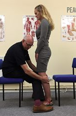
note that she has plenty of ankle dorsiflexion, more on the right. this is due to her right leg being anatomically longer and has to travel through a greater range of motion

look at the knee and the hip articulations to assess hip extension. It should match ankle dorsiflexion, no?


Dr Ivo Waerlop, one of The Gait Guys


#gait #gaitguys #thegaitguys #hipextension #LLD #quadratuslumborum #internaltibialtorsion #anklerocker #ankledorsiflexion

1st MTP Pain? The Biomechanics of the Big Toe...

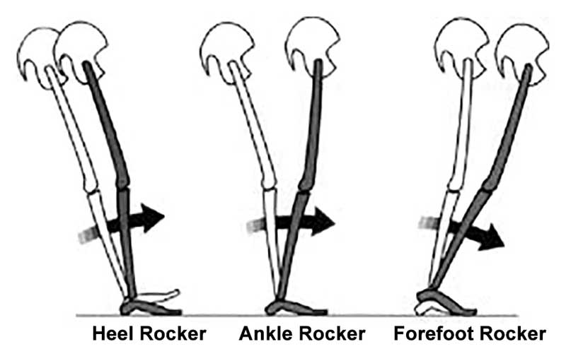
Remember the rockers? We have done a series on this in the past. Remember there are three: heel, ankle and forefoot. We are going to concentrate on the forefoot today.

As a reminder, forefoot rocker occurs at the 1st metatarsal phalangeal joint (big toe knuckle) as the tibia progresses over the forefoot during forward movement. You NEED 50 degrees to do this competently; you SHOULD have 65 degrees. When you don’t, you have a condition called hallux limitus. This could be from a number of reasons, from overpronation in the mid foot, to a bunion, to faulty firing patterns of the muscles which help to descend the 1st ray (the extensor hallucinations brevis, the peroneus longs and the short flexors off the toes). Pretty much, ANYTHING that causes a dorsal and posterior shift of the 1st MTP axis will cause limited forefoot rocker.

So, the question is, “Do you know where 1st 1st MTP pain may be coming from? How familiar are you with the mechanics of that joint?”

Take a few minutes to review it in this video with Dr Ivo Waerlop of The Gait Guys.

#gait, #gaitanalysis, #1stmtp, #forefootrocker, #thegaitguys,

Rockered shoes...they're for MORE than hallux limitus...

In other words, footwear with more "drop" in the front lessens the need for forefoot rocker (otherwise known as 1st metatarsophalangeal joint extension, or "the ability to bend your big toe backward)

“Most people have to wear MBTs a little at a time until they gain strength and stamina, so we recommend wearing them an hour a day for the first few days and to increase gradually until they feel strong enough to wear them for a full day,”

...sounds an awful lot like our mantra "skill, endurance, strength"...

A great read here. Keep this one around for reference...

https://lermagazine.com/article/rocker-bottom-footwear-effects-on-balance-gait

Dr Ivo, one of The Gait Guys

#rockeredshoes, #gait, #thegaitguys, #gaitanalysis, #gaitabnormaility, #rockerbottom

Images illustrate different types of rockers, with fulcrum locations indicated by vertical red lines. (Images courtesy of Arnie Davis, CPed.) via LER magazine: https://lermagazine.com/article/rocker-bottom-footwear-effects-on-balance-gait

Images illustrate different types of rockers, with fulcrum locations indicated by vertical red lines. (Images courtesy of Arnie Davis, CPed.) via LER magazine: https://lermagazine.com/article/rocker-bottom-footwear-effects-on-balance-gait

Try THIS at home...

Screen Shot 2018-02-07 at 11.23.24 AM.png

Cool test, results you can see and some literature to back it up. If you are interested at all in proprioception, this is an interesting read.

So, the question for us is: "Does ankle dorsiflexion actually create more stability, like is purported?"

“The point is that if I make their ankle rigid this way, then they can more effectively use the balance mechanisms at their knees, hips, and proximally, because they’re on a stable base. My proposition is that their balance is actually normal, apart from that distal segment. When their ankle is stabilized, they use their knees more effectively, and they become less dependent on their eyesight to maintain their balance.”

http://lermagazine.com/article/afos-and-balance-issues-in-peripheral-neuropathy

Your Gait Changes when you text....

GettyImages-534572749web-57a3087d3df78c3276b9fc80-2.jpg

Does texting alter your gait? It sure seems to slow you down, and according to this study, alter firing patterns of muscles about your ankle. Perhaps you are trying to preserve ankle rocker and maintain stability? It is interesting that ankle dorsiflexion actually increased and plantar flexion decreased.

"Young adults showed, overall, small gait modifications that could be mainly ascribable to gait speed reduction and a modified body posture due to phone handling. We found no significant alterations of ankle and knee kinematics and a slightly delayed activation onset of the left gastrocnemius lateralis. However, we found an increased co-contraction of tibialis anterior and gastrocnemius lateralis, especially during mid-stance. Conversely, we found a reduced co-contraction during terminal stance."


link to FREE FULL TEXT: https://www.ncbi.nlm.nih.gov/pmc/articles/PMC4579642/

More Foot Rocker Pathology Clues.

Is ankle rocker normal and adequate or is it limited ?  Is it limited in early midstance or late midstance ? How about at Toe off?  Is it even possible to distinguish this ? Well, we are splitting hairs now but we do think that it is possible. It is important to understand the pathologies on either end of the foot that can impact premature ankle rocker. 

Look at the photo above. You can see the clinical hint in the toe wear that this runner may have a premature heel rise. However, this is not solid evidence that every time you see this you must assume pathologic ankle rocker. The question is obviously, what is the cause.

Considerations:

1- weak anterior compartment, which is quite often paired with the evil neuroprotective tight calf-achilles posterior complex to offer the necessary sagittal protection at the ankle mortise.  This will cause premature heel rise from a posterior foot aspect.

2- rigid acquired blocked ankle rocker from something like “Footballer’s ankle”. This will also cause premature heel rise from a relatively posterior foot aspect.

3- there are multiple reasons for late midstance ankle rocker pathology. The client could completely avoid the normal pronation/supination phase of gait because of pain anywhere in the foot. For example, they could have plantar fascial pain, sesamoiditis, a weak first ray complex from hallux vaglus, they could have a painful bunion, they could be avoiding the collapse of a forefoot varus. There are many reasons but any of them can impair the timely pronation-supination phase in attempting to gain a rigid lever foot to toe off the big toe-medial column in “high gear” fashion. And when this happens the preparatory late midstance phase of gait can be delayed or rushed causing them to move into premature heel rise for any one of several reasons.  Rolling off to the outside and off of the lesser toes creates premature heel rise.  

4- And now for one anterior aspect cause of premature heel rise. This is obviously past the midstance phase but it can also cause premature heel rise. Turf toe, Hallux rigidus/limitus or even the dreaded fake out, the often mysterious Functional Hallux limitus (FnHL) can cause the heel to come up just a little early if the client cannot get to the full big toe dorsiflexion range.  

We could go on and on and include other issues such as altered Hip Extension Patterning, loss of hip extension range of motion, weak glutes, or even loss of terminal knee extension (from things like an incompleted ACL rehab, Osteoarthritis etc) but these are things for another time. Lets stay in the foot today.

All of these causes, with their premature heel rise component, will rush the foot to the forefoot and likely create Metatarsal head plantar loading and could cause forces appropriate enough to create stress responses to the bone. This abrupt forefoot loading thrust will often cause a reactive hammer toe effect.  Quite often just looking at the resting nature of a clients toes while they are lying down will show the underlying increase in neuro-protective hammering pattern (increased long toe flexor and short toe extensor activity paired with shortness of the opposing pairs which we review here in this short video link).  The astute observer will also note the EVA foam compressing of the shoe’s foot bed, and will also note the distal displacement of the MET head fat pad rendering the MET head pressures even greater osseously. 

Premature ankle rocker and heel rise can occur for many reasons. It can occur from problems with the shoe, posterior foot, anterior foot, toe off, ankle mortise, knee, hip or even arm swing pathomechanics.  

When premature heel rise and impaired ankle rocker rushes us to the front of the foot we drive the front half of the shoe into the ground as the foot plantarflexion is imparted into the shoe.  The timing of the normal biomechanical events is off and the pressures are altered.  instead of rolling over the forefoot and front half of the shoe after our body has moved past the foot these forces are occurring more so as our body mass is still over the foot. And the shoe can show us clues as to the torture it has sustained, just like in this photo case.

You must know the normal biomechanical gait events if you are going to put together the clues of each runner’s clinical mystery.  If you do not know normal how will you know abnormal when you see it ? If all you know is what you know, how will you know when you see something you don’t know ?

Shawn and Ivo, The Gait Guys … .  stomping out the world’s pathologic gait mechanics one person at a time. 

Ankle Rocker Revisited....

How many times have we talked about ankle rocker and its importance? So how are YOU measuring ankle rocker? Are you looking at it on the table? On the ground? Weight bearing? Knee flexed or extended (or both?). The knee is extended at initial contact, flexes through midstance, extends at terminal stance and pre swing and flexes again during swing phase until extending at the end of terminal swing for initial contact again.

What you see on the table may not (and many times doesn't)  translate to real life. Someone with limited ankle dorsiflexion non weight bearing may have normal amounts during gait and vice versa. With gravity in place and a functioning (or malfunctioning) vestibular system, things can change rapidly. Remember that the vestibular system drives the extensors and if inhibited, you will often have flexor dominance. Talk about a tight gastroc/soleus group!


"These findings indicate that nonweightbearing and weightbearing measurements of ankle DF PROM with knee extension should not be used interchangeably and that weightbearing ankle DF PROM with the knee extended is more appropriate for estimating ankle DF during gait."

Kang MH, Oh JS. Relationship Between Weightbearing Ankle Dorsiflexion Passive Range of Motion and Ankle Kinematics During Gait. J Am Podiatr Med Assoc. 2017 Jan;107(1):39-45. doi: 10.7547/14-112.


"There is no relationship between a static diagnosis of ankle dorsiflexion at 0° with dorsiflexion during gait. On the other hand, those subjects with less than -5° of dorsiflexion during static examination did exhibit reduced ankle range of motion during gait."


Gatt A, De Giorgio S, Chockalingam N, Formosa C. A pilot investigation into the relationship between static diagnosis of ankle equinus and dynamic ankle and foot dorsiflexion during stance phase of gait: Time to revisit theory? Foot (Edinb). 2017 Mar;30:47-52. doi: 10.1016/j.foot.2017.01.002. Epub 2017 Feb 6.


More on Rockered footwear and Hallux Limitus

Rockered footwear is for more than Hallux Limitus..

To go along with yesterdays post on Hallux Limitus... In case you missed it, click here

In other words, footwear with more "drop" in the front lessens the need for forefoot rocker (otherwise known as 1st metatarsophalangeal joint extension, or "the ability to bend your big toe backward)

“Most people have to wear MBTs a little at a time until they gain strength and stamina, so we recommend wearing them an hour a day for the first few days and to increase gradually until they feel strong enough to wear them for a full day,”

...sounds an awful lot like our mantra "skill, endurance, strength"...

A great read here. Keep this one around for reference...

http://lermagazine.com/article/rocker-bottom-footwear-effects-on-balance-gait

#rockeredshoe #rocker #footwear

You better keep that Hallux Dorsiflexion

Geee....Looks like forefoot rocker really IS IMPORTANT, eh?

...and what have we been saying about being able to dorsiflex your big toe? Watch how well you or your client can descend the 1st ray (remember that if the head of the 1st does not go down and anchor, its axis of rotation moves dorsally and posterior, limiting dorsiflexion of the joint)

This article should make you look at the "toe break" in a shoe...

"They found that increasing bending stiffness assisted with propulsion during running, reducing the metabolic cost of running by about 1%. However, at a certain level, the increased elasticity began to interfere with the natural flexion of the first MTP joint, reducing the contribution of ankle joint torque to push-off and counteracting the metabolic benefits. Within the study population, the threshold of bending stiffness for optimal energetics varied significantly from one runner to the next, suggesting running shoe design may need to be tuned to an individual runner’s needs." 

http://lermagazine.com/news/in-the-moment-sports-medicine/stiffer-shoes-improve-running-energetics-as-long-as-first-mtp-flexion-is-preserved

The buck DOES NOT stop here...

The buck doesn't stop here..

image from: http://www.aofas.org/footcaremd/treatments/pages/triple-arthrodesis.aspx

image from: http://www.aofas.org/footcaremd/treatments/pages/triple-arthrodesis.aspx

One of the most pervasive problems following a ankle arthrodesis, particularly a triple arthrodesis which involves fusing the subtalar (talocalcaneal), calcaneocuboid , and talonavicular joints results in a loss of ankle rocker. The "buck" needs to be passed somewhere and this usually will mean proximally in the lower kinetic chain.

Seeing adjacent joints with osteoarthritic changes following hypomobility have another joint is nothing new; you probably see it all the time in practice. Remember that it is not always have to be a "fusion". Simple longstanding pathomechanics or longstanding hypomobility will often cause the same problems.

A nice, full text referenced review 1 of her favorite journals. Some nice side discussions as well. Enjoy : )

"Altered biomechanics after ankle arthro­desis often increase stress on the adjacent joints in the foot, which can cause or exacerbate osteoarthritic degeneration in those joints. Clinicians and researchers are working to better understand this process and how to minimize patients’ risk."

http://lermagazine.com/…/adjacent-joint-arthritis-after-ank…
#anklemobility #anklerocker #triplearthrodesis #hypomobility


Achilles Tendonitis/Tendinopathy and NeedlingAchilles pain. You can’t live with it and you can’t live with it. Can needling help? The obvious answer is yes, but there is more as well.There appears to be sufficient data to support the use of needling…

Achilles Tendonitis/Tendinopathy and Needling


Achilles pain. You can’t live with it and you can’t live with it. Can needling help? The obvious answer is yes, but there is more as well.

There appears to be sufficient data to support the use of needling for achilles tendon problems . Perhaps it is the “reorganization” of collagen that makes it effective or a blood flow/vascularization phenomenon. The mechanism probably has something to do with pain and the reticular formation sending information down the cord via the lateral cell column (intermediolateral cell nucleus) or pain (nociceptive) afferents sending a collateral in the spinal cord to the dysfunctional muscle, affecting the alpha receptors and causing vasodilation. 

Loss of ankle dorsiflexion is a common factor that seems to contribute to achilles tendinopathies . It would seem that improving ankle rocker would be most helpful. In at least one study, needling restored ankle function and in another it improved strength. 

And don’t forget to go north of the lower leg/foot/ankle complex. The gluteus medius can many times the culprit as well. During running, the gluteus medius usually fires before heel strike, most likely to stabilize the hip and the pelvis. In runners with Achilles Tendonitis, its firing is delayed which may affect the kinematics of knee and ankle resulting in rear foot inversion. Perhaps the delayed action of the gluteus medius allows an adductory moment of the pelvis, moving the center of gravity medially. This could conceivably place additional stress on the achilles tendon (via the lateral gastroc) to create more eversion of the foot from midstance onward.

Similarly, in runners with achilles tendoinopathy, the gluteus maximus does not fire as long and activation is delayed. The glute max should be the primary hip extensor and decreased hip extension might be compensated by an increased ankle plantarflexion which could potentially increase the load on the Achilles tendon. 

So, in short, yes, needling will probably help, for these reasons and probably many more. Make sure to needle all the dysfunctional muscles up the chain, beginning at the foot and moving rostrally.

Effectiveness of Acupuncture Therapies to Manage Musculoskeletal Disorders of the Extremities: A Systematic Review. Cox J, Varatharajan S, Côté P, Optima Collaboration. J Orthop Sports Phys Ther. 2016 Jun;46(6):409-29. doi: 10.2519/jospt.2016.6270. Epub 2016 Apr 26

Acupuncture’s role in tendinopathy: new possibilities. Speed C. Acupunct Med. 2015 Feb;33(1):7-8. doi: 10.1136/acupmed-2014-010746. Epub 2015 Jan 9.

The effect of electroacupuncture on tendon repair in a rat Achilles tendon rupture model.  Inoue M, Nakajima M, Oi Y, Hojo T, Itoi M, Kitakoji H. Acupunct Med. 2015 Feb;33(1):58-64. doi: 10.1136/acupmed-2014-010611. Epub 2014 Oct 21.

KIishmishian B, Selfe J, Richards J A Historical Review of Acupuncture to the Achilles Tendon and the development of a standardized protocol for its use Journal of the Acupuncture Association of Chartered Physiotherpists Spring 2012,  69-78

Acupuncture for chronic Achilles tendnopathy: a randomized controlled study. Zhang BM1, Zhong LW, Xu SW, Jiang HR, Shen J. Chin J Integr Med. 2013 Dec;19(12):900-4. doi: 10.1007/s11655-012-1218-4. Epub 2012 Dec 21.

The effect of dry needling and treadmill running on inducing pathological changes in rat Achilles tendon. Kim BS, Joo YC, Choi BH, Kim KH, Kang JS, Park SR. Connect Tissue Res. 2015 Nov;56(6):452-60. doi: 10.3109/03008207.2015.1052876. Epub 2015 Jul 29.

Tendon needling for treatment of tendinopathy: A systematic review.
Krey D, Borchers J, McCamey K. Phys Sportsmed. 2015 Feb;43(1):80-6. doi: 10.1080/00913847.2015.1004296. Epub 2015 Jan 22. Review.

Acupuncture increases the diameter and reorganisation of collagen fibrils during rat tendonhealing.
de Almeida Mdos S, de Freitas KM, Oliveira LP, Vieira CP, Guerra Fda R, Dolder MA, Pimentel ER. Acupunct Med. 2015 Feb;33(1):51-7. doi: 10.1136/acupmed-2014-010548. Epub 2014 Aug 19.

Electroacupuncture increases the concentration and organization of collagen in a tendon healing model in rats.
de Almeida Mdos S, de Aro AA, Guerra Fda R, Vieira CP, de Campos Vidal B, Rosa Pimentel E. Connect Tissue Res. 2012;53(6):542-7. doi: 10.3109/03008207.2012.710671. Epub 2012 Aug 14.

Changes in blood circulation of the contralateral Achilles tendon during and after acupunctureand heating.Kubo K, Yajima H, Takayama M, Ikebukuro T, Mizoguchi H, Takakura N. Int J Sports Med. 2011 Oct;32(10):807-13. doi: 10.1055/s-0031-1277213. Epub 2011 May 26.

Microcirculatory effects of acupuncture and hyperthermia on Achilles tendon microcirculation. Kraemer R, Vogt PM, Knobloch K.
Eur J Appl Physiol. 2010 Jul;109(5):1007-8. doi: 10.1007/s00421-010-1442-6. Epub 2010 Mar 28.

Effects of acupuncture and heating on blood volume and oxygen saturation of human Achilles tendon in vivo. Kubo K, Yajima H, Takayama M, Ikebukuro T, Mizoguchi H, Takakura N. Eur J Appl Physiol. 2010 Jun;109(3):545-50. doi: 10.1007/s00421-010-1368-z. Epub 2010 Feb 6.

 Insertional achilles tendinopathy associated with altered transverse compressive and axial tensile strain during ankle dorsiflexion. Chimenti RL, Bucklin M, Kelly M, Ketz J, Flemister AS, Richards MS, Buckley MR.
J Orthop Res. 2016 Jun 16. doi: 10.1002/jor.23338. [Epub ahead of print]

Forefoot and rearfoot contributions to the lunge position in individuals with and without insertionalAchilles tendinopathy. Chimenti RL, Forenza A, Previte E, Tome J, Nawoczenski DA.Clin Biomech (Bristol, Avon). 2016 Jul;36:40-5. doi: 10.1016/j.clinbiomech.2016.05.007. Epub 2016 May 11.

Ankle Power and Endurance Outcomes Following Isolated Gastrocnemius Recession for AchillesTendinopathy. Nawoczenski DA, DiLiberto FE, Cantor MS, Tome JM, DiGiovanni BF. Foot Ankle Int. 2016 Mar 17. pii: 1071100716638128. [Epub ahead of print]

 In vivo quantification of the shear modulus of the human Achilles tendon during passive loading using shear wave dispersion analysis.
Helfenstein-Didier C, Andrade RJ, Brum J, Hug F, Tanter M, Nordez A, Gennisson JL. Phys Med Biol. 2016 Mar 21;61(6):2485-96. doi: 10.1088/0031-9155/61/6/2485. Epub 2016 Mar 7.

Changes of gait parameters and lower limb dynamics in recreational runners with achillestendinopathy. Kim S, Yu J. J Sports Sci Med. 2015 May 8;14(2):284-9. eCollection 2015 Jun.

Gastrocnemius recession for foot and ankle conditions in adults: Evidence-based recommendations. Cychosz CC, Phisitkul P, Belatti DA, Glazebrook MA, DiGiovanni CW. Foot Ankle Surg. 2015 Jun;21(2):77-85. doi: 10.1016/j.fas.2015.02.001. Epub 2015 Feb 26. Review.

Limited ankle dorsiflexion increases the risk for mid-portion Achilles tendinopathy in infantry recruits: a prospective cohort study. Rabin A, Kozol Z, Finestone AS. J Foot Ankle Res. 2014 Nov 18;7(1):48. doi: 10.1186/s13047-014-0048-3. eCollection 2014.

Perry J. Gait Analysis: Normal and Pathological Function. Thorofare, NJ: Slack 1992.

Chan YY, Mok KM, Yung PSh, Chan KM. Sports Med Arthrosc Rehabil Ther Technol. 2009 Jul 30;1:14. doi: 10.1186/1758-2555-1-14.

Bilateral effects of 6 weeks’ unilateral acupuncture and electroacupuncture on ankle dorsiflexors muscle strength: a pilot study. Zhou S, Huang LP, Liu J, Yu JH, Tian Q, Cao LJ. Arch Phys Med Rehabil. 2012 Jan;93(1):50-5. doi: 10.1016/j.apmr.2011.08.010. Epub 2011 Nov 8.

Franettovich Smith MM1, Honeywill C, Wyndow N, Crossley KM, Creaby MW. : Neuromotor control of gluteal muscles in runners with achilles tendinopathy.
Med Sci Sports Exerc. 2014 Mar;46(3):594-9.

tumblr_o8tlm8zPXq1qhko2so1_1280.jpg
tumblr_o8tlm8zPXq1qhko2so2_1280.jpg
tumblr_o8tlm8zPXq1qhko2so3_1280.jpg
tumblr_o8tlm8zPXq1qhko2so4_r1_1280.jpg

Wow!  Can you figure out why this person at the distal end of her first metatarsal under her medial sesamoid.

She recently underwent surgery for a broken fibula (distal with plate fixation) and microfracrure of the medial malleolus. You are looking at her full range of dorsiflexion which is improved from approximately 20° plantarflexion. She is now at just under 5°.

She has just begun weight-bearing and developed pain over the medial sesamoid.

The three rockers, depicted above from Thomas Michauds book, or necessary for normal gait.  This patient clearly has a loss of ankle rocker. Because of this loss her foot will cantilever forward and put pressure on the head of the first metatarsal.  This is resulting in excessive forefoot rocker.  Her other option would have been to pronate through the midfoot. Hers is relatively rigid so, as Dr. Allen likes to say, the “buck was passed to the next joint. ”

There needs to be harmony in the foot in that includes each rocker working independently and with in its normal range. Ankle rocker should be at least 10° with 15° been preferable and for footlocker at least 50° with 65 been preferable.

 If you need to know more about rockers, click here.

How are your hammy’s?Another tool for you, in addition to making sure the gluten are on line, to improve ankle rocker and hip extension.“This study concludes that neural mobilization techniques are a useful adjunct to static stretching, withou…

How are your hammy’s?

Another tool for you, in addition to making sure the gluten are on line, to improve ankle rocker and hip extension.

“This study concludes that neural mobilization techniques are a useful adjunct to static stretching, without any risk of adverse events or injuries. Athletes or trainers can consider using one or both types of neural mobilization techniques to enhance muscular flexibility. Dosage of the neural mobilization as well as the proposed working mechanism behind the increase in hamstring flexibility can be found in the full text of the article.”

http://www.ncbi.nlm.nih.gov/pubmed/26482098

Phys Ther Sport. 2016 Jan;17:30-7. doi: 10.1016/j.ptsp.2015.03.003. Epub 2015 Mar 17.
Short term effectiveness of neural sliders and neural tensioners as an adjunct to static stretching of hamstrings on knee extension angle in healthy individuals: A randomized controlled trial.
Sharma S, Balthillaya G2, Rao R, Mani R .

Sounds like a bad ideaOrthotics, can be useful adjunct to care. They can be used to give people biomechanics that they do not have while you were trying to improve them and help to make up for ranges of motion which do not seem attainable.From the g…

Sounds like a bad idea

Orthotics, can be useful adjunct to care. They can be used to give people biomechanics that they do not have while you were trying to improve them and help to make up for ranges of motion which do not seem attainable.

From the gate cycle we know that after initial contact and loading response the calcaneus should start to evert. The calcaneus will continue to evert until it encounters something (like the lateral heel counter of the shoe). At mid stance it should be fully everted and as the opposite leg comes in to swing, begin to invert. The lateral heel counter assists in the inversion/supination process.

To our knowledge, flip-flops, even if they have an increased arch, do not have a lateral heel counter and therefore will promote further lateral excursion of the calcaneus while the medial longitudinal arch is collapsing  (i.e.: midfoot pronation). Go ahead and place your foot into inversion and see what happens to your heel. It’s slides laterally.

It’s also well-established that flip-flops, through flexion of the distal toes and engagement of the long flexor tendons, inhibits ankle rocker. It is often necessary to engage these muscles to keep the flip-flop from coming off. Lack of ankle rocker usually will inhibit hip extension and that can cause a constellation of problems.

Though engagement of the long flexors of the toes will have a partial anti-pronatory effect, this is not enough to counter the excessive heel  eversion which is happening.

We generally do not think the flip-flops are a great idea and telling someone that it’s “OK” to wear flip-flops as long as it has appropriate arch support, is silly.

Can you guess why this person has left-sided plantar fasciitis?This question probably seem somewhat rhetorical. Take a good look at these pedographs which provide us some excellent clues.First of all,  note how much pressure there is over the metata…

Can you guess why this person has left-sided plantar fasciitis?

This question probably seem somewhat rhetorical. Take a good look at these pedographs which provide us some excellent clues.

First of all,  note how much pressure there is over the metatarsal heads. This is usually a clue that people are lacking ankle rocker and pressuring these heads as the leg cantilevers forward.  This person definitely have a difficult time getting the first metatarsal head down to the ground.

Notice the overall size of the left foot compared to the right (right one is splayed or longer). This is due to keeping the foot and somewhat of a supinated posture to prevent excessive tension on the plantar fascia.

The increase splay of the right foot indicates more mid foot pronation and if you look carefully there is slightly more printing at the medial longitudinal arch. This is contributing to the clawing of the second third and fourth toes on the right. Stand up, overpronate your right foot and notice how your center of gravity (and me) move medially.The toes will often clench in an attempt to create stability.

The patient’s pain is mostly at the medial and lateral calcaneal facets, and within the substance of the quadratus plantae with weakness of that muscle and the extensor digitorum longus. She has 5° ankle dorsiflexion left and 10 degrees on the right and hip extension which is similar.

The lack of ankle rocker and hip extension or causing her to pronate through her midfoot, Tensioning are plantar fascia at the insertion. The problem is worse on the left and therefore that is where the symptoms are.

Pedographs can be useful tool in the diagnostic process and provide clues as to biomechanical faults in the gait cycle.

tumblr_o80wf8tU9u1qhko2so1_400.jpg
tumblr_o80wf8tU9u1qhko2so2_1280.jpg

Do you know where your rocker is?

At 1st pass, some articles may seem like a sleeper, but there can be some great clinical pearls to be had. I recently ran across one of these. It was a presentation from the  42nd annual American Academy of Orthotists and Prosthetists meeting in Orlando, March 2016 entitled “ Shifting Position of Shoe Heel Rocker Affects Ankle Mechanics During Gait”. The title caught my eye.

They looked at ankle kinematics while keeping the toe portion of rocker constant at 63% of foot length, angled at 25 degrees and shifting the base of a rockered shoe from 1cm behind the medial malleolus, directly under it and 1cm anterior to it. Knee and hip kinematics did not differ significantly, however ankle range of motion did.

The more forward the ankle rocker, the less plantarflexion but more ankle dorsiflexion at midstance. So, the question begs, why do we care? Lets explore that further…

  • Think about the “average” heel rocker in a shoe. It largely has to do with the length of the heel and heel flare (base) of the shoe. The further back this is (ie; the more “flare”) the more plantar flexion at heel strike and less ankle dorsiflexion (and thus ankle rocker, as described HERE) you will see. Since loss of ankle dorsiflexion (ie: rocker) usually means a loss of hip extension (since these 2 things should be relatively equal during gait (see here), and that combination can be responsible for a whole host of problems that we talk about here on the blog all the time. Picking a shoe with a heel rocker based further forward (having less of a flare) would stand to promote more ankle dorsiflexion.
  • Having a shoe with a greater amount of “drop” from heel to toe (ie: ramp delta) is going to have the same effect. It will move the calcaneus forward with respect to the heel of the shoe and effectively move the rocker posteriorly.
  • Lastly, look a the shape of the outsole of the shoe. The toe drop is usually clear to see, but does it have a heel rocker (see the picture above)?

These are  a few examples of what to look for in a clients shoe when examining theirs or making a recommendation, depending on whether you are trying to improve or decrease ankle rocker. We can’t think of why you would want to decrease ankle rocker, but with conditions like rigid hallux limitus, where the person has limited or no dorsiflexion of the great toe, you may want to employ a rockered sole shoe. We would recommend one with the rocker set more forward.

yet another cause of impaired ankle rocker. Be sure to do a thorough exam!“Many pathogenic manifestations of equinus occur due to the center of pressure displacement that is seen in diseased states. Typically, the center of pressure on the foo…

yet another cause of impaired ankle rocker. Be sure to do a thorough exam!

“Many pathogenic manifestations of equinus occur due to the center of pressure displacement that is seen in diseased states. Typically, the center of pressure on the foot can be measured 6 cm anterior to the ankle during gait, but with equinus, it is shifted distally and laterally. The pull of the Achilles tendon cannot adequately compensate for the new distal and lateral center of pressure and, as a result, an overall pronatory force remains.”


http://lermagazine.com/article/equinus-its-surprising-role-in-foot-pathologies