A novel way to look at functional internal rotation of the hips

As clinicians (and coaches) we are often trying to figure out different ways to functionally assess internal rotation of the hips. How many times does the patient/client “appear“ to have appropriate internal rotation on the table only to find out that they don’t functionally and vice versa.

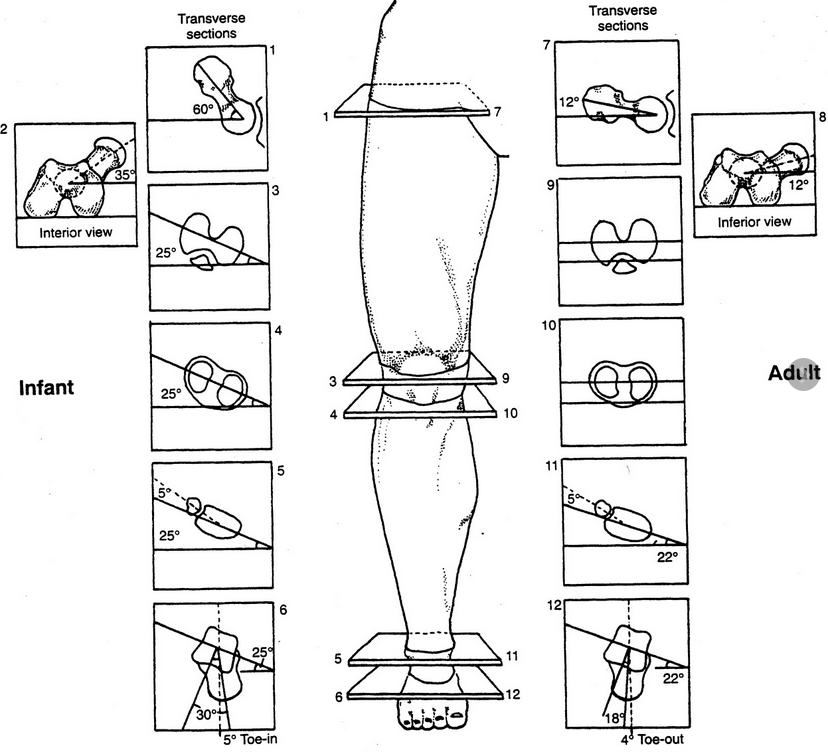
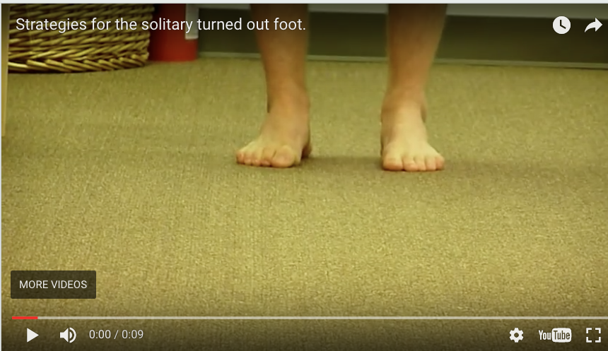
Take a look at it this gentleman who is a ski instructor. We are trying to simulate the standard side to side ski motion in a way that would be functionally appropriate. Keep in mind that he has bilateral internal tibial torsion and bilateral femoral retro torsion. When he began care at our office he had 5° external rotation on the right and about 8° external rotation on the left as his FULL AMOUNT OF INTERNAL ROTATION AVAILABLE to him bilaterally.

Treatment consisted largely of hip mobilization, Therapeutic exercises to emphasize internal rotation of the hips such as hip helicopters, airplanes and supine “chairs“ with internal rotation and adduction utilizing a ball between the knees; we also did acupuncture/needling of the hip capsules as well as anterior fibers of the gluteus medius and minimus. He now has about 5° internal rotation on the right now and a little less than 5 on the left. Note how the motion is clearly visualized in this video below.

Do you have other novel ways to test internal rotation of the hips functionally? Leave a comment or drop us a line and let us know

Feel like you want more? Join us this Wednesday evening on onlinece.com for Biomechanics 326: 6 MST

Dr Ivo Waerlop, one of The Gait Guys.

#functionaltesting #functionalmovement #hip #internalrotation #femoralretrotorsion #femoralretroversion #thegaitguys

The Hip "Airplane"

Here’s another great hip strengthening exercise for the gluteus medius, minimus, deep six external rotators as well as proprioceptive components about the hip. It is often used in conjunction with the hip helicopter exercises.

Dr Ivo Waerlop, one of The Gait Guys

#hipairplaneexercise #hippain #deep6extrenalrotators #gaitguys #proprioception #proprioceptiveexercises #thegaitguys


https://vimeo.com/371217385

The top 6 reasons we like hills for training ankle rocker and hip extension

image source: https://commons.wikimedia.org/wiki/File:Caer_Caradoc_hill.jpg

image source: https://commons.wikimedia.org/wiki/File:Caer_Caradoc_hill.jpg

1. Hills do not cost money and are almost always readily available : )

2. Being outside is good for your health

3. Hills do not pull the hip into extension and place a stretch (pull) on the anterior hip musculature including the rectus femoris, iliopsoas and iliacus. This causes a slow stretch of the muscle, activating the muscle spindles (Ia afferents) and causing a muscle contraction via the stretch reflex. This will inhibit the posterior compartment of hip extensors (especially the glute max) through reciprocal inhibition, making it difficult to fire them.

4. A hill does not force your knee into extension, eliciting a stretch reflex in the hamstrings like a treadmill does

5. A hill naturally puts the ankle into dorsiflexion, and, along with active pulling up of the toes, helps you to get more into your anterior compartment and eliminates the tendency of the ankle being pulled into dorsiflexion (like with a treadmill) which would initiate a stretch reflex in the gastroc/soleus and long flexors.

6. The increased hip flexor requirement of going uphill gives you more opportunity to engage the abs before the psoas and rectus femoris/TFL and on the stance phase leg, you can get an increased stretch of those muscles

Tips for picking the right hill and using it to your advantage

  • When just starting out, try and pick an incline that does not exceed the ankle dorsiflexion available to the patient/client

  • It’s OK if it’s uncomfortable, but not if its painful

  • Concentrate on pulling up the toes and dorsiflexing the ankle

  • Squeeze your glute at heel strike and toe off

  • leave your stance phase heel on the ground as long as possible

  • Place your hands on your abs and concentrate on activating them PRIOR to flexing your hip

Dr Ivo Waerlop, one of The Gait Guys

#walkinghills #traininganklerocker #thegaitguys # increasinghipextension


Metatarsalgia happens...

So a patient presents with forefoot pain, worse in the am upon awakening, with 1st weight bearing that would improve somewhat during the day, but would again get worse toward the end of the day and with increased activity. It began insidiously a few months ago (like so many problems do) and is getting progressively worse. Rest, ice and ibuprofen can offer some relief. You may see a dropped metatarsal head and puffiness and prominence in that area on the plantar surface of the foot, maybe not. Maybe you do a diagnostic ultrasound and see a lesion of the plantar plate as well? How did it get there? 

image courtesy of Tom Michaud: with permission

image courtesy of Tom Michaud: with permission

Lets look at the anatomy of the short flexors of the foot, as well as some biomechanics of the foot, ankle and hip. 

The flexor digitorum brevis (FDB) is innervated by the medial plantar nerve and arises from the medial aspect of the calcaneal tuberosity, the plantar aponeurosis (ie: plantar fascia) and the areas bewteen the plantar muscles. It travels distally, splitting at the metatarsal phalangeal articulation (this allows the long flexors to travel forward and insert on the distal phalanges); the ends come together to divide yet another time and each of the 2 portions of that tendon insert onto the middle of the middle phalanyx (1) 

As a result, in conjunction with the lumbricals, the FDB is a flexor of the metatarsophalangeal and proximal interphalangeal joints. In addition, it moves the axis of rotation of the metatasophalangeal joints dorsally, to counter act the function of the long flexors, which, when tight or overactive, have a tendency to drive this articulation anteriorly .Do you see any subtle extension of the metatarsophalangeal joint and flexion of the proximal interphalangeal joints on your exam?

We know that the FDB contracts faster than the other intrinsic muscles (2), playing a role in postural stability (3) and that the flexors temporally should contract earlier than the extensors (4), assumedly to move this joint axis posteriorly and allow proper joint centration. When this DOES NOT occur, the metatarsal heads are driven into the ground, causing irritation and pain.

If there is also a loss of ankle rocker this problem is made (much) worse. Why? Because, with the loss of one rocker, another must make up for the loss: ankle rocker decreases, forefoot rocker has to increase; this equals increased metatarsal head pressure. 

If you have been with us for any length of time, you know that ankle rocker and hip extension are intimately related, as one should equal the other, something we call “The “Z” angle”, that you have probably (hopefully?) read about here before. 

So what is the fix? Getting the FDB back on line for one. 

  • How about the toe waving exercise? 

  • How about the lift spread reach exercise? 

  • How about retraining ankle rocker and improving hip extension?

  • How about an orthotic with a metatarsal pad in the short term? 

  • How about some inflammation reducing modalities, like acupuncture, ice laser and pulsed ultrasound. 

  • Maybe some herbal or enzymatic anti inflammatories?


Dr Ivo Waerlop, one of The Gait Guys.

#gait #footpain #metatarsalgia #metatarsalpain #anklerocker #hipextension #thegaitguys


1. http://en.wikipedia.org/wiki/Flexor_digitorum_brevis_muscle

2. Tosovic D1, Ghebremedhin E, Glen C, Gorelick M, Mark Brown J.The architecture and contraction time of intrinsic foot muscles.J Electromyogr Kinesiol. 2012 Dec;22(6):930-8. doi: 10.1016/j.jelekin.2012.05.002. Epub 2012 Jun 27

3.Okai LA1, Kohn AF. Quantifying the Contributions of a Flexor Digitorum Brevis Muscle on Postural Stability.Motor Control. 2014 Jul 15. [Epub ahead of print]

4. Zelik KE1, La Scaleia V, Ivanenko YP, Lacquaniti F.Coordination of intrinsic and extrinsic foot muscles during walking.Eur J Appl Physiol. 2014 Nov 25. [Epub ahead of print]


Got hip extension?

Because she sure could use some...

we have see this gal before… yesterday in fact

  • left plantar plate lesion (yes, conformed on ultrasound)

  • left sided anatomical leg length discrepany

  • bilateral internal tibial torsion

  • incompetent L quadratus lumborum

  • adequate hip extension and ankle dorsiflexion available to her

  • lack of endurance in her abs

yep, lots more, but that is enough for now


note that she has plenty of ankle dorsiflexion, more on the right. this is due to her right leg being anatomically longer and has to travel through a greater range of motion

look at the knee and the hip articulations to assess hip extension. It should match ankle dorsiflexion, no?


Dr Ivo Waerlop, one of The Gait Guys


#gait #gaitguys #thegaitguys #hipextension #LLD #quadratuslumborum #internaltibialtorsion #anklerocker #ankledorsiflexion

An often overlooked culprit...In hip and hamstring insertional pain


The "Deep 6". In order from proximal to distal; the piriformis, obturator internus, gemelli superior, obturator externus, gemelli inferior and quadratus femoris. They are primarily external rotators but have a small footprint and act primarily as stabilizers. Here is what we think and what we have to say about them...


Dr Ivo Waerlop, one of The Gait Guys


#deepsix, #gait, #thegaitguys, #hipexternalrotators, #hipstabilizers, #running


Do you know your Torsions? If so, then you here is what you need to know about twisted people...

Are you twisted? Are your patients/clients twisted? You know about tibial torsions from yesterday but do you know about femoral torsions?

To go along with yesterdays post, here is some more info on femoral torsions. If you missed it, click here

The degree of version is the angle between an imaginary line drawn through the condyles of the femur and an imaginary line drawn through the head and neck of the femur. This is often referred to as the femoral neck angle or FNA.

IMAGE SOURCE: Michael T Cibulka; Determination and Significance of Femoral Neck Anteversion, Physical Therapy, Volume 84, Issue 6, 1 June 2004, Pages 550–558, https://doi.org/10.1093/ptj/84.6.550

IMAGE SOURCE: Michael T Cibulka; Determination and Significance of Femoral Neck Anteversion, Physical Therapy, Volume 84, Issue 6, 1 June 2004, Pages 550–558, https://doi.org/10.1093/ptj/84.6.550

Beginning about the 3rd month of embryological development (Lanz and Mayet 1953) the femoral neck angle reaches 60 degrees and decreases, with growth, to about 40 degrees (with an average of 30-60 degrees) at birth. It then decreases 25-30 degrees by adulthood to 8-20 degrees with males being at the lower and females at the upper end of the range.

The FNA angle, therefore, diminishes about 1.5 degrees a year until about 15 years of age. Femoral neck anteversion angle is typically symmetrical from the left side to the right side.

What causes torsion in the first place? By the sixth month in utero, the lumbar spine and hips of the fetus are fully flexed, so perhaps it is positional. Other sources say it coincides with the degree of osteogenesis. There is a growing consensus that muscular forces are responsible, particularly the iliopsoas or possibly the medial and lateral hip rotators.

Additional changes can occur after birth, particularly with sitting postures. “W” sitting or “cross legged” sitting have been associated with altering the available range of motion and thus the FNA, with the range increased in the direction the hip was held in; W sitting causing increased internal rotation and antetorsion and cross legged causing external rotation and retro torsion.

image source: T Michaud, with permission

image source: T Michaud, with permission

As discussed previously, there are at least 3 reasons we need to understand torsions and versions, They can alter the progression angle of gait, they usually affect the available ranges of motion of the limb and they can alter the coronal plane orientation of the limb.

  1. fermoral torsions often alter the progression angle of gait. In femoral antetorsion torsion, the knees often face inward, resulting in an intoed gait and a decreased progression angle of the foot. This can be differentiated from internal tibial torsion (ITT) by looking at the tibia and studying the position of the tibial tuberosity with respect to the foot, particularly the 2nd metatarsal. In ITT, the foot points inward while the tibial tuberosity points straight ahead. In an individual with no torsion, the tibial tuberosity lines up with the 2nd metatarsal. If the tibial tuerosity and 2nd met are lined up, and the knees still point inward, the individual probably has femoral ante torsion. Remember that a decreased progression angle is often associated with a decreased step width whereas an increased angle is often associated with an increased step width.

  2. Femoral torsions affect available ranges of motion of the limb. We remember that the thigh leg needs to internally rotate the requisite 4-6 degrees from initial contact to midstance (most folks have 40 degrees) If it is already fully internally rotated (as it may be with femoral retro torsion), that range of motion must be created or compensated for elsewhere. This, much like internal tibial torsion, can result in external rotation of the affected lower limb to create the range of motion needed.

  • Femoral retro torsion results in less internal rotation of the limb, and increased external rotation.

  • Femoral ante torsion results in less external rotation of the limb, and increased internal rotation.

          3. femoral torsions usually do not effect the coronal plane orientation of the lower limb,      since the “spin” is in the transverse or horizontal plane.

The take home message here about femoral torsions is that no matter what the cause:

  • FNA values that exist one to two standard deviations outside the range are considered “torsions”

  • Decreased values (ie, less than 8 degrees) are called “retro torsion” and increased values (greater than 20 degrees) are called “ante torsion”

  • Retro torsion causes a limitation of available internal rotation of the hip and an increase in external rotation

  • Ante torsion causes an increase in available internal rotation of the hip and decrease in external rotation

  • Femoral ante torsion will be perpetuated by “W” sitting (sitting on knees with the feet outside the thighs, promoting internal rotation of the femur)

  • Femoral antetorsion will be perpetuated by sitting cross legged, which forces the thigh into external rotation.

Michael T Cibulka; Determination and Significance of Femoral Neck Anteversion, Physical Therapy, Volume 84, Issue 6, 1 June 2004, Pages 550–558, https://doi.org/10.1093/ptj/84.6.550

http://www.clinicalgaitanalysis.com/faq/torsion.html

Souza AD, Ankolekar VH, Padmashali S, Das A, Souza A, Hosapatna M. Femoral Neck Anteversion and Neck Shaft Angles: Determination and their Clinical Implications in Fetuses of Different Gestational Ages. Malays Orthop J. 2015;9(2):33-36.

The Sartorius: insertional tendinitis and medial knee pain?

We all see folks with medial knee pain, many times women, with the pain located just below the medial tibial plateau. It often results from running, but sometimes with jumping sports like basketball as well. It has been our experience that these people are often diagnosed with an MCL type injury, but when you examine them further, they do not really fit the bill. All the ligaments are stable and there is no tenderness at the joint line. The is often tenderness at the pes anserine, but who is driving the bus here?

image source: https://commons.wikimedia.org/wiki/File:Muscles_and_tendons_of_the_legs_and_feet;_écorch_́figur_Wellcome_V0008276.jpg

image source: https://commons.wikimedia.org/wiki/File:Muscles_and_tendons_of_the_legs_and_feet;_écorch_́figur_Wellcome_V0008276.jpg

The sartorius originates from the anterior compartment of the thigh. During an ideal gait cycle, the sartorius fires from toe off through nearly terminal swing (1)

We remember that the abdominals should initiate thigh flexion with the iliopsoas, rectus femoris, tensor fascia lata and sartorius perpetuating the motion. Sometimes, when the abdominals are insufficient, we will substitute other thigh flexors, often the psoas and/or rectus femoris, but sometimes sartorius, especially in people with excessive midfoot pronation. Think about all of the medial rotation occurring at the knee during excessive midfoot pronation and when overpronation occurs, the extra compensatory external rotation that must occur to try and bring the knee back into the sagittal plane. The sartorius is positioned perfectly for this function, along with the semitendinosus which assists and external rotation in closed chain. This is why it is often implicated as the culprit in many cases of pes anserine bursitis (or as we like to say “sartorius insertional tendinitis” (2-3)

Some other things you may find interesting is that it is utilized more in crossing or cutting maneuvers while changing directions while running (4). This makes sense, given its anatomical course and origin/insertion. It can often be overlooked in adductor strains. It can also be avulsed during sprints, particularly in adolescents (5) and because of the course of the lateral femoral cutaneus nerve beneath it, can be the cause of meralgia paresthetica (6). It is proprotionally smaller in females (along with the gracilis and short head of the biceps femoris) (7). And during vertical jumping, is considered an internal rotator, along with the semimembranosis, semitendinosis, gracilis, and popliteus (8).

The sartorius is superficial in the anterior thigh, just under the skin, running from the ASIS, coursing lateral to medial and inserting at the pes anserine at its most superior aspect, just overlying the gracilis. Since it is an external rotator, knee flexor and assists in thigh abduction, you can easliy locate it by placing the patient in a "figure 4" position and having them resist as you pull downward on the leg. Be careful if you are needling this muscle because of the subsartorial canal (ie Hunters canal) lying just beneath it in the middle 1/3 of the thigh, from the apex of the femoral triangle to the adductor hiatus in the adductor magnus. It houses the femoral artery and vein, as well as the saphenous nerve and nerve to the vastus medialis.


  1. Michaud T: in Human Locomotion: The Conservative Management of Gait-Related Disorders 2011

  2. Imani F, Rahimzadeh P, Abolhasan Gharehdag F, Faiz SH. Sonoanatomic variation of pes anserine bursa. Korean J Pain. 2013;26(3):249-54. 

  3. Gupta, Aman & Saraf, Abhinesh & Yadav, Chandrajeet. (2013). ISSN 2347-954X (Print) High-Resolution Ultrasonography in PesAnserinus Bursitis: Case Report and Literature Review. 1. 753-757. 

  4. Rand MK, Ohtsuki T. EMG analysis of lower limb muscles in humans during quick change in running directions. Gait Posture. 2000 Oct;12(2):169-83.

  5. Manning CJ, Singhai S, Marshall P. Synchronised sartorius avulsions in adolescent sprinter. BMJ Case Rep. 2016 Jul 13;2016.

  6. Hsu CY, Wu CM, Lin SW, Cheng KL. Anterior superior iliac spine avulsion fracture presenting as meralgia paraesthetica in an adolescent sprinter. J Rehabil Med. 2014 Feb;46(2):188-90. doi: 10.2340/16501977-1247.

  7. Behan FP, Maden-Wilkinson TM, Pain MTG, Folland JP. Sex differences in muscle morphology of the knee flexors and knee extensors. PLoS One. 2018 Jan 23;13(1):e0190903.

  8. Cleather DJ. An important role of the biarticular hamstrings is to exert internal/external rotation moments on the tibia during vertical jumping. J Theor Biol. 2018 Oct 14;455:101-108

Is there a need for "Gait Retraining'?...We think so

photo source: https://commons.wikimedia.org/wiki/File:Severe_(Tönnis_grade_3)_osteoarthritis_of_the_hip.jpg

photo source: https://commons.wikimedia.org/wiki/File:Severe_(Tönnis_grade_3)_osteoarthritis_of_the_hip.jpg

There seems to be some controversy with regards to gait retraining. Some folks seem to believe that it should be “left to itself” and they are fully compensated already (1). Perhaps this is true…or not. We have not seen any studies that compare gait retraining vs non gait retraining as a whole, but there seems to be plenty for specific conditions (2). We all see folks AFTER THE FACT and seek to correct the problems and reverse, halt or slow the progression of further pathology. That seems to be what many of us do.

This recent study (3) looks ate altered loads and muscle recruitment patterns in patients with osteoarthritis. they conclude:

“This study documents alterations in hip kinematics and kinetics resulting in decreased hip loading in patients with hip OA. The results suggested that patients altered their gait to increase medio-lateral stability, thereby decreasing demand on the hip abductors. These findings support discharge of abductor muscles that may bear clinical relevance of tailored rehabilitation targeting hip abductor muscles strengthening and gait retraining.”

There is substantial evidence that hip pathomechanics lead to osteoarthritis (4, 5). Wouldn’t it make sense to assist in altering motor patterns and correct those biomechanical faults before it becomes a problem? Lets change our focus (if we haven’t already) and concentrate on skill, endurance and strength, in that order for the betterment of ourselves, our patients and humanity.

  1. Nigg BM, Baltich J, Hoerzer S, Enders H. Running shoes and running injuries: mythbusting and a proposal for two new paradigms: “preferred movement path” and “comfort filter” Br J Sports Med. 2015 Jul; doi: 10.1136/bjsports-2015-095054. bjsports - 2015-095054. 

  2. Davis IS, Futrell E. Gait Retraining: Altering the Fingerprint of Gait. Physical medicine and rehabilitation clinics of North America. 2016;27(1):339-355. doi:10.1016/j.pmr.2015.09.002. FREE FULL TEXT

  3. Meyer CAG, Wesseling M, Corten K, Nieuwenhuys A, Monari D5, Simon JP, Jonkers I, Desloovere K. Hip movement pathomechanics of patients with hip osteoarthritis aim at reducing hip joint loading on the osteoarthritic side. Gait Posture. 2018 Jan;59:11-17. doi: 10.1016/j.gaitpost.2017.09.020. Epub 2017 Sep 22.

  4. Christian Egloff, Thomas Hügle, Victor Valderrabano: Biomechanics and pathomechanisms of osteoarthritis Swiss Med Wkly. 2012;142:w13583 FREE FULL TEXT

  5. https://www.the-rheumatologist.org/article/get-out-of-your-oa-box/?singlepage=1&theme=print-friendly

Podcast 138 (for real). Are you fighting your own gait/running neurology?

Topics:
1. Running with the extensors. Convergence and divergence of neurons.
2. Fighting your gait neurology. The lies about the Bird dog rehab exercise.
3. ACL and ACL rehab. Surgery or no sugery. Wise? Risks ? How social media discussions might just be getting it wrong.
4. Cross over gait and lateral heel strike and ensuing problems at great toe off. A failure to medial foot tripod high gear toe off ?
5. Are the hip flexors actually hip flexors in gait ? what are your high knee drills doing? Anything good?

Key words: acl, analysis, cross, extensor, flexors, gait, heel, hip, instability, knee, over, plri, pools, problems, running, strike, surgery

Links to find the podcast:

iTunes page: https://itunes.apple.com/us/podcast/the-gait-guys-podcast/id559864138?mt=2

Direct Download:http://traffic.libsyn.com/thegaitguys/pod_138_real_-_82818_2.12_PM.mp3

Permalink URL:http://thegaitguys.libsyn.com/podcast-138-for-real

Libsyn URL: http://directory.libsyn.com/episode/index/id/6978817

Our Websites:
www.thegaitguys.com

summitchiroandrehab.com

doctorallen.co

shawnallen.net

Our website is all you need to remember. Everything you want, need and wish for is right there on the site.
Interested in our stuff ? Want to buy some of our lectures or our National Shoe Fit program? Click here (thegaitguys.com or thegaitguys.tumblr.com) and you will come to our websites. In the tabs, you will find tabs for STORE, SEMINARS, BOOK etc. We also lecture every 3rd Wednesday of the month on onlineCE.com. We have an extensive catalogued library of our courses there, you can take them any time for a nominal fee (~$20).

Our podcast is on iTunes and just about every other podcast harbor site, just google "the gait guys podcast", you will find us.

The Adductor Magnus; Not just for adduction anymore...

add mag.png
brunkine6_ch12_f0018-2.png


Yet another paper (1) to support the notion that the adductors (particularly the adductor magnus, in this paper), act as external rotators (especially eccentrically), not internal rotators as is commonly purported in many anatomy texts (2) . Remember that the lower extremity is internally rotating (as a whole) from initial contact to midstance and externally rotating from midstance to pre swing. SOMETHING needs to help attenuate some of that internal rotation (and pronation) that occurs during the 1st part of stance phase and assist in external rotation (and supination); now you can add the adductor magnus to the popliteus, deep six external rotators, anterior and posterior compartments of the lower leg to the hamstrings and quads.

"This study suggests that adductor magnus has at least two functionally unique regions. Differences were most evident during rotation. The different direction-specific actions may imply that each segment performs separate roles in hip stability and movement. These findings may have implications on injury prevention and rehabilitation for adductor-related groin injuries, hamstring strain injury and hip pathology."


1. Benn ML, Pizzari T, Rath L, Tucker K, Semciw AI1 . Adductor magnus: An emg investigation into proximal and distal portions and direction specific action. Clin Anat. 2018 Mar 9. doi: 10.1002/ca.23068. [Epub ahead of print]

2. Leighton RD. A functional model to describe the action of the adductor muscles at the hip in the transverse plane.Physiother Theory Pract. 2006 Nov;22(5):251-62.


add mag.png

How is your foot is connected to your butt....?

EVD-marcha-075.jpg

If you have been following us for any length of time, you have heard us talk about how the lower kinetic chain is connected, how ankle rocker effects hip extension and how important hallux (great toe) extension is. 

What can we conclude from this study?

toe spreading exercises are important for reducing navicular drop (and thus mid foot pronation, at least statically)
In addition to increased abductor hallucis recruitment in ascending and descending stairs, when hip external rotation exercises were added along with toe spreading exercises folks had more recruitment of the vastus medialis (a closed chain external rotator of the leg and thigh)
 
Keep in mind:

the exercises given were all non weight bearing and open chain for the external rotators. Imagine what might have happened if they were both closed chain AND weight bearing!
They concentrated on the effects of toe spreading (AKA  lift/spread/reach) on the abductor hallucis. It also has far reaching effects on the dorsal interossei, long and short extensors of the toes. 

Abstract: The purpose of the present study was to examine the effects of toe-spread (TS) exercises and hip external rotator strengthening exercises for pronated feet on lower extremity muscle activities during stair-walking. [Subjects and Methods] The participants were 20 healthy adults with no present or previous pain, no past history of surgery on the foot or the ankle, and no foot deformities. Ten subjects performed hip external rotator strengthening exercises and TS exercises and the remaining ten subjects performed only TS exercises five times per week for four weeks. [Results] Less change in navicular drop height occurred in the group that performed hip external rotator exercises than in the group that performed only TS exercises. The group that performed only TS exercises showed increased abductor hallucis muscle activity during both stair-climbing and -descending, and the group that performed hip external rotator exercises showed increased muscle activities of the vastus medialis and abductor hallucis during stair-climbing and increased muscle activity of only the abductor hallucis during stair-descending after exercise. [Conclusion] Stair-walking can be more effectively performed if the hip external rotator muscle is strengthened when TS exercises are performed for the pronated foot.

Goo YM, Kim DY, Kim TH. The effects of hip external rotator exercises and toe-spread exercises on lower extremity muscle activities during stair-walking in subjects with pronated foot. J Phys Ther Sci. 2016 Mar;28(3):816-9. doi: 10.1589/jpts.28.816. Epub 2016 Mar 31. 
link to  FREE FULL TEXT: https://www.ncbi.nlm.nih.gov/pmc/articles/PMC4842445/

So, what attaches to that hip capsule anyway....

I was trying to figure to which muscles attached to the labrum of the hip, as I see many folks where theres has gone south. I had always wondered if the iliopsoas attached, since many people with labral pathology have hip flexor dysfunction, where they use their psoas and iliacus as hip flexion initiators (or sometimes the rectus femoris, TFL and sartorius), instead of the abdominals. It turns out that NO MUSCLES attach to the labrum, but some attach to the capsule. 

Screen Shot 2018-03-26 at 9.18.31 AM.png

Have you noticed that many of the muscles on the list below (not the obturator internus) are internal rotators AND work during the 1st part of stance phase? Remember "glide and roll"? With internal rotation of the hip comes posterior translation of the femoral head. If these are dysfunctional, you may get capsular "pinching". Think about it with the next patient with hip joint pain from initial contact to midstance. 

"An updated knowledge of the intricate relationship of the pericapsular and capsular structures is essential in guiding our treatment of the hip. Following dissection the authors were able to discern that the iliocapsularis, indirect head of the rectus, conjoint tendon (of the psoas and iliacus),  obturator externus and gluteus minimus all have consistent capsular contributions whereas the piriformis did not have a capsular attachment."


Walters BL, Cooper JH, Rodriguez JA New findings in hip capsular anatomy: dimensions of capsular thickness and pericapsular contributions.
Arthroscopy. 2014 Oct;30(10):1235-45. doi: 10.1016/j.arthro.2014.05.012. Epub 2014 Jul 23.

Building a better Bridge: Part 2

Along the same vein as our last post, consider abducting the leg 30 degrees, which increases gluteus maximus activity, lessens anterior pelvic tilt and lessens erector spinae activity. Of course, pelvic tilt should have clued you in to a weak core in the 1st place : )

PURPOSE: To investigate how the erector spinae (ES) and gluteus maximus (GM) muscle activity and the anterior pelvic tilt angle change with different hip abduction angles during a bridging exercise.

METHODS: Twenty healthy participants (10 males and 10 females, aged 21.6 ± 1.6) voluntarily participated in this study. Surface electromyography (EMG) signals were recorded from the ES and GM during bridging at three hip abduction angles: 0°, 15°, and 30°. Simultaneously, the anterior pelvic tilt angle was measured using Image J software.

RESULTS: The EMG amplitude of the GM muscle and the GM/ES EMG ratio were greatest at 30° hip abduction, followed by 15° and then 0° hip abduction during the bridging exercise. In contrast, the ES EMG amplitude at 30° hip abduction was significantly lesser than that at 0° and 15° abduction. Additionally, the anterior pelvic tilt angle was significantly lower at 30° hip abduction than at 0° or 15°.

CONCLUSIONS: Bridging with 30° hip abduction can be recommended as an effective method to selectively facilitate GM muscle activity, minimize compensatory ES muscle activity, and decrease the anterior pelvic tilt angle.

Kang SY1, Choung SD2, Jeon HS3. Modifying the hip abduction angle during bridging exercise can facilitate gluteus maximus activity. Man Ther. 2016 Apr;22:211-5. doi: 10.1016/j.math.2015.12.010. Epub 2016 Jan 2.


A return to "the Kickstand Effect". So your foot is turned out, externally rotated ?

Amputee War Veteran Sergeant Christopher Melendez Became a Pro WrestlerRead more at http://www.craveonline.com/mandatory/1053779-standing-tall-how-amputee-war-veteran-christopher-melendez-beca#XeD2LrZ2xmtXQ6um.99

Amputee War Veteran Sergeant Christopher Melendez Became a Pro Wrestler
Read more at http://www.craveonline.com/mandatory/1053779-standing-tall-how-amputee-war-veteran-christopher-melendez-beca#XeD2LrZ2xmtXQ6um.99

Why is my foot turned out ?  A 3rd return to the solitary externally rotated foot.

Below you will find our 2 prior articles on this topic, but this is a relatable concept to other thing which we have embedded in many of our blog posts and podcasts over the last decade of sharing what we know.

In the photo above the brave Army Veteran Sergeant Melendez one can see the concept brilliantly as he only has one limb.  One can see the concept in full play, he must balance his body mass over one point, not two like the rest of us lucky folk.  In trying to balance over one point, if the foot is straight forward (if one is blessed with close to neutral torsional bone alignment) one will have good stability in the sagittal plane (forward /back) but will be at risk to fall, drift or sway into the frontal plane. Here Sergeant Melendez displays the foot and limb turn out into the frontal plane so that he can use the quadriceps to help him protect into that frontal plane, plus, by situating his base posture in more of an externally rotated position (likely losing internal rotation capability over time, unless forcibly maintained through specific exercises) he can more fully and skillfully engage all 3 divisions of the gluteus maximus and medius, and perhaps hamstrings and adductors and who knows what else, to maintain a more stable and likely less fatiguable posture. Go ahead, try it for yourself, this is easier to balance and maintain that a straight sagittal foot posturing. The one trouble he might have, is not deviating too much, or too often, into a frontal plane drift hip-pelvis posture. This will put much aberrant compressive load onto the roof of the femoral head-acetabular interval, where most of us begin a degenerative hip arthritis journey, unfortunately. 

Side note:   So you might think your client has FAI ?  Maybe start here, our thinking might lead you done a helpful path to get started. Search our blog for FAI as well.

here are the 2 prior articles on the topic, with video.  Watch for this one, it is everywhere out in the world, walking amongst us.  
Thank you for your service Sergeant Melendez.  Here is the article written by K. Thor Jensen, on Crave Online.  

https://thegaitguys.tumblr.com/post/14262793786/gait-problem-the-solitary-externally-rotated

https://thegaitguys.tumblr.com/post/40617674450/a-return-to-the-solitary-externally-rotated-foot

Shawn & Ivo, The Gait Guys

An often overlooked culprit in hip pain...

We often find clinically that the quadratus femoris as becoming the 1st dysfunctional muscle of the deep 6 external rotators (1) and its pain referral pattern can mimic the piriformis (2) and piriformis syndrome (3) as well as hamstring insertional tendinitis.  It has also been implicated in some cases of femoroacetabular impingement (4)  as well as ishiofemoral impingement (5). It is active during walking stance phase, and moreso during stance while running as well as with a clamshell exercise with external rotation (6). It appears to be maximally lengthened with flexion and adduction or abduction, with internal rotation ( a great position of you need to stretch this muscle), and is deducted to be strongest going from a 60-90 degree flexed position into extension (ie: it has the with the largest moment arms observed for extension in the deduced force-length efficient range of 60-90° flexion)(7).

Needling this muscle can sometimes pose a challenge. Here is a demo of one way to accomplish it I often employ while needling some of the other surrounding hip musculature.

Consider the QF the next time you have someone with hamstring insertional pain, or diffuse hip pain that you are having a difficult time localizing.

  1. Personal observation
  2. Janet G. Travell , M.D., and David G. Simons, M.D., Myofascial Pain and Dysfunction: The Trigger Point Manual, The Lower Extremities vol. 2 (Baltimore: Williams & Wilkins, 1992) pp. 186-193.
  3. Dalmau-Carolà J Myofascial pain syndrome affecting the quadratus femoris Pain Pract. 2010 May-Jun;10(3):257-60. doi: 10.1111/j.1533-2500.2009.00347.x. Epub 2010 Feb 11

  4.  Diamond LEVan den Hoorn WBennell KLWrigley TVHinman RSO'Donnell JHodges PW. Coordination of deep hip muscle activity is altered in symptomatic femoroacetabular impingement.  J Orthop Res. 2016 Aug 11. doi: 10.1002/jor.23391. [Epub ahead of print]

  5. http://radsource.us/ischiofemoral-impingement-syndrome/
  6. Semciw, Adam I. et al. Quadratus femoris: An EMG investigation during walking and running Journal of Biomechanics , Volume 48 , Issue 12 , 3433 - 3439

  7. Vaarbakken KSteen HSamuelsen GDahl HALeergaard TBStuge B .Primary functions of the quadratus femoris and obturator externus muscles indicated from lengths and moment arms measured in mobilized cadavers. Clin Biomech (Bristol, Avon). 2015 Mar;30(3):231-7. doi: 10.1016/j.clinbiomech.2015.02.004. Epub 2015 Feb 11.

Things often work better in pairs… Especially with Exercise

You have heard us always talk about how the lower kinetic chain is connected, how ankle rocker effects hip extension and how important hallux (great toe) extension is.

What can we conclude form this study?

  • toe spreading exercises are important for reducing navicular drop (and thus mid foot pronation, at least statically)
  • In addition to increased abductor hallucis recruitment in ascending and descending stairs, when hip external rotation exercises were added along with toe spreading exercises folks had more recruitment of the vastus medialis (a closed chain external rotator of the leg and thigh)

Keep in mind:

  • the exercises given were all non weight bearing and open chain for the external rotators. Imagine what might have happened if they were both closed chain AND weight bearing!
  • They concentrated on the effects of toe spreading (AKAlift/spread/reach) on the abductor hallucis. It also has far reaching effects on the dorsal interossei, long and short extensors of the toes.


Goo YM, Kim DY, Kim TH. The effects of hip external rotator exercises and toe-spread exercises on lower extremity muscle activities during stair-walking in subjects with pronated foot. J Phys Ther Sci. 2016 Mar;28(3):816-9. doi: 10.1589/jpts.28.816. Epub 2016 Mar 31.

link toFREE FUL TEXT: https://www.ncbi.nlm.nih.gov/pmc/articles/PMC4842445/