Compressing the Distal Tib-Fib joint. Really ?!
If you do not know your biomechanics, this could be a road to problems for your client. 

Many who viewed this video will not know the “Caveat emptor” we will expand upon below. 

Here is the meat of today’s blog post: The distal tibia-fibular syndesmosis is supposed to separate during ankle dorsiflexion to accommodate for the wider ankle mortise anteriorly, compressing the two bones could nullify this range.

There are new ideas on the web every day; through new ideas come new innovations. These bands (some call them Voodoo bands) are all the rage right now, and they may have value depending on their use (and the wisdom of the user), but from what we can tell even the innovators are at a bit of a loss as to what the heck is truly happening with their use. All we have right now is theories, but understandably things have to start somewhere. We propose some logical thoughts on this video implementation here today.  
Some people are using the bands over larger muscle masses, perhaps using the compression and shear between tissue layers to act as a kind of “flossing” or “active release” to the muscle and myofascia. The goal seems mostly to gain more joint range and thus greater function through that joint. Some think the vascular/ischemic challenge is creating partial effects. Others appear to be using the band to change joint compression to change stability to change mobility. We suspect others are using them to manage joint shear, we hope they realize they are impacting that component. One must keep in mind that a joint that is cylindrically compressed (such as in the video) and then subsequently loaded may still undergo motion and shear at the opposing joint surfaces. The question is, are you getting what you want?  The even bigger question is have you the earned stability on that new mobility if you are about to go add activity on this new range? New “borrowed” range, meaning mobility that has been acquired but not been earned via the muscles that were supposed to achieve and maintain it, may be a recipe for injury risk. One must also not forget that with a primary motion there is an accessory motion (ie. roll and glide are in opposite directions for concave/convex joint relationships). This is a concept of joint centration, and when there is excessive uncontrolled shear there cannot be adequate centration. Too much axial compression may limit primary motion while minimizing shear but could impact accessory motion, a mulit-edged sword. There is really no good and certain way to do all this except intrinsically via the muscles surrounding the cylinder. Any extrinsic attempts must be met with the realization and understanding of the true mechanics of compression, shear, primary motion and accessory motions. So just keep in mind that things like impingement, shear and peripheral joint loading (as opposed to centrated loading) are risk factors when these components are not well understood.

That all said, we bring these concerns to light today in regards to the above mobilization video.  Many who viewed this video will not know the “Caveat emptor” we have eluded to above and will expand upon below. That caveat should have (in our opinion) been mentioned. We are not trying to pick on folks, trolling or being pricks, we know everyone is just trying to help contribute to the mass knowledge base here on the web but one has to understand biomechanics in order to deliver a clean honest method without tipping the risk reward scale. We think our caveat is very much worth mentioning so that the knowledge is available to everyone. We bet the doctor in the video knows all about what we are writing here today, but many others will not, and so by debating and critiquing we all raise the bar, for the good of all mankind. In turn, we expect the same critique should happen to our material, after all, the collective mind is more powerful than the individual mind.


Here is the meat of today’s blog post:

The distal tibia-fibular syndesmosis is supposed to separate during ankle dorsiflexion to accommodate for the wider ankle mortise anteriorly. More gently stated, as dorsiflexion progresses at the ankle mortise complex, the distal tibia-fibula must be able to change to accept the wider anterior mortise engagement. This is normal ankle biomechanics, for everyone ! So, why would you want to compress the distal tib-fib and attempt at arresting or limiting the normal spreading process during ankle dorsiflexion? What about the ligamentous structures that depend upon clean terminal dorsiflexion and congruent tib-fib-mortise orchestration ? Cylindrical compression could impair or limit terminal dorsiflexion range.  Wasn’t this the purpose of the mobilization in the first place?  This mobilization is just not something that we will be recommending you start tossing out in your rehab or training room unless you can justify on a case by case basis a reason for possibly working against normal clean biomechanics, unless of course you are sure to stay within reasonable compression limits.  If you compress these bones too much, you are potentially creating mid or end range joint impingement.   We are sure the argue point will be that the band is not applied tightly enough to create sufficient compression to limit this normal range. This may be true. But, 

  • “Think of how stupid the average person is, and realize half of them are stupider than that.” -George Carlin

All joking aside now, but for the “average Joe” who might think that more is better, our caveat is worthy in our opinion especially when you see the volume of band used in some other videos.  

SIDEBAR: Dear Gait Guys brethren, as members of those on the web who are supposed to know better, we all have a responsibility to act and portray truth and accuracy to those that are not in our lines of profession and knowledge. There are videos on line demonstrating a cavalier approach to using these bands (we are not at all referring to today’s video), we beg you to think about who is seeing these videos, possibly herds of runners and athletes looking for quick answers to their problems.  Know that you may be the first line of intervention to help direct these folks to an informed way to implement self-treatment.


Back to the video for one final point.

We see that after some cylinder compression is rendered by the band, as terminal ankle dorsiflexion is mobilized we see end range mobilizing of internal and external rotation. Remember, if the compression is too much (and again, it may not be in this case if band tension application is reasonable), as dorsiflexion is attempted we will have more closed pack-type joint compression binding mid-range, and this may mean risk to articular cartilage. Just something to keep in mind. Listen to your client feedback when they do this or you instruct them, pain is obviously not a welcome outcome when you are performing potentially impinging therapies.


Here on The Gait Guys we previously shared our mnemonic , “anterior strength achieves posterior length to drive ankle dorsiflexion range”. That does not in any way mean that mobilizations are not worthy efforts at any time during a treatment.  As a clinician, sometimes you have to address the tissue length of the posterior compartment tissues, but if that is not the primary cause of loss of dorsiflexion you are commissioned to look elsewhere.  Also, remember that ankle dorsiflexion can be disguised through foot pronation and this in itself can enable pathology. This is perhaps one of the biggest omissions in ankle dorsiflexion mobilization videos across the board.


Make no mistake, you can mobilize all you want but at the end of the day you must improve skill, endurance and strength (S.E.S.) as well as functional stability and capacity on these new patterns of mobility if you are to do your client justice. Failure to do these things will result in loss of the gained mobility and risk for injury. Almost anyone can gain more joint motion, we have all been doing this various ways for decades. Can you earn enough capacity to keep the new mobility on a clean and correct motor pattern without corruption is the bigger question. Remember, just because you force a joint range, as opposed to earning it, doesn’t mean it is wise. Try this logic on any adhesive capsulitis shoulder patient, you will surely receive a five finger death punch in return. 


There is much in the way of innovation and free thinking out there today and everyday the internet opens our collective eyes and minds to new ideas and possibilities. We all must keep in mind that many of these new things are in their infancy, some will survive with validation and some will wither away without it. It is up to the practitioner to take their client’s case to heart, do the best they can with the knowledge they have, accept when their scope of knowledge and practice has been met, and always first “do no harm”.  Most things can be fixed, or at the very least improved upon but the tough cases often require deep wells of knowledge and experience. Sometimes we have to tread into uncharged waters, we just have to make sure we do no harm and try to work around a framework of science based knowledge.


We talk about this concept and video in greater depth in Podcast 90. Feel free to listen in,

here is that link


Dr, Shawn Allen


… . just two guys trying to provide logic to things we do not have complete answers for at this time.


For your reading pleasure, here is another fella who has gone through some  similar internal dialogue trying to find answers regarding this Voodoo band stuff. We respect his thoughts and dialogue, very much so.  He covers many thoughts and theories, it is worth your time if you are using this type of therapy. 


http://valeohealthclinic.com/voodoo-floss-really-voodoo/

Podcast 91: Gait, Vision & some truths about leg length discrepancies

Show sponsors:
www.newbalancechicago.com

A. Link to our server:
http://traffic.libsyn.com/thegaitguys/pod_91f.mp3

Direct Download:
http://thegaitguys.libsyn.com/91

Other Gait Guys stuff

B. iTunes link:
https://itunes.apple.com/us/podcast/the-gait-guys-podcast/id559864138

C. Gait Guys online /download store (National Shoe Fit Certification and more !) :
http://store.payloadz.com/results/results.aspx?m=80204

D. other web based Gait Guys lectures:
Monthly lectures at : www.onlinece.com   type in Dr. Waerlop or Dr. Allen,  ”Biomechanics”

Our Book: Pedographs and Gait Analysis and Clinical Case Studies

electronic copies available here:

Amazon/Kindle:

http://www.amazon.com/Pedographs-Gait-Analysis-Clinical-Studies-ebook/dp/B00AC18M3E

Barnes and Noble / Nook Reader:

http://www.barnesandnoble.com/w/pedographs-and-gait-analysis-ivo-waerlop-and-shawn-allen/1112754833?ean=9781466953895

https://itunes.apple.com/us/book/pedographs-and-gait-analysis/id554516085?mt=11

Hardcopy available from our publisher:

http://bookstore.trafford.com/Products/SKU-000155825/Pedographs-and-Gait-Analysis.aspx

Show notes:

Gait and vision: Gaze Fixation
What’s Up With That: Birds Bob Their Heads When They Walk
http://www.wired.com/2015/01/whats-birds-bob-heads-walk/
 
Shod vs unshod
 
Short leg talk:
11 strategies to negotiate around a leg length discrepancy

From a Reader:

Dear Gait Guys, Dr. Shawn and Dr. Ivo,  I was referred to this post of yours on hip IR…http://thegaitguys.tumblr.com/post/14262793786/gait-problem-the-solitary-externally-rotated   I am impressed by the level of details of your understanding of the gait and biomechanics. Although I am still trying to understand all of your points in this post, I would like to ask you:  What if my IR is limited due to a structural issue? The acetabular retroversion of the right hip in my case. 

I.e. if I am structurally unable to rotate the hip internally.
What will happen? 
What would be a solution to the problem in that case? 

Single-leg drop landing movement strategies 6 months following first-time acute lateral ankle sprain injury - Doherty - 2014 - Scandinavian Journal of Medicine & Science in Sports
http://onlinelibrary.wiley.com/doi/10.1111/sms.12390/abstract

Hey Gait Guys,

I understand that 1st MP Joint dorsiflexion, ankle rocker, and hip extension are 3 key factors for moving in the sagittal plane from your blog and podcasts so far. I really love how you guys drill in our heads to increase anterior strength to increase posterior length to further ankle rocker. I’ve seen the shuffle gait and was curious if you had a good hip extension exercise to really activate the posterior hip extensors and increase anterior length. 

From Abby Road to Vivaldi; the Sensory information in has a corresponding motor output.

Going for a run or a workout? What you listen to has an impact on your motor output, but why ? Functional MRI of the listening brain found that different regions become active when listening to different types of music and instrumental versus vocals.
From Abby Road to Vivaldi; the Sensory information in has a corresponding motor output. The brain shifts the sound to different areas depending on the music. This is why ACDC is likely a better sound track for your next run than Vivaldi.
“Computer algorithms were used to identify specific aspects of the music, which the researchers were able to match with specific, activated brain areas. The researchers found that vocal and instrumental music get treated differently. While both hemispheres of the brain deal with musical features, the presence of lyrics shifts the processing of musical features to the left auditory cortex.” - Allie Wilkinson

A 60 second podcast.
http://www.scientificamerican.com/podcast/episode/different-brain-regions-handle-different-music-types/

A case of gait spasticity.

We have been saying it for a long time now. Gait is a huge window into the function of the human brain and nervous system. It is often the first presenting sign that something is wrong. In the case below, a 48 year old woman presented because of a gradually developing spastic gait. 
Lab testing showed mild increases of transaminase and total bilirubin. Blood manganese level was markedly elevated. 
MRI showed high signal intensities at the globus pallidus and cerebral peducles, and bilateral deep white matter, posterior limbs of the internal capsule and right upper cervical spinal cord. 
A diagnosis of Idiopathic portal hypertension was made. 
According to this article:

“Hepatic diseases often show high signal intensities at the basal ganglia on T1-weighted images, and this seemed to be due to accumulation of manganese in our case. Because demyelination or axonal injury of the spinal cord are found in hepatic disease, we speculate that the high signal intensities at the spinal cord on T2-weighted images of our case reflect hepatic myelopathy, which may also be caused by high blood levels of manganese.”


The Gait Guys say once again, “gait changes in a client may be the first clinical presentation of other pathology, not all the time, but enough that you should be looking for anomalies.

Rinsho Shinkeigaku. 2002 Sep;42(9):885-8.
[A case of idiopathic portal hypertension (IPH) with hypermanganemia presenting as spastic gait].
Obama R1, Tachikawa H, Yoshii F, Takeoka T, Shinohara Y.

Lets make a resolution…Or not…
Cool guy, cool picture, cool scenery. Motivational? He is the fitness guru the developed the “Insanity” workout series, amongst others.
But looks may be deceiving.  
For reference, draw a line…

Lets make a resolution…Or not…

Cool guy, cool picture, cool scenery. Motivational? He is the fitness guru the developed the “Insanity” workout series, amongst others.

But looks may be deceiving.  

For reference, draw a line from the philitrium, interpec interval, symphisis pubis to area bisected between feet. 

Did you notice the following?

  • crossing arms across midline; look how far that left arm is abducted.
  • look at pelvis list to left. If you bring that arm in, you need to compensate somewhere
  • did you notice the hip hike on the left? That may have something to do with the excessive internal rotation of the thigh on the left. Is that because of the pelvis shift to the left (to compensate) or is he making up for limited internal rotation of the right hip?
  • what about the subtle head tilt to the right? is that driving the compensation or is it another compensation?

Questions, questions, questions…

We are choosing to make a resolution without him for the time being : ) More on this photo another day.

Sometimes it is easy and straight forward.
HISTORY: A 56 YO 200 # male construction worker presents with pain at the bottom of his right foot, worse in the am, getting better as the day goes on till midday, then getting worse again. Better with rest…

Sometimes it is easy and straight forward.

HISTORY: A 56 YO 200 # male construction worker presents with pain at the bottom of his right foot, worse in the am, getting better as the day goes on till midday, then getting worse again. Better with rest and ice. More supportive shoes and a heel gel pad offer him some relief. Past history of plantar fascitis. 

OBJECTIVE:           Tenderness at medial calcaneal facet right side;  tenderness also in the arch and over the flexor hallucis longus tendon and short flexors of the toes. Ankle dorsiflexion is less than 5 degrees on the right, and 15 on the left.  Hip extension was less than 10 degrees bilaterally. He has mild bi-lat. external tibial torsion.

Gait evaluation reveled an increased progression angle right greater than left.  Very limited ankle dorsiflexion noted bi-lat (decreased ankle rocker). 

There is weakness of the short flexors (FDB) and long extensors (EDL) of the toes on the right. Poor endurance of the intrinsic musculature of the arch as well as interossei musculature during standing arch test.

PEDOGRAPH FINDINGS: 

ASSESSMENT:       From history and exam, plantar fascitis.

PLAN:           He was given the following exercises:  lift/spread/reach, the one leg balancing, shuffle walks and toes up walking. These were filmed via ipad and sent to him.  We are going to build him a medium heel cup, full length orthotic made out of acrylic.  We will see him again later this week.  We will do some symptomatic treatment utilizing manual stimulation techniques, pulsed ultrasound and additional exercises aimed at improving dorsiflexion as well as hip extension. 


"Postures must have integrity. Patterns must have economy."

We love Gray Cook’s memes.

“Postures must have integrity.  Patterns must have economy.”

This one is a keeper…….we would like to add that “patterns must have economy AND capacity”.

We have talked about central fatigue here on FB and our blog, and it has alluded to the fact that neuromuscular motor patterns are driven centrally from the CPG’s (central pattern generators in a few areas of the brain). Metabolic capacity problems can alter motor patterns, so fatigue can come centrally as well as peripherally at the muscle, which we typically think of when we think of fatigue. The brain has a metabolic demand as well, and if it hits a “fuel” limitation (cerebral hypometabolism) the movement driven from that path will be corrupt. Craig Liebenson refers to muscle “amnesia”, perhaps this is what he is alluding to, it is a central fuel capacity fatigue issue to be more precise. Here at The Gait Guys we like to say you better have S.E.S. (skill, endurance, strength). The endurance is a local and a central fuel endurance thing. Thanks Gray ! Move well, move often.

Shawn and Ivo

the gait guys

_______

“Human muscle fatigue does not simply reside in the muscle”.

So you like to “activate” clients muscles huh? Its the big flashy trend right now done by some folks who know very little about what they are doing and perhaps adding risk to athletes right before an event or practice.
How much do you really know what you are doing ?
Have you heard of “central fatigue” and the neural mechanisms underlying it? Do you think that merely “activating” your client will make them safe and perform better on the field ? What if it added even more risk to their system ? If you are only driving the changes at the end organ, the muscles and their receptors, you may not even be half way there. Read on … .

“Muscle fatigue is an exercise-induced reduction in maximal voluntary muscle force. It may arise not only because of peripheral changes at the level of the muscle, but also because the central nervous system fails to drive the motoneurons adequately. Much data suggest that voluntary activation of human motoneurons and muscle fibers is suboptimal and thus maximal voluntary force is commonly less than true maximal force. Hence, maximal voluntary strength can often be below true maximal muscle force. The technique of twitch interpolation has helped to reveal the changes in drive to motoneurons during fatigue. Voluntary activation usually diminishes during maximal voluntary isometric tasks, that is central fatigue develops, and motor unit firing rates decline.Transcranial magnetic stimulation over the motor cortex during fatiguing exercise has revealed focal changes in cortical excitability and inhibitability based on electromyographic (EMG) recordings, and a decline in supraspinal "drive” based on force recordings. Some of the changes in motor cortical behavior can be dissociated from the development of this “supraspinal” fatigue. Central changes also occur at a spinal level due to the altered input from muscle spindle, tendon organ, and group III and IV muscle afferents innervating the fatiguing muscle. Some intrinsic adaptive properties of the motoneurons help to minimize fatigue. A number of other central changes occur during fatigue and affect, for example, proprioception, tremor, and postural control. Human muscle fatigue does not simply reside in the muscle.“

Hopefully stuff like this ruffles some feathers, raises eyebrows and questions, starts deeper meaningful dialogues, forces people to understand their scope and pay grade, and forces us all to ask harder questions especially when things seems easy and too good to be true. There is no finger pointing here dear brethren, so no need to retaliate or raise up arms to defend a position. Just read the research and ask yourself the tough questions…… “am i part of the solution, or part of the problem”? We can all do better, lets all raise up and step up, and elevate the professions together. It can only make it better for those that need it, our clients and patients.

Physiol Rev. 2001 Oct;81(4):1725-89.
Spinal and supraspinal factors in human muscle fatigue.
Gandevia SC


Podcast 90: The brain: A deeper look at gait and motor control

Show sponsors:
www.newbalancechicago.com


Other Gait Guys stuff

A. Server links to the podcast:

http://directory.libsyn.com/episode/index/id/3302518

http://traffic.libsyn.com/thegaitguys/pod_90f.mp3

http://thegaitguys.libsyn.com/90

B. iTunes link:
https://itunes.apple.com/us/podcast/the-gait-guys-podcast/id559864138

C. Gait Guys online /download store (National Shoe Fit Certification and more !) :
http://store.payloadz.com/results/results.aspx?m=80204

D. other web based Gait Guys lectures:
Monthly lectures at : www.onlinece.com   type in Dr. Waerlop or Dr. Allen,  ”Biomechanics”

Our Book: Pedographs and Gait Analysis and Clinical Case Studies

electronic copies available here:

Amazon/Kindle:
http://www.amazon.com/Pedographs-Gait-Analysis-Clinical-Studies-ebook/dp/B00AC18M3E

Barnes and Noble / Nook Reader:
http://www.barnesandnoble.com/w/pedographs-and-gait-analysis-ivo-waerlop-and-shawn-allen/1112754833?ean=9781466953895

https://itunes.apple.com/us/book/pedographs-and-gait-analysis/id554516085?mt=11

Hardcopy available from our publisher:
http://bookstore.trafford.com/Products/SKU-000155825/Pedographs-and-Gait-Analysis.aspx


Show notes:

Researchers Identify Important Control Mechanisms for Walking
http://neurosciencenews.com/spinal-cord-activation-neurology-walking-1698/
 “Using statistical methods, we were able to identify a small number of basic patterns that underlie muscle activities in the legs and control periodic activation or deactivation of muscles to produce cyclical movements, such as those associated with walking

People watching: Different brain pathways responsible for person, movement recognition
http://medicalxpress.com/news/2015-01-people-brain-pathways-responsible-person.html
Each time you see a person that you know, your brain seemingly effortlessly and immediately recognizes that person by his or her face and body. Just as easily, your brain understands a person’s movements, allowing you to perform critical skills such as interpreting social cues, detecting threats and determining the difference between skipping and jumping.

Remapping the brian.
http://news360.com/article/273798702/#
Published in the Journal of Neuroscience, 

How Rotation gets you dorsiflexion: Easy solutions for ankle mobility
 http://thenominalist.wordpress.com/2014/12/16/how-rotation-gets-you-dorsiflexion-easy-solutions-for-ankle-mobility/

Wedges: http://www.footfoundation.com/

Its hard to change the neurology of engrained habits……..
Neuromuscular Exercise Post Partial Medial Meniscectomy: Randomized Controlled Trial.
Hall M1, Hinman RS, Wrigley TV, Roos EM, Hodges PW, Staples MP, Bennell KL.
Journal Med Sci Sports Exerc. 2014 Dec 23. [Epub ahead of print]

Doc Martins Boots: Tobias and Curtis
http://www.journeys.com/product.aspx?id=243165&green=157F2C73-F732-5ACF-AC09-89894E6EE1F1
Tobias and Curtis

Relationship between physical impairments and movement patterns during gait in patients with end-stage hip osteoarthritis
Joseph Zeni, Jr.1,2  et al
http://onlinelibrary.wiley.com/doi/10.1002/jor.22772/abstract;jsessionid=E0C246BA281C71A4124C03FEB608C474.f02t02

VAncouver Gait course: http://twinbridgesphysiotherapy.com/courses-events/foot-and-gait-course/

Prof. Ted Carrick and the eyes, and some cursory thoughts on gait and brain function as a whole.  We have been blessed to learn from this man and those from his institute, come listen and find out why.

The movements of the eyes are keys to human brain function and movement such as gait.  What kind of eye stuff you ask ? 

How are your clients eye pursuits, saccades, VOR, vergence, OPK or fixation abilities ? All 6 of these are necessary for normal eye and brain function. Without these working properly gait can also be impaired and muscles will not function correctly if they are tied directly to the gait and movement systems.

This is just the tip of the ice berg however.  What about the function of your client’s basal ganglia, thalamus, cerebellum, mesencephalon, cortex, or the vestibular system, as a small sampling. What about the tracts that feed and interconnect all of this stuff, like the corticospinal, vestibulocerebellar, spinocerebellar, rubrospinal, recticulospinal, or vestibulospinal tracts, to name a few ? What about the lobes of the brain, the frontal, parietal, occipital, temporal ?  

Dear gait brethren, you must see that human function is about the nervous system. Nothing happens to the end organ receptors, the muscles, joints, motor patterns and others without proper orchestration of the central, peripheral and autonomic systems. Gait is nothing short of a miraculous event bringing all of the nervous system’s amazing parts into a beautiful symphony of timed and rhythmic events, arm swing, balance, vision, proprioception, postural restrain from gravity and so much more. 

Don’t get too caught up in the latest greatest treatment fad or exercise on the web without understanding that safe, effective, efficient, pain free human locomotion is a product of the orchestra’s grand conductor, the brain.  
The brain is organized beautifully. Do you find yourself over and over again activating your client’s proximal flexors ? You are plugging into the rubrospinal pathways, and perhaps that is not where the golden honey and buscuits are found.  And if you find yourself delving into your client’s distal extensors ? Well, you are plugging into their recticulospinal pathways. How about their proximal extensors ? … . lateral vestibulospinal pathways.  Treatment cannot, and should not, be random. There is a recipe and a right way.  You are either part of your client’s solution or part of their problem. 

Thank you for your brilliance Dr. Ted Carrick, you have changed our lives and those that want the deeper answers as to why and how.  When you know these answers, you don’t need to dip into the latest greatest super double chocolate fudge brownie ice cream “exercise” of the week, when cool and calculated pure Vanilla bean at the right place and the right time will serve as the best answer … .  if you know what you are dealing with, and if you have the right tools.

More on this fun stuff another time. Have a great week gait brethren !

Shawn and Ivo

the gait guys

tumblr_nhdevsLYyN1qhko2so3_400.jpg
tumblr_nhdevsLYyN1qhko2so4_r1_250.jpg
tumblr_nhdevsLYyN1qhko2so1_1280.jpg
tumblr_nhdevsLYyN1qhko2so2_250.jpg

Keeping it Objective.

For clinicians and some die hard foot geeks, we often like to keep things objective. What could be more objective than an angular measurement? A few important measurements when examining or radiographing feet can give us information about clinical decision making (not that we suggest radiographs for mensuration purposes unless you are a surgeon, but when they are already available, why not put them to good use ?). When things fall outside the accepted range, or appear to be heading that way, these numbers can help guide us when to intervene. 

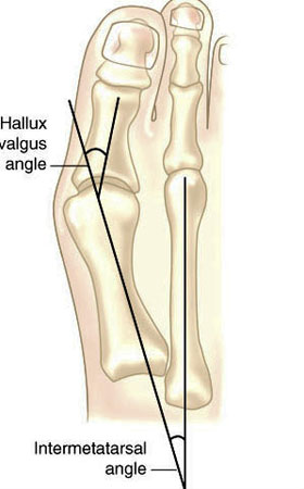
Hallux valgus refers to the big toe headed west (or east, depending on the foot and your GPS). In other words, the proximal and distal phalanyx of the great toe (hallux) have an angle with the 1st metatarsal shaft of typically > 15 degrees. This angle, called the Hallux Valgus Angle (HVA above) is used to judge severity, often for surgical intervention purposes but can guide conservative management as well. 

Metatarsus Primus Varus (literally, varus deformity of the 1st metatarsal) often accompanies Hallux Valgus. It describes medial deviation of the 1st metatarsal shaft, greater than 9 degrees. This angle is called the intermetatarsal angle and is measured by the angle formed by lines drawn parallel along the long axis of the 1st and 2nd metatarsal shafts. 

One other measurement is the Distal Metatarsal Articular Angle, which measures the angle between the metatarsal shaft and the base of the distal articular cap (ie, where the cartilage is) of the 1st metatarsal. This typically should be less than 10 degrees, preferably less than 6 degrees. Remember, these are static angles, things can change with movement, engagement, weight bearing strategies and shoes. What you see statically does not always predict dynamic angles and joint relationship.s

Are you doing surgery? Perhaps, as a last resort. Hallux valgus and metatarsus primus varus can be treated conservatively.

How do you do that?

The answer is both simple and complex.

The simple answer is: anchor the head of the 1st ray and normalize foot function. This could be accomplished by:

  • EHB exercises to descend the head of the 1st metatarsal
  • exercise the peroneus longus, to assist in descending the head of the 1st metatarsal
  • short flexor exercises, such as toe waving, to raise the heads of the lesser metatarsals relative to the 1st
  • work the long extensors, particularly of the lesser metatarsals to create balance between the flexors and extensors
  • consider using a product like “Correct Toes” to normalize the pull of the muscles and physically move the proximal and distal phalanyx of the hallux
  • wear shoes with wide toe boxes, to allow the foot to physically splay
  • consider using an orthotic with a 1st ray cut out, to help descend the head of the 1st metatarsal

This is by no means an exhaustive list and you probably have some ideas of your own. 

The complex answer is that in the above example, we have only included conservative interventions for the foot and have not moved further up the kinetic (or neurological chain). Could improving ankle rocker help create more normal mechanics? Would you accomplish this by working the anterior leg muscles, the hip extensors, or both? Could a weak abdominal external oblique be contributing? How about a faulty activation pattern of the gluteus medius? Could a congenital defect or genetic be playing a role? We have not asked “What caused this to occur in the 1st place?”

Examine your patients and clients. Understand the biomechanics of what is happening. Design a rehab program based on your findings. Try new ideas and therapies. it is only through our failures that we can truly learn.

The Gait Guys

references used:

http://www.bjjprocs.boneandjoint.org.uk/content/90-B/SUPP_II/228.3

http://www.slideshare.net/ANALISIS/hallux-valgus-2008-pp-tshare

http://www.orthobullets.com/foot-and-ankle/7008/hallux-valgus

http://www.slideshare.net/bahetisidharth/hallux-valgus-31768699?related=1

To met pad or not to met pad; that is the question. 

We use these in practice all the time. Think of it as a “helper” while you are training your patient or client to have better foot mechanics. 

“It’s all about the stress transfer, so what you’re trying to do is unload a certain spot on the foot—generally the met head with a met pad—and transfer it to a more proximal region,” 

Here is a nice evidence based review:

http://lermagazine.com/cover_story/evidence-based-use-of-metatarsal-pads
Social media reader comments and questions:
Reader: You demonstrated a peroneal exercise a few days back. In addition to the use of MT pads, is it effective in supporting transverse metatarsal arch?
  • The Gait Guys It can be. In our opinion, the exercises are key and the pad supports the exercises
  • The Gait Guys the goal is to restore foot function so that you do not need the pad. If your client is patient and willing to work on the exercises you likely won’t need a met pad, they may help speed things up, but remember, it is a crutch and crutches can be helpful but one can get lazy in using them and not do the supportive work. We only try to use them sparingly and only when necessary.

Following up on the peroneal topic of the week. HEre is a trademark exercise of ours.
http://thegaitguys.tumblr.com/post/63377772517/trade-secret-proper-calf-raise-we-are-selling

  • From a Reader: Just used the exercise on myself and a patient. Really like it. 2 questions. Can you get a better effect if performed on the edge of a step? Secondly, do you want to start in a tripod position and perhaps with short foot?
  • The Gait Guys We are assuming you mean letting the heel drop lower than the step/forefoot: and based on that assumption……if you feel that your client is ready for eccentric strength you can opt to do a slow decent into a negative heel. However, if there is ankle rocker limitation it would be wise to keep in mind that some of that end range dorsiflexion one might think they are getting from the ankle mortise joint might be from arch/midfoot collapse dorsiflexion , rear foot eversion and strain the plantar structures. Perhaps as they progress and gain still and control perhaps it is a good option. AWESOME question. As always, just because someone can do something doesnt mean they can do it safely. As for the tripod question…….well, they will be in the forefoot bipod mostly……bc the heel is off the ground……but this brings up technical issues if they are forefoot valgus or FF varus…..esp if they are rigid deformities……..it is doubtful many people are even considering this……..so once again, bravo Sir ! Is is quite often the small things that are the defining issues of the success or failure of a methodology. Bravo. !!!!! So glad you are part of this brethren David !

The Fudge Factor

The Fudge Factor

It is often easier to accomplish a task faster, rather than slower (like an exercise or skiing) because of the cortex “interpolating” or making its “best guess” as to what (based on experience) is going to happen. There is a certain amount of guess work (or what we call “the fudge factor”) involved.

Walking at a slower speed (or performing an exercise at a slower speed for that matter) has increased muscular demands, than doing it more quickly. Here is one study that exemplifies that.

“These findings may reflect a relatively higher than expected demand for peroneus longus and tibialis posterior to assist with medio-lateral foot stability at very slow speeds”

Gait Posture. 2014 Apr;39(4):1080-5. doi: 10.1016/j.gaitpost.2014.01.018. Epub 2014 Feb 6.
Electromyographic patterns of tibialis posterior and related muscles when walking at different speeds.
Murley GS1, Menz HB2, Landorf KB2.

The effect of walking speed on superficial lower limb muscles, such as tibialis anterior and triceps surae, is well established. However, there are no published data available for tibialis posterior - a muscle that plays an important role in controlling foot motion. The purpose of this study was to characterise the electromyographic timing and amplitude of selected lower limb muscles across five walking speeds. Thirty young adults were instructed to walk barefoot while electromyographic activity was recorded from tibialis posterior and peroneus longus via intramuscular electrodes, and medial gastrocnemius and tibialis anterior via surface electrodes. At faster walking speeds, peak electromyographic amplitude increased systematically during the contact and midstance/propulsion phases. Changes in the time of peak amplitude were also observed for tibialis posterior, tibialis anterior and peroneus longus activity; however, these were muscle and phase specific. During contact phase, peak electromyographic amplitude for tibialis posterior and peroneus longus was similar across very slow to slow walking speeds. During midstance/propulsion phase, peak electromyographic amplitude for tibialis posterior and medial gastrocnemius was similar across very slow to slow walking speeds. These findings may reflect a relatively higher than expected demand for peroneus longus and tibialis posterior to assist with medio-lateral foot stability at very slow speeds. Similarly, peak amplitude of medial gastrocnemius was also relatively unchanged at the very slow speed, presumably to compensate for the reduced forward momentum. The data presented in this study may serve as a reference for comparing similarly matched participants with foot deformity and/or pathological gait.

Copyright © 2014 Elsevier B.V. All rights reserved.

‪#‎gait‬
‪#‎thegaitguys‬

So you think you are an iliotibial band syndrome guru ?  This study has some interesting provoking thoughts about the mechanics we have all previously assumed. It is good to challenge established teachings, for it is only through interrogating old w…
So you think you are an iliotibial band syndrome guru ?  This study has some interesting provoking thoughts about the mechanics we have all previously assumed. It is good to challenge established teachings, for it is only through interrogating old ways that we may see the true light of things.
The iliotibial band (ITB) syndrome is a common overuse injury that is commonly misunderstood. It has been regarded as a friction syndrome where the ITB rubs against he lateral femoral epicondyle because of its previously assumed variable function, below 30 degrees knee extension it has been though to act as an extensor of the knee, and above 30 degrees (ie more knee flexion) it has been thought to act as flexor.  It is thought to be a culprit (with the biceps femoris) of the shift phenomenon in the “pivot shift test” for posterolateral rotatory instability of the knee (PLRI).  Here is an interesting perspective from a 2006 journal article.
“In all cadavers, the ITB was anchored to the distal femur by fibrous strands, associated with a layer of richly innervated and vascularized fat. In no cadaver, volunteer or patient was a bursa seen. The MR scans showed that the ITB was compressed against the epicondyle at 30° of knee flexion as a consequence of tibial internal rotation, but moved laterally in extension. MR signal changes in the patients with ITB syndrome were present in the region occupied by fat, deep to the ITB. The ITB is prevented from rolling over the epicondyle by its femoral anchorage and because it is a part of the fascia lata. We suggest that it creates the illusion of movement, because of changing tension in its anterior and posterior fibres during knee flexion. Thus, on anatomical grounds, ITB overuse injuries may be more likely to be associated with fat compression beneath the tract, rather than with repetitive friction as the knee flexes and extends.”

We found this article interesting because it challenges many thoughts about its actual movement, (“it creates an illusion of movement”) because of changing of tension in the anterior and posterior fibres. As this article suggests, it is unlikely that there is any forward and backward motion of the band over the epicondyle during flexion and extension, rather the illusion of movement is from a gradual shifting of load to and from the anterior and posterior fiber bundles during flexion/extension. It is also an interesting article to us because it suggests and challenges that the clinical phenomenon is associated with fat compression rather than friction over the epicondyle.  The authors go into discussion of how the fat beneath the distal ITBand at the knee level is well vascularized and that Pacinian corpuscles can be present in adipose tissue supporting the view that fat compression may have a proprioceptive role and a roll in pain production when the corpuscles undergo hypertrophy in such a clinical setting.
Just remember what we have been saying all along when treating what you think are lateral chain problems, the ITBand receives most of the tendon of the gluteus maximus so do not forget to examine the hip and pelvis function, but so not forget the critical contribution that impaired foot and ankle function can have proximally at the knee.
This study has some interesting provoking thoughts about the mechanics we have all previously assumed. It is good to challenge established teachings, for it is only through interrogating old ways that we may see the true light of things.
If you are looking for more of our thoughts on this topic, we discussed a clinical case in our last podcast (link here). 
Shawn and Ivo, 
the gait guys

Fairclough J, Hayashi K, Toumi H, et al. The functional anatomy of the iliotibial band during flexion and extension of the knee: implications for understanding iliotibial band syndrome. Journal of Anatomy 2006;208(3):309-316. doi:10.1111/j.1469-7580.2006.00531.x

CAI: More on Chronic Ankle Instability.

More peroneii action! In folks with chronic ankle instability, it contracts earlier, longer (throughout stance phase) but not stronger…This article looks at activation times and patterns of folks with chronic ankle instability. 

One should never wonder why repeated ankle sprains occur. We have hit this topic hard in the past.  Chronic Ankle Instability (CAI) clients exhibit prioprioceptive and postural control challenges. According to this article, additionally, CAI clients have gait. 

Time of activation for all muscles tested occurred earlier in the CAI group than in the control group. The peroneus longus was activated for a longer duration across the entire stride cycle in the CAI group.”

Individuals with CAI demonstrated neuromuscular-activation strategies throughout the lower extremity that were different from those of healthy control participants. ”

Did you see our trademark “goto” exercise in yesterday’s social media Facebook blog post ?  It is a keeper if you ask us.  Don’t ignore chronic peroneal challenges, they will come back to haunt you.

_________

Lower Extremity Muscle Activation in Patients With or Without Chronic Ankle Instability.  Mark A. FegerMEd, ATCLuke DonovanMEd, ATCJoseph M. HartPhD, ATCJay HertelPhD, ATC, FNATA, FACSM Department of Kinesiology, The University of Virginia, Charlottesville

http://www.natajournals.com/doi/abs/10.4085/1062-6050-50.2.06 

Results:  Time of activation for all muscles tested occurred earlier in the CAI group than in the control group. The peroneus longus was activated for a longer duration across the entire stride cycle in the CAI group (36.0% ± 10.3%) than the control group (23.3% ± 22.2%; P = .05). No differences were noted between groups for measures of electromyographic amplitude at either preinitial or postinitial contact (P > .05).

Conclusions:  We identified differences between the CAI and control groups in the timing of muscle activation relative to heel strike in multiple lower extremity muscles and in the percentage of activation time across the entire stride cycle in the peroneus longus muscle. Individuals with CAI demonstrated neuromuscular-activation strategies throughout the lower extremity that were different from those of healthy control participants. Targeted therapeutic interventions for CAI may need to be focused on restoring normal neuromuscular function during gait.

More research on the peronei and chronic ankle instability.

More peroneii action! In folks with chronic ankle instability, it contracts earlier, longer (throughout stance phase) but not stronger…This article looks at activation times and patterns of folks with chronic ankle instability. 

One should never wonder why repeated ankle sprains occur. We have hit this topic hard in the past. Chronic Ankle Instability (CAI) clients exhibit prioprioceptive and postural control challenges. According to this article, additionally, CAI clients have measurable gait changes. 

“Time of activation for all muscles tested occurred earlier in the CAI group than in the control group. The peroneus longus was activated for a longer duration across the entire stride cycle in the CAI group.”
“Individuals with CAI demonstrated neuromuscular-activation strategies throughout the lower extremity that were different from those of healthy control participants. ”

Did you see our trademark “goto” exercise in yesterday’s social media Facebook blog post ? It is a keeper if you ask us. Don’t ignore chronic peroneal challenges, they will come back to haunt you.

_________

Lower Extremity Muscle Activation in Patients With or Without Chronic Ankle Instability. Mark A. Feger, MEd, ATC; Luke Donovan, MEd, ATC; Joseph M. Hart, PhD, ATC; Jay Hertel, PhD, ATC, FNATA, FACSM Department of Kinesiology, The University of Virginia, Charlottesville

http://www.natajournals.com/doi/abs/10.4085/1062-6050-50.2.06

Podcast 89: 2015 Shoe Talk, Foot Beds, and shoe stuff you need to know.

A. server links

http://traffic.libsyn.com/thegaitguys/pod_89f.mp3

http://thegaitguys.libsyn.com/89

B. iTunes link:
https://itunes.apple.com/us/podcast/the-gait-guys-podcast/id559864138

C. Gait Guys online /download store (National Shoe Fit Certification and more !) :
http://store.payloadz.com/results/results.aspx?m=80204

D. other web based Gait Guys lectures:
Monthly lectures at : www.onlinece.com   type in Dr. Waerlop or Dr. Allen,  ”Biomechanics”

E. Our Book: Pedographs and Gait Analysis and Clinical Case Studies

electronic copies available here:

Amazon/Kindle: 

http://www.amazon.com/Pedographs-Gait-Analysis-Clinical-Studies-ebook/dp/B00AC18M3E

Barnes and Noble / Nook /iTunes Readers:

http://www.barnesandnoble.com/w/pedographs-and-gait-analysis-ivo-waerlop-and-shawn-allen/1112754833?ean=9781466953895

https://itunes.apple.com/us/book/pedographs-and-gait-analysis/id554516085?mt=11

 Hardcopy available from our publisher:

http://bookstore.trafford.com/Products/SKU-000155825/Pedographs-and-Gait-Analysis.aspx


Show notes:

 
Running Shoes : Alex and Blaise ?
 
And on that same topic, Foot beds, sock liners and orthotics:
 
super feet
what you put in your shoe can change the way the shoe was designed to work……careful what you put in the shoe 
 
 
and … 
Effect of rocker shoes on plantar pressure pattern in healthy femal… - PubMed
http://www.ncbi.nlm.nih.gov/pubmed/24370440
 
Why Running Shoes do not work:
Vastus lateralis. Closed chain internal rotator of the thigh. Stimulation of this point (ST34) improves gait in elderly individuals (who we assume have compromised mechanoreceptors in the muscles and joints). Think about incorporating this muscle in…

Vastus lateralis. Closed chain internal rotator of the thigh. Stimulation of this point (ST34) improves gait in elderly individuals (who we assume have compromised mechanoreceptors in the muscles and joints). Think about incorporating this muscle into your rehab protocol, particularly in older folks. 

Arch Phys Med Rehabil. 2011 Jan;92(1):7-14. doi: 10.1016/j.apmr.2010.09.023.
Stimulation of acupoint ST-34 acutely improves gait performance in geriatric patients during rehabilitation: A randomized controlled trial.
Hauer K1, Wendt I, Schwenk M, Rohr C, Oster P, Greten J.

“CONCLUSIONS:
Study results showed that a 1-time administration of a specific acupoint stimulation regimen statistically significantly improved gait performance during geriatric ward rehabilitation. If sustainability of effects can be documented, acupuncture may prove to be an inexpensive intervention that may mildly improve motor performance in frail geriatric patients.”

http://www.ncbi.nlm.nih.gov/pubmed/21187200

Commentary on this topic copied from our social media:

  • Reader: The abstract doesn’t give much away. I suspect using the acupuncture technique DECREASED activity of the VL leading to a more balanced muscular response to gait…kinda like massaging out an overactive muscle - which I don’t like doing but people do…
  • The Gait Guys Could be. We like to think of it as it created homeostasis of the muscular system. The effects of inserting a needle are not just local but global.
  • Reader:Hmm. I am not sure there is evidence to support that statement.  Something changed. Homeostasis was not necessarily achieved. Their gait improved. For how long is unknown. Also, it is unlikely that the people applying the acupuncture were blinded so that is a source of bias. It is interesting for sure, but drawing conclusions is difficult.
  • The Gait Guys agreed. you bring up some good points. The folks doing the acupuncture were not blinded
We think it has to do with reciprocal inhibition and increased long flexor activity, which was eluded to but not discussed at length in the article. “As previously mentioned, few studies have investigated the effects of thong style flip-flops …

We think it has to do with reciprocal inhibition and increased long flexor activity, which was eluded to but not discussed at length in the article. 

“As previously mentioned, few studies have investigated the effects of thong style flip-flops on gait dynamics, one of which was the initial study done by the authors at Auburn University in which several gait kinematic and kinetic measures differed between two types of footwear illustrating that walking in flip-flops alters one’s gait when compared to sneakers.”


http://lermagazine.com/article/flip-flops-fashionable-but-functionally-flawed

Commentary on this article from our social media sites:

  • Reader: It’s great to see a study being done trying to quantify the effects that flip flops may have. Thanks for sharing.
  • The Gait Guys agreed. there are several in the article
  • Reader: Russ Brandt When I had mild shin splints from running I noticed that wearing flip flops inflamed my shins