Tibial torsion and the effect on progression angle

more tibial torsion = a change in progression angle.

How does tibial torsion impact the development of the foot progression angle?

Stratifying the data by Foot Progression Angle (FPA) revealed there were significant differences in tibial torsion among the groups and provided evidence that tibial torsion influences the direction and magnitude of the FPA. Offsetting torsions between the tibia and femur were more common in people with higher and lower FPA and had clearer patterns where the tibia tended to follow the direction of the FPA.

So, got that? The foot progression angle follows the tibial torsion...

Why do we care?

the greater the “kickstand” angle to the foot, the more we progress through the mid foot (rather than from the lateral aspect of the heel, up the lateral column, across the transverse metatarsal arch and through the 1st ray). This causes more mid foot pronation and more medial knee fall, resulting in gait inefficiency and often times in our experiences, increased knee pain.

Gait Posture. 2016 Sep;49:426-30. doi: 10.1016/j.gaitpost.2016.08.004. Epub 2016 Aug 3.
The rotational profile: A study of lower limb axial torsion, hip rotation, and the foot progression angle in healthy adults.
Hudson D1.

tumblr_o471xcV05d1qhko2so1_1280.jpg
tumblr_o471xcV05d1qhko2so3_1280.jpg
tumblr_o471xcV05d1qhko2so2_1280.jpg
tumblr_o471xcV05d1qhko2so4_1280.jpg

Holy twisted tibias Batman! What is going here in this R sided knee pain patient?

In the 1st picture note this patient is in a neutral posture. Note how far externally rotated her right foot is compared to the left. Note that when you drop a plumbline down from the tibial tuberosity it does not pass-through or between the second and third metatarsals. Also note the incident left short leg
In the next picture both of the patients legs are fully externally rotated. Note the large disparity from right to left. Because of the limited extra rotation of the right hip this patient most likely has femoral retro torsion. This means that the angle of her femoral head is at a greater than 12° angle. We would normally expect approximately 40° of external Rotation. 4 to 6° is requisite for normal gait and supination.

In the next picture the patients knees are fully internally rotated you can see that she has an excessive amount of internal rotation on the right compare to left, confirming her femoral antetorsion.

When this patient puts her feet straight (last picture), her knees point to the inside causing the patello femoral dysfunction right greater than left. No wonder she has right-sided knee pain!

Because of the degree of external tibial torsion (14 to 21° considered normal), activity modification is imperative. A foot leveling orthotic with a modified UCB, also inverting the orthotic is helpful to bring her foot somewhat more to the midline (the orthotic pushes the knee further outside the sagittal plane and the patient internally rotate the need to compensate, thus giving a better alignment).

a note on tibial torsion. As the fetus matures, The tibia then rotates externally, and most newborns have an average of 0- 4° of internal tibial torsion. At birth, there should be little to no torsion of the tibia; the proximal and distal portions of the bone have little angular difference (see above: top). Postnatally, the tibia should twist outward (externally) a total of 15 degrees until adult values are reached between ages 8 and 10 years of 23° of external tibial torsion (range, 0° to 40°). more cool stuff on torsions here

Wow, cool stuff, eh?

Change the foot, change the knee (and vice versa). A video case of External Tibial Torsion.

Here is a perfect example of external tibial torsion. Are you treating and training people and messing with their orthotics, squat knee-foot posturing or making gait/running/jumping changes or recommendations? If you are doing all of this and you do not know about tibial torsions, then shame on you, go apologize to these people right now. You could be causing them mechanical grief. Go buy them ice cream (even if they are “paleo”), that fixes most unintentional human mistakes. 

This is a classic presentation of external tibial torsion. This is an anatomic problem, you cannot fix this intrinsically, but you can help extrinsically. You teach these people about this issue and why the foot and the knee cannot cooperate. You teach them why their feet are spun out (increased foot progression angle) while their knee tracks straight forward sagittally. You teach them why they might heel strike far laterally and why their pronation phase might be abrupt. As in this video, you teach them why they might fashionably choose to narrow the foot progression angle (foot turned in) while at the same time having to bear weight on the lateral foot (in supination to externally spin the tibia) to keep the knee tracking sagittally. You teach them why this will be impossible to do in pumps (inversion sprain ouch) and why over time this will anger many joints and tendons. You teach them that without this accommodation they will track the knee inside the sagittal plane (as seen in the video).  You teach them why they might be at greater risk of having foot prontation issue pathologies, why they might have limited internal hip rotation, why orthotics likely do not do much for them (yes, there are exceptions), why certain shoes are a challenge for them while others are magical and why over time their once beautiful arch has begun to “fall” and be less prominent as they attenuate the plantar tissues.  

As you get good with this gait and biomechanics stuff, you should readily see and understand all of the issues discussed here today in a mere flash of instant brilliance so you know what to offer your client, in understanding and remedy options. As you have seen in this video, when left to their own devices, they naturally allow the knee to find the sagittal plane in a nice forward hinge. In this posture the foot is excessively progressed outward. Again, this is because of the tibial long bone torsion. This is their anatomy, this is not functional in this case. You cannot fix this, you help them manage this, first with their awareness, then with your brilliance.  You may implement exercises and gait strategies to help them become aware of mechanical issues and how to protect the foot-ankle, the knee and the hip. You teach them why they might have a tendency towards anterior pelvis posturing or sway back type postures. You teach them why, in some cases, they choose knee hyperextension as a comfortable yet lazy stance postural habit. You teach them why some shoes are “happy” shoes for them, and why others are pure evil.

A foundational principle we teach here at The Gait Guys is that the knee is a simple hinge between two multiaxial joints on either side of the knee, the hip and the foot-ankle complex. The knee really can only flex and extend, and when the mechanics above and below are challenged the knee has little depth to its abilities to tolerate much of anything except simple sagittal hinging. You can see that the foot posturing and tibial torsion rule the roost here in this video. You should learn in time that managing this case above and below the knee is where the pot of gold is found. You will learn in time that taping the knee is often futile, yet a worthy experiment both for you and the client in the discovery process, but that a life time of taping is not logical. External tibial torsion, although affording the knee that sagittal hinge plane, can narrow its range of safe sagittal mechanics and it is up to you to  help them learn and discover that razor’s edge safely and effectively when the torsion is large.  You should also discuss with them that as they plastically tissue adapt over the years (ie. pronate more and lose more arch integrity), this razor’s edge may widen or narrow for the knee mechanics as well as the hip and foot-ankle complex.  

For your reading pleasure, a classic example of how to interrogate a safe sagittal knee progression was discussed in this blog video piece we wrote recently, linked here.

Look and you shall find, but only if you know what you are looking for.

* Please now know that you should never off the cuff tell someone to turn inwards their outwardly spun foot. But if you do, have ice cream on hand, just in case.

Need more to spin your head ? Think about whether their IT band complex is going to be functioning normally.  Oy, where is that ice cream !

Shawn Allen, one of the gait guys

tumblr_nxo5gvpjDh1qhko2so1_1280.jpg
tumblr_nxo5gvpjDh1qhko2so2_1280.jpg

Now THERE”S some internal tibial torsion!

So, this gent came in to see us with L sided knee pain after it collapsed with an audible “pop” during a baseball game. He has +1/+2 laxity in his ACL on that side. He has subpatellar and joint line pain on full flexion, which is limited slightly to 130 (compared to 145 right)

 We know he has internal torsion because a line drawn from the tibial tuberosity dropped inferiorly does not pass through or near the plane of the 2nd metatarsal (more on tibial torsions here)

What would you do? Here’s what we did:

  • acupuncture to reduce swelling
  • took him out of his motion control shoes (which pitch him further outside the saggital plane)
  • gave him propriosensory exercises (1 leg balance: eyes open/ eyes closed; 1 legged mini squats, BOSU ball standing: eyes open/eyes closed)
  • potty squats in a pain free range
  • ice prn
  • asked him to avoid full flexion

Is it any wonder he injured his knee? Imagine placing the FOOT in the saggital plane, which places the knee FAR outside it; now load the joint an twist, OUCH!

tumblr_nfzg0nM4pC1qhko2so1_1280.jpg
tumblr_nfzg0nM4pC1qhko2so2_1280.jpg
tumblr_nfzg0nM4pC1qhko2so3_1280.jpg
tumblr_nfzg0nM4pC1qhko2so4_1280.jpg
tumblr_nfzg0nM4pC1qhko2so5_1280.jpg
tumblr_nfzg0nM4pC1qhko2so6_1280.jpg

What would you do? This is what we did.

History:

This 7 year old girl is brought in by her mother because of knee misalignment while skiing, L > R. No history of trauma; normal term birth with no complications. No knee pain. Of incidental note, she is deaf in the left ear.

Exam findings:

She has bi-lat. external tibial torsion, left much worse than right (40 degrees transmallolear angle vs 22 degrees. for info on measuring torsions, click here). remember, you should be able to draw a line from the tibial tuberosity down through the 2nd metatarsal head. 

She has a 5mm anatomical leg length deficiency on the right (see top above left).

She has femoral antetorsion right side with very little external rotation, approximately 10 degrees,  internal rotation is in excess of 50.  Left side has normal femoral versions (for a review of femoral versions and torsions, click here).  See last 2 pictures which are full internal and external rotation respectively.

She has a mild uncompensated forefoot varus (cannot really see from the pictures, you will need to take our word for it) with a relatively cavus arch to her foot(see center and last picture on right.

Neurologically, she appeared to have integrity with respect to sensation, motor strength and deep tendon reflexes in the lower extremities.

Assessment:

Pathomechanical alignment as described.  Severe left external tibial torsion. MIld to moderate right. Femoral antetorsion right.

Plan:

We are going to build her a medium heel cup full length modified UCB orthotic inverting the cast bi-lat. left greater than right.  We gave her  balance and coordination exercises, heel walking, lift/spread/reach and one leg balancing. She will follow up for a dispense.  Her mother will try to get a better fitting ski boot as the one she has currently is two sizes too big. She will return for a dispense. She should consider wearing the orthotics in everyday footwear as well. We will do a follow up post in a few weeks. 

The Gait Guys. Teaching you something new in each and every post. Like this post? Tell and share it with a friend. Don’t like this post? Let us know!

This is part 2 of a 2 part post; with the video from the case previously discussed

please note the following in the video:

  • body lean to left during left stance phase (to clear right longer leg)
  • circumduction of right lower extremity  (to clear right longer leg)
  • lack of arm swing bilaterally (cortical involvement)
  • patient looking down while walking (decomposition of gait)
  • shortened step length (decomposition of gait)
  • increased tibial varum bilaterally

ASSESSMENT:  This patient’s short leg and internal tibial torsion impediments to her full recovery. She has increased tibial varum noted which is complicating the picture. This is causing pathomechanics and an abductory moment not only at the knee but also in the lumbar, thoracic and cervical spines.

WHAT DID WE DO?:                    

  • We attempted to do the one leg standing exercise. She needed to hold on and did not feel stable on the left hip while performing this.  This is probably more of confidence rather than ability issue. 
  • We gave her the stand/sit exercise to try to improve gluteal recruitment.
  • We also gave her the lift/spread/reach exercise to attempt to strengthen her feet.
  • A full-length 5 mm lift was cut for the left shoe  She felt more stable when walking on this.
  • She was treated with IC, PIR and manipulative therapy and neuromuscular stim of the knee as well as left hip area above, below and at the joint line of the knee as well as gluteus medius and minimus.   
  • We may need to consider building a more aggressive orthotic with a forefoot varus post depending upon her progress and response to care  

 The Gait Guys. Making it real, each and every post here on the blog.

special thanks to SZ for allowing us to publish her case, so others can learn

tumblr_nc9grebaIQ1qhko2so1_1280.jpg
tumblr_nc9grebaIQ1qhko2so2_1280.jpg
tumblr_nc9grebaIQ1qhko2so3_1280.jpg

Wow! What would you do?

This is part 1 of a 2 part post. Look for the other one a few minutes after this one with a video up top for the conclusion

PRESENTING PROBLEM: This 54 YO female patient presents with with left sided knee pain.  She had a total knee replacement (TKR) done in 2011.  She’s had a significant amount of discomfort on the medial aspect of the knee since then. She had an MRI of the hip done thinking the problem was there, and found nothing.   She is walking with a bad limp, left leg is half inch shorter than the right.  Pain is worse at night, changes with weather. 

She has knee pain on the lateral aspect (points to tibial plateau and joint line) with swelling that goes down to the ankle left side.  She has been wearing a “Good Feet” OTC orthotic on the left side which she states helps quite a bit.

Generally speaking, stretching and analgesics make the discomfort better.    Ibuprofen 400 mg. b.i.d. can take the edge off  Soft sided brace (neoprene sleeve) makes a difference as well. The hard sided brace gives her difficulty.

WORK HISTORY: She works for a preschool.  Her job involves standing and getting up and down a lot.  

FAMILY HISTORY:  She has left sided lid ptosis, this evidently is familial.  

PHYSICAL EXAM:  She stood 5’ 1” and weighed approx. 150 pounds.

Viewing the knees bi-lat., the left knee is markedly externally rotated.

She does have a left short leg; tibial and femoral.  She has bilateral tibial torsion (look at the tibial tuberosities and drop a line straight down; it should pass through the 2nd metatarsal head) and marked internal tibial torsion on the left side (>60 degrees) with femoral retrotorsion (less than 8 degree angle of femoral head with the shaft) on this side.  There is no rotation of the thigh or leg past zero degrees midline. .  She had 10 degrees of tibial varum on the left hand side.  Her Q-angle is 10 degrees on that side.  There is plantar flexion inversion of the foot.  Left lower extremity has less sensation secondary to the her TKR  surgery.

Gait evaluation reveals a fair amount of midfoot pronation noted on the left hand side in addition to an intoed gait.  She has to lean her body over to the left to get the right leg to clear.

Some mild weakness noted of hip abduction musculature left hand side gluteus medius, middle and anterior fibers. Knee stability tests were negative.

Neurologically, otherwise, she had full integrity with respect to sensation, motor strength and deep tendon reflexes in the upper and lower extremities.

Please see part 2 of this post for additional info including our assessment and what WE did.

 The Gait Guys. Making it real, each and every post here on the blog.

special thanks to SZ for allowing us to publish her case, so others can learn

tumblr_n9y3v5nRcH1qhko2so1_1280.jpg
tumblr_n9y3v5nRcH1qhko2so2_1280.jpg
tumblr_n9y3v5nRcH1qhko2so3_1280.jpg
tumblr_n9y3v5nRcH1qhko2so4_1280.jpg
tumblr_n9y3v5nRcH1qhko2so5_1280.jpg

More on the “little guy”

We have been following this little guy for some time now. If you have not been keeping up, perhaps you should read herehere and here 1st. 

So, what do we see in these latest pictures?

Top left: neutral view.

  • He enjoys flip flops; probably not the best thing for a developing kiddo, in light of the excessive engagement of the posterior compartment (and reciprocal inhibition of the anterior compartment)
  • he has some tibial varum (ie bowleggedness) L > R
  • he has some developmental genu valgum whnich appears to be improving (need a Q angle review? click here)
  • no tibial torsion present on L: for a review on torsions, click here
  • still some external tibial torsion present on R (see section below on middle shots)

Top right and bottom: full internal rotation of R thigh: compare with bottom: full internal rotation of L thigh

  • he has adequate internal rotation (4 degrees needed) but not as great as left side (see bottom shot); this represents some improvement since we started
  • he has generous internal rotation of the left thigh

Middle Left: full external rotation of right thigh

  • note the position of the knee and the position of the foot; external tibial torsion is present. for a review of torsions, click here.
  • he has limited external rotation of the right thigh (compared with the left. The knee should fall more outside the saggital plane

Middle right: full external rotation of the left thigh

  • note the position of the knee and the position of the foot; internal tibial torsion is present. 
  • he has generous external rotation of the left thigh (compared with the left)


of other significant note: most of his calcaneal valgus has resolved; longitudinal arches are improved.

What now?

  • He continues to develop normally and continues to improve since his original presentation to the office
  • Having the child continue to walk barefoot
  • Continue to wear shoes with little torsional rigidity, to encourage additional additional intrinsic strength to the feet
  • He should continue to limit “W” sitting, as this will tend to increase the genu valgus present
  • We reviewed 1 leg balancing “games” and encouraged continuing agility activities, like balance beam, hopping, skipping and jumping on each leg individually
  • added in using a push and pedal bike
  • added in heel walking exercises

Ivo and Shawn. Bald. Good looking. Extraordinary Gait Geeks. Taking the world of gait literacy by storm with each and every post.

tumblr_n9y3ea9yyg1qhko2so4_r1_1280.jpg
tumblr_n9y3ea9yyg1qhko2so3_1280.png
tumblr_n9y3ea9yyg1qhko2so2_1280.jpg
tumblr_n9y3ea9yyg1qhko2so1_1280.jpg

How do you measure tibial torsion anyway?

With all the talk on the Crossfit blog about the knees out debate, we though we would shed some light on measuring torsions, beginning with tibial torsion, since this does not seem to have been taken account of in the discussion and we feel it is germane. 

Yo may have seen some of our other posts in tibial torsion here or here; this post will serve to help you measure it. 

Looking at the top left picture: we can see that the axis of the tibial plateau and the transmalleolar axis (an imaginary line drawn through the medial and lateral malleolus) are parallel at birth (net angle zero) and progress to 22 degrees at skeletal maturity, resulting from the outward rotation of the tibia of about 1-1.5 degrees per year. This results in a normal external tibial version of about 17-18 degrees (you subtract 5 degrees for the talar neck angle, talked about in the link above). Note that this is the normal or ideal angle we would expect (hope?) to see. Go 2 standard deviations in either direction and we have external and internal tibial torsions.

You can go about taking this measurement in may ways; we will outline 2 of them. 

  1. In the upper left picture, we see an individual who has their knee flexed to 90 degrees over the side of a table while seated. This represents the tibial plateau angle. You the use a protractor to measure the angle between the tibial plateau and an imaginary line drawn through the medial and lateral malleoli. This is the transmalleolar angle. You then subtract 5 degrees from this number (remember the talar neck angle?) to get the angle of tibial version (or torsion).
  2. In the lower left and right pictures, we have the patient supine with the knees pointed upward and tibial plateau flat on the table. Then, working from inferiorly, use a goniometer to measure the angle of the transmalleolar axis. Again, we subtract 5 degrees for the talar neck.

We would encourage you to read up on torsions. This post, which we wrote over a year ago, is probably one of the most important ones on tibial torsions. 

Torsions. Important stuff, especially when you are talking about the axis of the knees in activities like a squat. Remember, the knee is a hinge between 2 multiaxial joints (hip and ankle) and will often take the brunt of the (patho)mechanics, as it has fewer degrees of freedom of movement. If you have external tibial torsion and you push your knees (angle your feet) out further, you are moving the knees outside the saggital plane. You have better have a very competent medial tripod! If you have internal tibial torsion, angling the feet out may be a good idea. Know your (or your patients/clients/athletes) anatomy!

The Gait Guys. Bald, Good Looking and Twisted. Here to help you navigate your way through better biomechanics. 

tumblr_n9ql3rlqGi1qhko2so2_r1_1280.jpg
tumblr_n9ql3rlqGi1qhko2so1_1280.gif
tumblr_n9ql3rlqGi1qhko2so3_r1_1280.gif
tumblr_n9ql3rlqGi1qhko2so4_r1_1280.gif

ETT and Hip Extension

Not Extra Terrestrial Tricks, but rather External Tibial Torsion. How it effects hip extension.

We received this question from Matthew P on our Facebook post from 8/1 (original post from here) which was based on this article, and thought it would make an excellent opportunity to teach. 

I looked at this yesterday and had actually first come across it a year or more ago when I was trying to find some resources for femoral torsion. You guys are about the only ones discussing at length the impact and implication of adult femoral torsion.

Re: tibial torsion and your post yesterday saying that > 30deg external torsion can affect both knee and hip extension, what I still don’t understand is the mechanism behind the hip limitation. How would that torsion translate through the leg to the hip?

There are a few things we need to remember to make sense of this:

  • tibial torsion is the angular difference between the tibial plateau and distal tibial malleoli and refers only to the tibia, not the entire lower extremity (see top photo)
  • pronation can occur in the rear foot, mid foot, and fore foot
  • pronation causes internal spin of the leg and thigh, due to plantar flexion, eversion and abduction of the talus (see middle photo)
  • internal spin of the hip causes posterior translation of the femoral head via the “glide and roll” phenomenon
  • these are appropriate mechanics during the 1st ½ of the gait cycle (initial contact to mid stance)
  • pronation is one of the 4 shock absorbing mechanisms (pronation, ankle dorsiflexion, knee flexion, hip flexion) of the lower extremity

now try this (yes, at home!)

  • flex your knee
  • internally rotate you leg
  • allow your arch to flatten
  • try and extend your hip

Remember these facts about supination

  • supination is initiated by the swing phase leg as it starts in early swing and continues to terminal swing (see third picture)
  • supination (from full pronation), should occur from midstance to pre swing
  • supination makes the foot into a “rigid lever” to transpose forces from above the foot into the foot and allow for propulsion (see third picture)
  • supination involves external rotation of the lower leg and thigh (see pictures 3 and 4)
  • external rotation of the hip is accompanied by anterior glide of the femoral head via the “glide and roll” phenomenon
  • this position puts the gluteal muscles (max and posterior fibers of the gluteus medius) at a mechanical advantage

now try this:

  • extend your knee
  • externally rotate your leg
  • you should have a full arch at this point
  • try and extend your hip

What did you (hopefully) learn?

  1. When the hip is in an externally rotated position it is easier to extend; the femur head moves anteriorly, the femoral joint capsule becomes tighter and stability is created
  2. when the hip is internally rotated, it is more difficult to extend
  • the femur head glides posteriorly, changing the axis of rotation of the joint
  • the gluteus maximus and posterior fibers of the gluteus medius are at a mechanical disadvantage

OK. Got it? We sure hope so! Excellent question, Matthew. Thanks for the opportunity to teach this concept.

The Gait Guys. Taking you closer to mastering the gait cycle with each post. 

Foot Progression Angle Exaggeration: External Tibial Torsion

Take a look at the tibial tuberosity and then where you think the 2nd metatarsal head would be. What do you see? The 2nd metatarsal is lateral to the tibial tuberosity. You are looking at external tibial torsion. 

Lets see how this external tibail torsion behaves during a knee bending. Observe the medial drift of the knee during weight bearing knee flexion. Many folks would say that the problem here is the increased foot prontation, but that is not where the problem lies, that is where many of the forces are funneling though. The client is pronating more because the external tibial torsion that is creating this appearance has put the knee inside the foot tripods region of stability.

In external tibial torsion there is an external torsion or a “twist” along the length of the tibia (diaphysis or long section). This occurs in this example to the degree that the ankle joint (mortise joint) can no longer cooperate with sagittal knee joint.  When taking a client with external tibial torsion and pre-postioning their foot in a relatively acceptable/normal foot progression angle there is a conflict at the knee, meaning that the knee cannot hinge forward in its usual sagittal plane. In this case with the foot progression angle smaller than what this client would posture the foot, the knee the knee will be forced to drift medially.

Are you looking for torsions of the lower limb in your clients ?

Are you forcing them into foot postures that look better to  you but that which are conflicting to your clients given body mechanics ?  Would you correct this client’s foot turn out (increased progression angle) ? IF you did you would likely cause them knee pain in time.  Would you put them into a stability shoe to try and control the pronation ? Again, you are likely to draw their knee outside the saggital knee hinge that is presently pain free. There is more to shoe fit that just looking at the foot. First do no harm is our mantra ! 

Remember, telling someone to turn their foot in or out because it doesn’t appear correct to your eyes can significantly impair either local or global joints , and often both. Torsions can occur in the talus, the tibia and the femur.

Furthermore, torsions can have an impact on foot posturing at foot strike and affect the limbs loading response, from foot to core and even arm swing can be altered.  Letting your foot fall naturally beneath your body does not mean that you have the clean anatomy to do so without a short term or long term cost. 

This is some of the toughest stuff you will deal with clinically. The fence is narrow, if you do to little correction you fall off the fence into the wrong yard and create problems. If you do to much correction you get the same result. These torsional issues are a delicate balancing act many times. You first have to know what you have, then you have to know where the fix is, and then how much is safe.  Tricky stuff. This is exactly why in some folks a stability shoe can be magic or tragic and in others dropping into zero drop minimalism can be magic or tragic.  

Want more on torsion and versions ?  Type the words into the search box on our blog. We have plenty of good info for you.

Shawn and Ivo, The Gait Guys

Podcast 52: Limb Dominance & Other Cool Stuff

A. Link to our server:

http://thegaitguys.libsyn.com/podcast-52-limb-dominance-other-cool-stuff

B. iTunes link:

https://itunes.apple.com/us/podcast/the-gait-guys-podcast/id559864138

C. Gait Guys online /download store (National Shoe Fit Certification and more !) :

http://store.payloadz.com/results/results.aspx?m=80204

D. other web based Gait Guys lectures:

www.onlinece.com   type in Dr. Waerlop or Dr. Allen,  ”Biomechanics”

________________________________________

* Today’s show notes:

Neuroscience:

1. The Potential Downside of Wearable Biomechanical Monitoring Devices for Running

http://runblogger.com/2014/01/downside-of-wearable-biomechanical-monitoring-devices-for-running.html

2. Google’s Next Crazy Project: Smart Contact Lenses | Entrepreneur.com
http://www.entrepreneur.com/article/230927
3. How Humans Burn Fewer Calories Than Other Mammals
http://www.runnersworld.com/weight-loss/how-humans-burn-fewer-calories-than-other-mammals
4. Update: I was listening to your podcast and you said Ice Bug was out of business. I sell them in my store in Fairbanks Alaska where we have snow and ice on the ground for 6-7 months a year. The US distributor is Ice Bug USA. I also carry a the Salomon Snow Cross studded shoe.
The effects of limb dominance and fatigue on running biomechanics
http://www.gaitposture.com/article/S0966-6362(13)00702-9/abstract
5. Email case:
Dear Gaitguys,
   I have been on a search since October to determine the cause of my chronic tendonitis in my knees and right hip. Through my search I was told that my femurs are rotated internally and my tibia are externally rotated. This is causing my patella to face inward.
  I was told by one PT that I will never be able to run long distances without developing tendonitis. I want to believe he is wrong because I love running. I came across something called Femoral Anterior Glide in my research and was wondering if you guys believe this is a real condition. Also is there a way a person can know if they have this problem?
Thank you for all of your great posts!
 Kate 
6. Blog reader:

My 11 year old son walks with a very noticeable external tibial torsion. We just recently noticed this and I came upon your site while trying to research it. I also read that many time it has to do with a problem with the patella. Both my husband and daughter have had patella problems but do not have the duck walk like my son. I did ask a a pediatric sports medicine specialist about the problem and he said some kids just walk that way. Where should I bring my son for help with this?

7. Blog reader asks:

About a year ago there was an article posted called: “A case of the non-resolving ankle sprain. Things to think about when the ankle and foot just do not fully come around after a sprain”. I am 15 weeks into an identical problem and I was wondering if there was any way you guys could follow up with “MR” to see if he was ever able to resolve his issue. It is such a unique and frustrating case (being able to walk but not run) and I haven’t found any other instance of it until now. Thank you.

8. Shoe Fit Certification program
Link:Gait Guys online /download store (National Shoe Fit Certification and more !) :

http://store.payloadz.com/results/results.aspx?m=80204

9. Second metatarsal osteotomies for metatarsalgia: A robotic cadaveric study of the effect of osteotomy plane and metatarsal shortening on plantar pressure - Trask - 2013 - Journal of Orthopaedic Research - Wiley Online Library

http://onlinelibrary.wiley.com/doi/10.1002/jor.22524/abstract;jsessionid=BCFFA5207512C41214E7F3D601729EFE.f01t01

 Master of your own physiology
You don’t need perfect mechanics to win. Look at these fine gents and take note.
On the left we have Kenensia “Canny” Bekele, world and Olympic 5,000m and 10,000m world record holder, who sat back as …

 Master of your own physiology

You don’t need perfect mechanics to win. Look at these fine gents and take note.

On the left we have Kenensia “Canny” Bekele, world and Olympic 5,000m and 10,000m world record holder, who sat back as Mo Farah and Haile Gebrselassie set the pace for most of the race, and then sprinted at the end and won by 1 second. Note the crossover and lack of space between his thighs. Note also the internal tibial torsion of the left tibia and slight head tilt to the right.

In the middle is Mo Farah, the current 10,000 meter Olympic and World champion and 5000 meter Olympic, World and European champion. look at the pelvic dip on the right..and the valgus angle of the left knee…and external tibail torsion of the left tibia…and the differing arm swing (right side abducted).

Finally, on the right,  we have Haile Gebrselassie, an Ethiopian like Bekele, who won two Olympic gold medals over 10,000 meters and four Wld Championship titles in the event. He won the Berlin Marathon four times consecutively and also had three straight wins at the Dubai Marathon.  At 40, he is the eldest of the group, with his right lower extremity external tibial torsion and subtle dip of the left pelvis on right sided weight bearing.

So What? All these great athletes have mastered their own physiology and overcome any biomechanical faults they may appear to have. Could they be faster? Maybe. We think so.

Your body will find a way to compensate. That does not mean you will be slower. It means, like each of these men, that you will probably be injured at some point.

In the words of Big Z from Surf’s Up “Winners find a way”. You can too and so can your clients and athletes. Skill, endurance and strength. The big 3. Make sure you an the folks you care for have them.

We are The Gait Guys. Teaching you more with each post we write and helping you sort through the sea of information out there.

Video case: The King’s Preference: Short and Sweet. A quick and easy case demonstrating the patellar tracking struggles with external tibial torsion.

Our favorite functional evaluation piece of equipment as well as our favorite piece of therapy equipment is the Total Gym.  Here we clearly demonstrate, to us and the client, in partial weight bearing load, the effects of external tibial torsion.  

Remember, the knee is sort of the King of all joints when it comes to the lower extremity.  The knee is a sagittal plane hinge, and so all it wants to do is hinge forward, freely without binding from deficits at the hip or knee. But we cannot ignore the simple fact that pre-pubescent kids the long bone derotation process is still undergoing, and in adults the process may have been corrupted or insufficient.  

In this case it should be obvious that the knee is sagittal and free to hinge when the foot is at a large foot progression angle.  This allows the knee to hinge cleanly. But when the foot is corrected to the sagittal plane, as you see in the second half of the video, the knee tracks inward and this can cause patellofemoral pain syndromes, swelling, challenges to the menisci (and possible eventual tears) and challenges to the ACL and other accessory restraints.  Additionally, this medial drift is a longer and more difficult challenge to the eccentric phase external rotators such as the gluteus maximius not to mention many of the other muscles and their optimal function.  

So, the next time you see a large foot progression angle in a client or in their walk (duck footed if you will) try to resist the natural urge to tell them to corrrect the foot angle. They are likely doing it to keep the King happy.  And furthermore, be careful on your coaching recommendations during squats, olympic lifts, lunges and running.  Just because you do not like the way the foot looks doesn’t mean you should antagonize the King of joints.  

External tibial torsion, its not something you want to see, but when you do see it, you have to know its degree, its effects at the knee, hip and foot as well as how it might impact hip extension, pelvic neutrality, foot strike, foot type, toe off and so many other aspects.

Whoever said gait analysis was easy was a liar. And if all they use is a video camera and fancy analysis software they have show up with only part of the team. And if they said they were an expert  in gait only a few years into practice, you had better also look for a jester’s hat somewhere hiding in the corner. After all, the King would want to know !

Shawn and Ivo, your court jesters for the last 3+ years.  Maybe we will get a promotion from the King someday soon !

Photo: Where is your knee joint hinge point ?  Say that 4 times fast. 
Here is a photo of 4 elite runners. We suspect it is an 800m race  because #100 is Ahmed Bile who is the son of Olympian and world champion Abdi Bile.
In this photo you can see t…

Photo: Where is your knee joint hinge point ?  Say that 4 times fast.

Here is a photo of 4 elite runners. We suspect it is an 800m race  because #100 is Ahmed Bile who is the son of Olympian and world champion Abdi Bile.

In this photo you can see that Ahmed #100 has a significantly large foot progression angle (large foot turn out) and this likely represents external tibial torsion or femoral antetorsion while #454 has a neutral foot turn out as does #232.  #46 has a modest foot progression angle. Grossly, #46 also has the patella right over the foot and so tibial torsion is not likely. Now, move up to observe their knee progression. All of them have a forward (sagittally) oriented knee progression. How can that be? Well, it is simple if you know your torsional issues. After all, the knee is a hinge and if you are running forward your knee pretty much should hinge forward as well.  Now, there is much room for conversation here and debate but we are just trying to make and observation and a point. To a large extent the knee rules the roost in the lower limb in terms of sagittal progression because it is the joint with the least number of tolerances. The knee only hinges in flexion and extension where as the hip and ankle/foot have frontal and axial planes they can notably tap into when the sagittal is challenged.  Again, look at #100 and our point is made.

Look at the 2 fellas in the middle (454 and 232). they have a internally (medially) postured knee/thigh yet their foot progression angle is mostly neutral and the knees are hinging forward.  Does #454 have internal tibial torsion? It could be (hint, look at his right trailing leg, specifically the patella and foot postures) but the left limb looks cleaner although adducted suggesting he might like the cross-over gait or it is more external tibial torsioned. Where as the 2 outer fellas, 100 and 46, are more neutrally oriented knees/thighs (one could make the case that #100 has a more externally oriented femur) yet increased progression foot progression angle in an environment of a forward hinging knee.

So what gives ? Torsions. Yes, we are soapboxing on torsions again. Torsions in the tibia, torsions in the femur. Versions are normal expressed angles, tibial torsions are abnormal.

Now, as life would have it, look over the right shoulder of #100. See the fella in the red headband? Ya, that guy losing.  He has the cleanest lines of the bunch. How is that for cruel irony ?  Sometimes it ain’t what you got, it is what you do with what you got.  Unless of course he is actually wincing in pain and trailing behind because he got spiked by #100 and that hideously frontal plane splayed foot !

Lastly, this wouldn’t be an official Gait Guys post if we did not preach to remember that “what you see is not the problem, what you see in a gait analysis is the person’s compensatory strategy around their deficits”. And here we see deficits. Our observations today are merely just that, observations. Now someone has to get them on a table and examine them and confirm our observations, prove them wrong and/or discover the joint, muscular and motor pattern deficits that created these observations.  Or, someone has to confirm that all parts are working and that they were at the end of the line when the straight long bones were first handed out.  

Today’s Lesson:  Get in line, and get in line early. (just kidding of course)

The Gait Guys.  Calling it they way we see it, but reserving the right to plead the 5th or change our minds after an examination.  We would suggest to everyone, when it counts and when your reputation is on the line, plead the 5th, until you have completed your hands on clinical examination.  "Seeing may be believing" but that still doesn’t always make it so.

Want to learn more about these kinds of things, foot beds, foot types, shoe anatomy and shoe function, proper shoe prescription etc ?  Our National Shoe Fit program will help you get smarter about this stuff. email us at : thegaitguys@gmail.com 

Gait Guys online /download store:http://store.payloadz.com/results/results.aspx?m=80204

*Photo courtesy of BIG EAST Conference

tumblr_mqf5re5X3Q1qhko2so1_1280.jpg
tumblr_mqf5re5X3Q1qhko2so2_1280.jpg
tumblr_mqf5re5X3Q1qhko2so3_1280.jpg

Trojan horses for knee menisci.

 Orthotics and internal tibial torsion. Good? Bad? or Ugly? It depends…

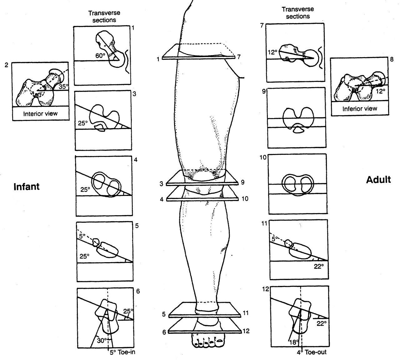
Hopefully you remember about torsions, especially internal tibial torsion (see above). Tibial torsions are deviations (in this case, in the transverse plane) of the long axis of the bone. The bone is basically twisted along its long axis, like wringing out a wet towel. They are measured by drawing an imaginary line through the medial and lateral malleoli, as well as through the two halves of the tibial plateau, and measuring the angle between them (see 2nd picture above). For a more complete review of torsions, click here.

 At birth there should be little to no angular difference between the proximal and distal tibia, and this changes to about 19-22 degrees in the adult; the shaft of the tibia rotates outward (externally) with growth resulting in a normal tibial external version (see 3rd picture above).  Sometimes, the angular difference is less than zero at birth and the tibia does not rotate outward (externally) resulting in internal tibial torsion.

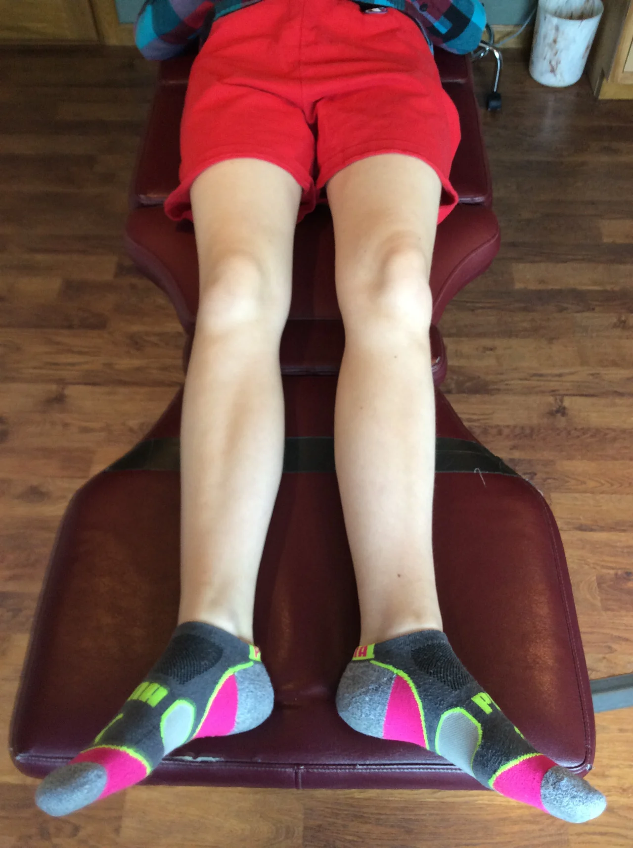
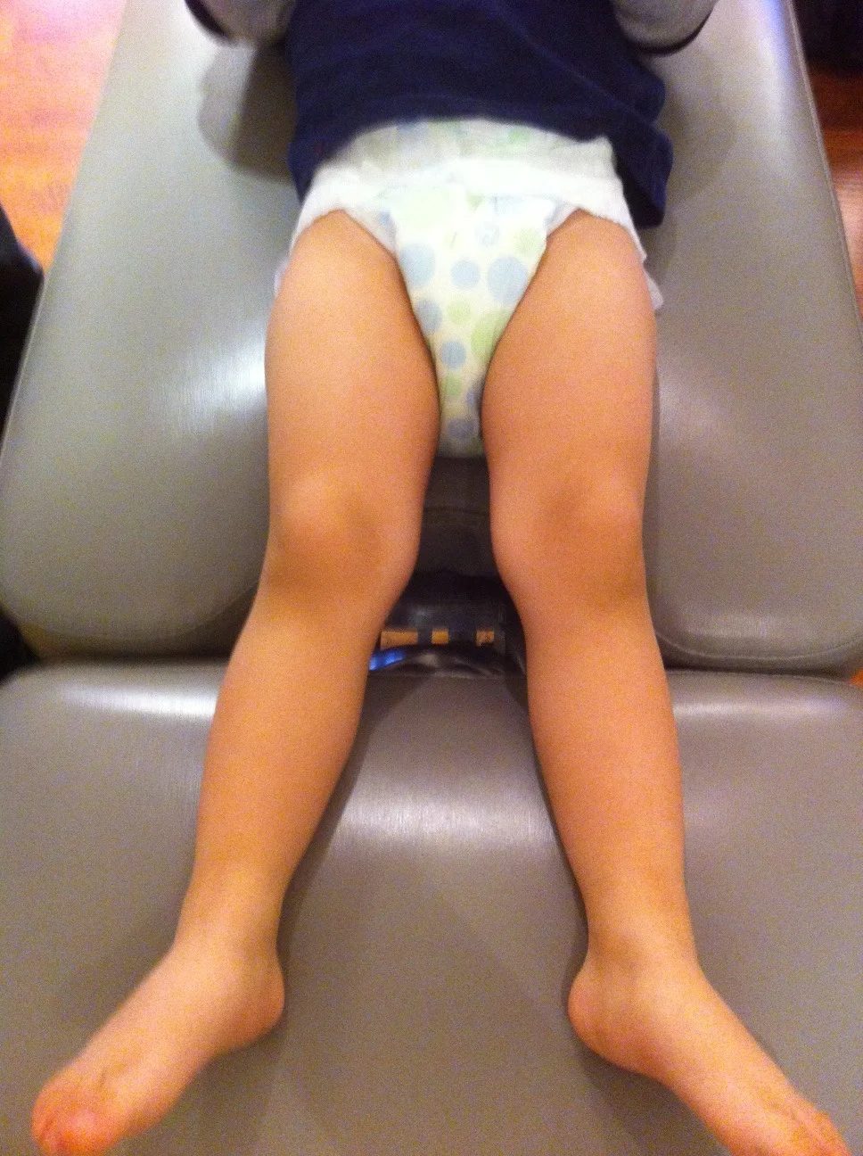
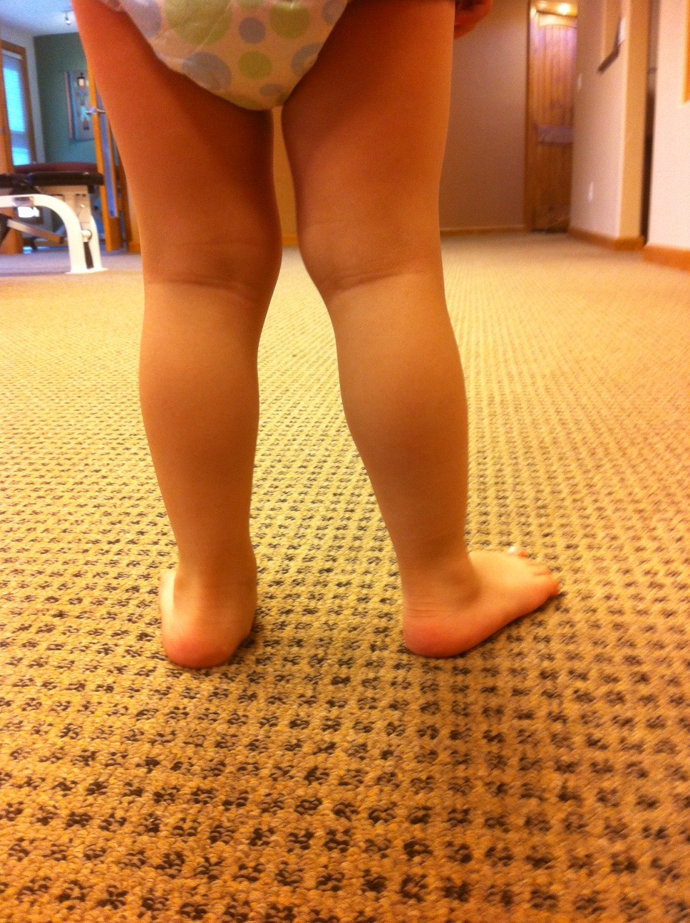
Internal tibial torsion usually results in a decreased progression angle (more on those here). This often causes a “toed in gait” and the foot remains in supination for a longer period of time (supination is adduction, inversion and plantar flexion), making the foot a rigid lever. When we examine the person in a standing position with the knees in the coronal plane, the feet point inward. When we move the feet to a more normal posture, the knees rotate outward from the coronal plane.

Folks with internal tibial torsion often have a forefoot varus (the forefoot is inverted with respect to the rear foot) because of the amount of supination they are in, which we talked about in the previous paragraph, (see also here). When folks have a forefoot varus, they have a tendency to pronate more through the forefoot, and when people pronate more other folks like to typically put them in orthotics to “get rid of that pronation”(because we all know that pronation is the scourge of humanity, and if there were less pronation in the world, there would probably be fewer wars, famine and poverty : )

 So what happens to the knee when we place an orthotic in the shoe? Most orthotics are designed to slow pronation of the midfoot, so they basically supinate the foot, causing the talus to dorsiflex, abduct and invert. This rotates the leg (and thus the knee) externally. With internal tibial torsion, often the knee is already externally rotated because your brain will not allow you to progress forward with your toes in too far, you would trip. So, the orthotic rotates the knee out further, bringing it outside the sagittal plane. This does not bode well long term, as it creates a rotational and friction conflict at the knee (remember the knee is basically a hinge between two ball and socket joints). Guess where the conflict manifests itself? At the meniscus. This, over time, is a great way to macerate a meniscus and create a problem.

Does this mean an orthotic is never indicated? No it does not. It means that if you use one, you should probably make sure the part of the orthotic anterior to the styloid of the 5th metatarsal has a valgus post built into it. This valgus moment will help to bring the knee back to the midline during the propulsive phase of gait. See our recent post here about forefoot valgus posting. Do you think this is ever considered in stores when dispensing foot beds for shoes ?  Not all foot beds are evil or a problem mind you, but we have seen some in stores that are real risky business if you ask us.

The bottom line? Know how to use the tools you have available, or someone is going to get hurt. When in doubt, exercise is usually a safer alternative and often has less likelihood of creating a Trojan Horse.  

Want to learn more about these kinds of things, foot beds, foot types etc ?  Our National Shoe Fit program will help you get smarter about this stuff. email us at : thegaitguys@gmail.com 

Gait Guys online /download store:http://store.payloadz.com/results/results.aspx?m=80204

The Gait Guys. Raising questions and providing answers and guidance, with each and every post.


all material copyright 2013 The Gait Guys/ The Homunculus Group. All rights reserved. Please ask before lifting our wares. 

What have we here?

Take a look at the tibial tuberosity and then where you think the 2nd metatarsal head would be. What do you see? The 2nd metatarsal is lateral to the tibial tuberosity. You are looking at external tibial torsion.

Lets see how this external tibail torsion behaves during a knee bend on a total gym. Observe the medial drift of the knee during weight bearing knee flexion. 

In external tibial torsion there is an external torsion or a “twist” along the length of the tibia (diaphysis or long section) (need a review? click here). This occurs in this example to the degree that the ankle joint (mortise joint) can no longer cooperate with sagittal knee joint.  When taking a client with external tibial torsion and pre-postioning their foot in a relatively acceptable/normal foot progression angle as seen here, there is a conflict at the knee, meaning that the knee cannot hinge forward in its usual sagittal plane. In this case with the foot progression angle smaller than what this client would posture the foot, you an see that as they bend the knee the knee is forced to drift medially and as soon as the heel is unloaded a pure “adductory twist” is noted (you can see the heel jump medially in an attempt to find a more tolerable sagittal knee bend).

Are you looking for torsions of the lower limb in your clients ?

Are you forcing them into foot postures that look better to  you but that which are conflicting to your clients given body mechanics ?  Remember, telling someone to turn their foot in or out because it doesn’t appear correct to your eyes can significantly impair either local or global joints , and often both. Torsions can occur in the talus, the tibia and the femur.

Furthermore, torsions can have an impact on foot posturing at foot strike and affect the limbs loading response, from foot to core and even arm swing can be altered.  Letting your foot fall naturally beneath your body does not mean that you have the clean anatomy to do so without a short term or long term cost. 

Want more on torsion and versions ?  Type the words into the search box on our blog. We have plenty of good info for you.

Shawn and Ivo, The Gait Guys

External Tibial Torsion as expressed during gait.

So, last week we watched this young lad doing some static ankle and knee bends, essentially some mini squats.  Here was what we found (LINK). It is IMPERATIVE that you watch this LINK first before watching today’s video above.

Now that you have watched that link here is what you should be seeing today.

You should see that the left foot is extremely turned out. We talked about why in the linked post from last week. It is because of the degree of external tibial torsion.  When it is present the knee rides inside the foot progression line (the knee bends into the forward / sagittal plane when the ankle bends into its more lateral /coronal / frontal plane (they all mean the same thing) ie. when the foot points outwards.

Remember, the knee has only one choice of motion, to hinge forward and backward. When the knee is asked to hinge in any other direction once the foot is locked to the ground there is torque placed upon the knee joint and thus shear forces.  Menisci do not like shear forces, nor does articular joint cartilage.

So, once again we see the rule of “you cannot beat the brain” playing out. The brain took the joint with the least amount of tolerance, the knee, and gave it the easy job.  The foot was asked to entertain another plane of motion as evidenced here in this video with significant increased foot progression angle. 

When the foot progression angle is increased but the knee still must follow the forward body progression (instead of following the foot direction) the motion through the foot will be directly through the medial longitudinal foot arch.  And as seen here, over time this arch will fail and collapse. 

Essentially this lad is hinging the ankle sagittally / forward through the subtalar and midtarsal joints, instead of through the ankle mortise joint where ankle hinging normally should occur.

This is a recipe for disaster. As you can see here.  You MUST also know and see here that there is an obvious limp down onto that left limb. It appears the left limb is shorter. And with this degree of external tibial torsion and the excessive degree of foot pronation, the limb will be shorter. You need to know that internal limb spin and pronation both functionally shorten the limb length.  This fella amongst other functional things is going to need a full length sole lift. We will start with 3mm rubber infused cork to do so. And let him accomodate to that to start.

We will attempt to correct as much foot tripod (anti-pronation) control as possible to help reduce leg shortness as well as to help reduce long term damage to the foot from this excessive pronation. We will also strengthen the left gluteus medius (it was very weak) to help him engage the frontal/lateral/coronal plane better. This may bring that foot in a little. But remember, the foot cannot come in so far that it drives the knee medially. Remember who is ruling the roost here !…… the knee.  It only has one free range, the hip and foot have 3 ! 

Shawn and Ivo

External Tibial Torsion: A Video showing the effects on the knee, foot and hip.

Tibial torsion is measured as the angular difference between the tibial plateau and the distal malleoli. Here we have put 2 blue dots on the tibia, one at the middle of the tibial plateau and one at the half-way bisection of the malleoli (“ankle bones”). You can see that the dots clearly do not rest on the same vertical plane, they are nowhere close in this case !  This is tibial torsion.  And since the lower dot is outside the upper dot, this is EXTERNAL TIBIAL TORSION.  It is one of the factors which determine the progression angle of the foot (see our post here).  It is easily seen here that, the foot will follow the lower dot because that is where the foot is attached to the ankle mortise joint.  This is thus what is referred to as an “increased or excessive” Foot Progression Angle.  Some will loosely, and humorously, refer to this as being “Duck Footed”.  (But we have never seen a duck with external tibial torsion so who knows how this came about ! :-)

Normally, the angle is 0 degrees in an infant leaving the feet straight or slightly “in-toed”, and the tibia “unwinds” with growth, leaving the angle in adults at approximately 22 degrees. Angles in excess of 25 degrees are considered external tibial torsion; angles less than 15 degrees, internal tibial torsion.

In this case video it is critical to note a few things:

1- at the beginning we coached the client to straighten the feet forward so we could see the effects of the tibial torsion on the knees. In External Tibial Torsion, as in this case, the knees will always drift inwards (this is why these clients will always turn out the feet so that the knees and patella can track forward in the  normal hinge progression that is necessary for gait.  A case of external tibial torsion like this case will never see them walking with the feet straight forward (0 degree foot progression angle) because they will knock the knees together and the patella will track incorrectly and develop knee tracking pain.

2- you should be able to see that the client cannot dorsiflex the ankle sufficiently at the start because of the binding of the ankle into the torsioned distal tibia-fibula ankle mortise joint.  The client gets “locked out” and cannot squat more than a few degrees.  Be sure to notice this.  These clients should not be pigeon-holed into how they do squats and lunges (“Straighten your feed lad!” should not be your recommendation, they just won’t be able to do much if they do. They will cheat !)

3. The second set of squats show them with the feet turned out excessively. They are able to get down further now, but the knees are now tracking too far outside and not forward.  This was too much accommodation for the external tibial torsion. 

4. The 3rd set are done (at 0:28 seconds into the video) with a more reasonable foot alignment.  Reasonable for this client but far too much for someone who does NOT have external tibial torsion. You can see that the single planar hinge joint knee (the joint with the least tolerance) now moves nicely forward towards the camera.  So, they will walk with the feet at this progression angle because this is where the external tibial torsion has left the knees to rest in the sagittal (forward) plane.  Here the client will have minimal if any knee issues. However, one can only imagine what their ankles and feet and hips will think of all this !

To discern tibial torsions from femoral torsions, observe the orientation of the tibial tuberosity (the upper blue dot) with respect to the foot; in tibial torsion, there is a large difference. If this angle is withing the 15-25 degree range, then the torsion lies in the femur (femoral retro torsion and ante torsion…the subject of another post).

So, if you are training, coaching or rehabing  a client are you aware of issues like these ? Are you attempting to drive skill, endurance and strength into your clients into a lower limb plane that is reasonable for their bony alignment?  Maybe you were not even aware of these issues at all ?  Lets hope not.  You just cannot pigeon-hole all of your clients into a similar paradigm. It just isn’t that simple.  Not if you do not want to injure someone.  We get alot of these cases, sadly.

We discuss this in more detail in our Shoe Fit program that is soon to launch, but we also presented several 1 hour slide presentations on www.onlineCE.com in the recent months if you wanted to take those lectures.

Shawn and Ivo, two twisted fellas.  Torsioned dudes.  One is internally torsioned, the other external……… we cancel eachother out !

tumblr_m1h0g09NlZ1qhko2so1_1280.jpg
tumblr_m1h0g09NlZ1qhko2so2_1280.jpg
tumblr_m1h0g09NlZ1qhko2so3_1280.jpg
tumblr_m1h0g09NlZ1qhko2so4_1280.jpg
tumblr_m1h0g09NlZ1qhko2so5_1280.jpg

Quiz: Let’s see how your blog reading has been going. 

These 5 photos are of a 2.5 year old child brought into your office for evaluation by his father. They have been seen by another practitioner who has given him orthotics with full length varus posts to wear.

  • What do you see?
  • What is your assessment?
  • What do you tell the parent?

In the standing views, what stands out?

  1. a moderate rearfoot (calcaneal) valgus (ie. rear foot medial  heel collapse)
  2. the flattened medial longitudinal arches of the foot ( ie. a little flat footed)
  3. the genu valgus (ie. knees are a little “knock knee’d”)
  4. he bears weight separately on each lower extremity as you can see from the pictures.  He never bears weight on both limb symmetrically, there is much weight bearing shifting meaning there is always a dominant limb bearing most of the weight.
  5. the knees face inward in the standing position
  6. the feet point outward (with the knees straight) in the supine position

did you see all of these?

What is your assessment?

1, 2)  pes planus and hyperpronation are the norm for children under 6 years of age

3) genu valgus is not abnormal in children, with many presenting maximally at age 3, and usually resolving by age 9 (see our post here)

4) he bears weight separately on each lower extremity (L>R from rear, R>L from front) so there probably is not a leg length discrepancy.  This is often a hip-core stability issue and as fatigue sets in weight bearing shift is automatized. 

5,6) This child has external tibial torsion. As seen in the supine photo, when the knees face forward, the feet have an increased progression angle (they turn out). We are born with some degree / or little to none, tibial torsion and the in-toeing of infants is due to the angle of the talar neck (30 degrees) and femoral anteversion (the angle of the neck of the femur and the distal end is 35 degrees).  The lower limbs rotate outward at a rate of approximately 1.5 degrees per year to reach a final angle of 22 degrees….. that is of course if the normal derotation that a child’s lower limbs go through occurs timely and completely.

What do you tell the parent?

1,2)  Although research shows that wearing arch supports (navicular wedge or “cookie”, not a full varus wedge as was the case here) can speed development of the the arches, they will in fact most often develop regardless of supportive footwear or support.  Many studies show that footwear impairs muscular development of the foot. One study showed that arch supports in children prevent derotation of the talar head and promote development of a Rothbart Foot Type. How about some flexible shoes (or no shoes) for the boy?  (need to review the Rothbart foot type ? click here for one of our very first blog posts on the topic)

3. We will measure the genu valgus and track it every 6 months to make sure it is regressing. If it persists or becomes worse, we may address it then. How about having your kid walk barefoot?

4. no worries, he is resting each side as the other fatigues. Endurance development takes time, just like marathon training. For gosh sake, the kid is 2.5 years old. Give him a break !

5,6) We will measure his progression angle and degree of torsion every 6 months (along with the genu valgus). This is normal up to reaching skeletal maturity.

Well, how did you do?

Corrective exercises are always nice, but when is too soon? Can their immature nervous system handle it ?  CAn they comprehend the exercise ? Sometimes turning them into a game and taking what you can get is good enough to help promote healthy limb derotation.  Walking with the toes up helps develop arch independence and helps to teach the brain about the foot tripod. But at 2.5 years old, good luck expecting more than that.  

The Gait Guys: two handsome bald guys (one by genetics, and one by aspiring choice) aging gracefully and promoting foot and gait literacy, one case at a time.