Invalid video embed.

What have we here?

Take a look at the tibial tuberosity and then where you think the 2nd metatarsal head would be. What do you see? The 2nd metatarsal is lateral to the tibial tuberosity. You are looking at external tibial torsion.

Lets see how this external tibail torsion behaves during a knee bend on a total gym. Observe the medial drift of the knee during weight bearing knee flexion. 

In external tibial torsion there is an external torsion or a “twist” along the length of the tibia (diaphysis or long section) (need a review? click here). This occurs in this example to the degree that the ankle joint (mortise joint) can no longer cooperate with sagittal knee joint.  When taking a client with external tibial torsion and pre-postioning their foot in a relatively acceptable/normal foot progression angle as seen here, there is a conflict at the knee, meaning that the knee cannot hinge forward in its usual sagittal plane. In this case with the foot progression angle smaller than what this client would posture the foot, you an see that as they bend the knee the knee is forced to drift medially and as soon as the heel is unloaded a pure “adductory twist” is noted (you can see the heel jump medially in an attempt to find a more tolerable sagittal knee bend).

Are you looking for torsions of the lower limb in your clients ?

Are you forcing them into foot postures that look better to  you but that which are conflicting to your clients given body mechanics ?  Remember, telling someone to turn their foot in or out because it doesn’t appear correct to your eyes can significantly impair either local or global joints , and often both. Torsions can occur in the talus, the tibia and the femur.

Furthermore, torsions can have an impact on foot posturing at foot strike and affect the limbs loading response, from foot to core and even arm swing can be altered.  Letting your foot fall naturally beneath your body does not mean that you have the clean anatomy to do so without a short term or long term cost. 

Want more on torsion and versions ?  Type the words into the search box on our blog. We have plenty of good info for you.

Shawn and Ivo, The Gait Guys

tumblr_miuvjyHzjf1qhko2so1_1280.jpg
tumblr_miuvjyHzjf1qhko2so2_250.jpg
tumblr_miuvjyHzjf1qhko2so3_400.gif
tumblr_miuvjyHzjf1qhko2so4_250.jpg

Twisted, Part 4


Hopefully you have been keeping us with us. If you missed the 1st 3 of this series, go back 3 weeks and start reading again, or do a search on the blog page for “torsion”.

The final chapter of developmental versions involves the femur. The degree of version is the angle between an imaginary line drawn through the condyles of the femur and an imaginary line drawn through the head and neck of the femur. This is often referred to as the femoral neck angle or FNA.

Beginning about the 3rd month of embryological development (Lanz and Mayet 1953) and reaches about 40 degrees (with an average of 30-60 degrees) at birth. It then decreases 25-30 degrees by adulthood to 8-20 degrees with males being at the lower and females at the upper end of the range.

The FNA angle, therefore, diminishes about 1.5 degrees a year until about 15 years of age. Femoral neck anteversion angle is typically symmetrical from the left side to the right side.

What causes torsion in the first place? By the sixth month in utero, the lumbar spine and hips of the fetus are fully flexed, so perhaps it is positional. Other sources say it coincides with the degree of osteogenesis. There is a growing consensus that muscular forces are responsible, particularly the iliopsoas  or possibly the medial and lateral hip rotators.

Additional changes can occur after birth, particularly with sitting postures. “W” sitting or “cross legged” sitting have been associated with altering the available range of motion and thus the FNA, with the range increased in the direction the hip was held in; W sitting causing increased internal rotation and antetorsion and cross legged causing external rotation and retro torsion.

As discussed previously, there are at least 3 reasons we need to understand torsions and versions, They can alter the progression angle of gait, they usually affect the available ranges of motion of the limb and they can alter the coronal plane orientation of the limb.

1. fermoral torsions often alter the progression angle of gait.  In femoral antetorsion torsion, the knees often face inward, resulting in an intoed gait and a decreased progression angle of the foot. This can be differentiated from internal tibial torsion (ITT) by looking at the tibia and studying the position of the tibial tuberosity with respect to the foot, particularly the 2nd metatarsal. In ITT, the foot points inward while the tibial tuberosity points straight ahead. In an individual with no torsion, the tibial tuberosity lines up with the 2nd metatarsal. If the tibial tuerosity and 2nd met are lined up,  and the knees still point inward, the individual probably has femoral ante torsion. Remember that a decreased progression angle is often associated with a decreased step width whereas an increased angle is often associated with an increased step width. See the person with external tibial torsion in the above picture?

2. Femoral torsions affect available ranges of motion of the limb. We remember that the thigh leg needs to internally rotate the requisite 4-6 degrees from initial contact to midstance (most folks have 40 degrees) If it is already fully internally rotated (as it may be with femoral retro torsion), that range of motion must be created or compensated for elsewhere. This, much like internal tibial torsion, can result in external rotation of the affected lower limb to create the range of motion needed.

Femoral retro torsion results in less internal rotation of the limb, and increased external rotation.

Femoral ante torsion results in less external rotation of the limb, and increased internal rotation.

3. femoral torsions usually do not effect the coronal plane orientation of the lower limb, since the “spin” is in the transverse or horizontal plane.

The take home message here about femoral torsions is that no matter what the cause:

  •  FNA values that exist one to two standard deviations outside the range are considered “torsions”

  • Decreased values (ie, less than 8 degrees) are called “retro torsion” and increased values (greater than 20 degrees) are called “ante torsion”
  • Retro torsion causes a limitation of available internal rotation of the hip and an increase in external rotation

  • Ante torsion causes an increase in available internal rotation  of the hip and decrease in external rotation
  • Femoral ante torsion will be perpetuated by “W” sitting (sitting on knees with the feet outside the thighs, promoting internal rotation of the femur)

  • Femoral antetorsion will be perpetuated by sitting cross legged, which forces the thigh into external rotation.


Stay tuned for a case tomorrow to test your learning over the last few weeks.


We remain: Bald, good looking and intelligent…The Gait Guys


All material copyright 2013 The Gait Guys/ The Homunculus Group. All rights reserved.  Please ask to use our stuff!

Step width alters iliotibial band strain during running.

More substantiation that “the cross over gait” is a pathologic process.

Did you get to hear podcast #23 yet ?  Here is the link (iTunes).  In podcast #23 we talked at length about the effects of step width in runners.  Reducing ones step width will result in a progression into what we have been referring to for years as “the cross over gait”.  We have been reducing this phenomenon in our runners, and many walkers, for over a decade now to reduce many of the lower limb pathologic processes that ensue when the cross over is left unchecked and worse yet, strength and endurance is loaded upon the faulty pattern.  Everyone’s gait in this realm will differ because of pelvis width, femoral and tibial torsion, genu posturing (knee valgum, varum)  and foot structure and type. All of these factors must be taken into account when deciding upon the degree of step width correction.  Ultimately the goal in a perfect world would be to have the foot and knee stack pristinely under the centrated hip joint proper, but we all know that ideal biomechanics are the unicorn when it comes to humans. Anatomic variation is the known norm and this must not be forgotten, this was pounded into all of our heads in medical school.
As this article from the Nov 2012 J. of Sports Biomechanics clearly states, iliotibial band strain and strain rate is significantly greater in narrow based gait scenarios and that increasing step width during running, particularly in those who tend towards the lazier narrower step width, may be beneficial in not only the treatment but the prevention of future lateral hip and knee biomechanical syndromes such as IT band syndrome.  So, if you are a slave to your foam roller and need your IT band foam roller fix daily, you might want to look a little deeper at your biomechanics and make some changes.  Our videos here will be helpful to you and our writings on the Cross Over gait  and link here will be helpful as well.
In  summary, there is just so much more to good running form than just following the mantra “let my feet fall under my body mass and everything will be just fine”.  We wish it was this easy, but it is not. Unfortunately, too many of the sources on the internet are maintaining that good running form is mostly just that simple. Sadly, we find it our mission to bring the bitter tasting truth to the web when it comes to these things.  One just cannot ignore the factors of pelvis
width, femoral and tibial version and torsion, genu posturing (knee valgum, varum) and foot structure and foot type (and we mean so much more than are you a pronator or supinator).  These factors will alter lower limb biomechanics and may drive even the runner with heightened awareness of foot strike and running form into less than optimal foot strike positioning and loading response. Furthermore, one needs to be acutely aware that merely taking the cooked down under-toned postulation of this journal article, that being increasing step width will resolve their IT band problems, may not resolve their problem. In fact, without taking the issues of pelvis width, torsion, version, foot type and the like into account, making these changes could bring about more problems.  Seeking the advise of a knowledgeable physician in this complicated field of human locomotion is paramount to solve your chronic issues.
There is more to clean running than just a
midfoot-forefoot strike under the body mass, a good forward lean and high cadence. And we are here to bring those other issues to light, for the sake of every injured and frustrated runner.  Remember, uninjured does not always mean efficient. And efficient does not always mean uninjured.

Shawn and Ivo, The Gait Guys

_______________________________
Sports Biomech.
2012 Nov;11(4):464-72.Meardon SA, Campbell S, Derrick TR.

Step width alters iliotibial band strain during running.

Abstract
excerpted:

“Greater ITB strain and strain rate were found in the narrower step width condition (p < 0.001, p = 0.040). ITB strain was significantly (p < 0.001) greater in the narrow condition than the preferred and wide conditions and it was greater in the preferred condition than the wide condition. ITB strain rate was significantly greater in the narrow condition than the wide condition (p = 0.020). Polynomial contrasts revealed a linear increase in both ITB strain and strain rate with decreasing step width. We conclude that relatively small decreases in step width can substantially increase ITB strain as well as strain rates. Increasing step width during running, especially in persons whose running style is characterized by a narrow step width, may be beneficial in the treatment and prevention of running-related ITB syndrome.”

Podcast #24: Chronic achilles issues, beer recovery drink and case studies.

podcast link: http://thegaitguys.libsyn.com/pod-24-the-chronic-achilles-beer-cases

iTunes link: https://itunes.apple.com/us/podcast/the-gait-guys-podcast/id559864138

Show notes:

Gait Guys online store:

http://store.payloadz.com/results/results.aspx?m=80204

Today’s show notes:

1. J Trauma Acute Care Surg. 2013 Mar;74(3):946-7. doi: 10.1097/TA.0b013e31828272ad.
Achilles’ death: Anatomical considerations regarding the most famous trauma of the Trojan War.
2. J Foot Ankle Surg. 2001 May-Jun;40(3):132-6.Saxena A, Bareither D.

Magnetic resonance and cadaveric findings of the “watershed band” of the achilles tendon.
3. http://www.sciencedaily.com/releases/2013/02/130212112019.htm


Fallout from Nuclear Testing Shows That the Achilles Tendon Can’t Heal Itself
4. K. M. Heinemeier, P. Schjerling, J. Heinemeier, S. P. Magnusson, M. Kjaer. Lack of tissue renewal in human adult Achilles tendon is revealed by nuclear bomb 14C. The FASEB Journal, 2013; DOI: 10.1096/fj.12-225599

5.  http://www.washingtontimes.com/news/2013/feb/10/scientists-suggest-beer-after-workout/#.USRSIq-QMnw.facebook

Scientists suggest beer after a workout
6. Sports Biomech. 2012 Nov;11(4):464-72.

Step width alters iliotibial band strain during running.
7. _http://skorarunning.com/confessions-of-an-overpronator

Over-Pronation

8. http://www.championseverywhere.com/why-gait-analysis-doesnt-work-future-of-the-shoe-industry

Why gait analysis doesn’t work (future of the shoe industry

tumblr_min5irxcLp1qhko2so1_1280.jpg
tumblr_min5irxcLp1qhko2so2_1280.jpg

Yet another IFGEC certified individual, and our 1st in Asia! Congrats, Andy! Here’s what he has to say:

My name is Andy Neo aka Dee. I work as the regional senior tech rep for a distributor shoe company managing Singapore, Malaysia, Thailand & Indonesia. Additionally I graduated with a Bachelor degree in Sports & Outdoor from Monash University and an avid distance runner.   

I’m a keen learner about exercise physiology & human movement but there is no course for shoe fitting since I started working in the footwear industry few years back. A lot of knowledge was self learnt through trial & error because there are no industrial standards across the running shoes market. Fortunately I happened to tumble The Gait Guy’s blog & Facebook and was a keen follower of their regular posts. I was overjoyed when Dr Ivo & Dr Shawn in 2012 announced the opportunity to participate the National Shoe Fit Certification Examination online especially for me coming from another continent. The extensive 3hr lecture did helps to bridge the gap between health science, sports science & footwear education (seriously lacking in modern shoe industry). The lecture video was downloaded and studied at my own pace was really beneficial for working adults like myself. My advice to future IFGEC candidates is to have a good read up of human anatomy, shoe anatomy and human biomechanics for better understanding because the extensive 3hours lecture use technical terminology that can be jargon but definitely worthy information.

The quality of  examination questions required critical thinking & hence raise the standard of the certificate. Passing the IFGEC examination would elevate my profession so now I can better impart the knowledge to my retail staffs across 4 countries for a holistic shoe-fit service.

I recommend representative from performance footwear company & medical healthcare professions to spend quality time to sign up for this level 1 course because the contents will narrow the gap between health science & shoe industry.”


For more information on IFGEC certification, please email us at thegaitguys@gmail.com

http://thegaitguys.tumblr.com/post/21713480315/the-chef-another-abnormal-gait-pattern-in
Last night we caught a DVR&rsquo;d season show of Bourdain&rsquo;s new show &ldquo;the Layover&rdquo; on the Travel Channel.  Great new show, we love his diatri…

http://thegaitguys.tumblr.com/post/21713480315/the-chef-another-abnormal-gait-pattern-in

Last night we caught a DVR’d season show of Bourdain’s new show “the Layover” on the Travel Channel.  Great new show, we love his diatribes, rants and command of the English language. It reminded us of this post we did last year where we looked in depth at his unique gait flaw, the circumducting foot.

Join us again for this great gait dissection, see the link at the top. Even if you have been with us for over a year and still somewhat remember this gait pattern and our explanation of it, you will likely pick up another layer of  understanding after our last year of teaching  here on The Gait Guys blog.

Shawn and Ivo, The Gait Guys

tumblr_miivgh6NpC1qhko2so1_1280.jpg
tumblr_miivgh6NpC1qhko2so2_1280.png

Yes, we are all twisted. Part 3 continued.

If you missed yesterdays post, this one will make more sense if you go back and read it.Today we talk about compensations for tibial torsions.

As discussed in previous posts, there are at least 3 reasons we need to understand  tibial torsions and versions:

1. They will often alter the progression angle of gait.  In internal tibial torsion, there will often be a decreased progression angle of the foot and with external, an increased angle of progression. A decreased progression angle is often associated with a decreased step width whereas an increased angle is often associated with an increased step width.

2. They affect available ranges of motion (ROM) of the limb. We remember that the lower leg needs to internally rotate the requisite 4-6 degrees from initial contact to midstance:

ROM changes that may occur with internal tibial torsion

  • If it is already fully internally rotated (as it may be with internal tibial torsion), that range of motion must be created or compensated for elsewhere.
  • This can result in external rotation of the affected lower limb to create the range of motion neede
  • Circumduction of the lower limb, because the foot is already in a supinated posture, and the decreased range of motion of the foot needs to be compensated for.
  • A shortened step length, due to increased compressive forces at the medial knee
  • And alteration of vertical and medial lateral ground reactive forces
  • A rolling off the lateral aspect of the foot, due to it being in a more supinated posture

ROM changes that may occur with external tibial torsion          

  • external tibial torsion often results in the increased midfoot pronation, through the deformity, because more range of motion is possible both at the hip and foot at the subtalar joint

3. They often can effect the coronal plane orientation of the lower limb.

In internal tibial torsion, due to the foot being more rigid and the deformity often being accompanied by increased tibial varum, the knee often falls outside the plane of the foot (rather than being “stacked”), resulting in a decreased step width and often a cross over gait pattern (click here for more info on crossover)

In external tibial torsion, the foot is often more pliable. This often results in an increased step width and well as the knee falling inside (or medially) to the plane of the foot. Because of the increased hip and foot ranges of motion available,  the foot is not an adequate lever, shortening step length and sometimes requiring increased pelvic motion to “get around” the stance phase leg.

Whew! This stuff can be tough, Thanks for hanging in there! Next stop: Femoral Torsions and Versions!

Ivo and Shawn; your torsioned friends : )


All material copyright 2013 The Gait Guys/ The Homunculus Group. All rights reserved.  Ask before you lift our stuff, Lee is watching……

tumblr_mic2p8EQnN1qhko2so1_1280.jpg
tumblr_mic2p8EQnN1qhko2so2_1280.png
tumblr_mic2p8EQnN1qhko2so3_1280.gif

Yes, we are all twisted; Part 3


In the last 2 posts we discussed the differences between torsions and versions, as well as talar version and torsion, 1 of the 3 major versional events that occur during normal development (missed out? Click here and here to re read them).

In this post we discuss tibial versions and torsions.

The tibia and femur are more prone to torsional defects, as they are longer lamellar (layered) bones as opposed to the cancellous bone that makes up the talus. These often present as an “in toeing” or “out toeing” of the foot with respect to the leg; changing the progression angle of gait (click here for more on progression angles).

Tibial versions and torsions can be measured by the “thigh foot angle” (the angulation of the foot to the thigh with the leg bent 90 degrees: above right) or the “transmalleolar angle” (the angle that a line drawn between the medial and lateral malleoli of the ankle makes with the tibial plateau: above left). 

At a gestational age of 5 months, the fetus has approximately 20° of internal tibial torsion. As the fetus matures, The tibia then rotates externally, and most newborns have an average of 0- 4° of internal tibial torsion. At birth, there should be little to no torsion of the tibia; the proximal and distal portions of the bone have little angular difference (see above: top). Postnatally, the tibia should twist outward (externally) a total of 15 degrees until adult values are reached between ages 8 and 10 years of 23° of external tibial torsion (range, 0° to 40°). 

Sometimes the rotation at birth is excessive. This is called a torsion. Five in 10,000 children born will have rotational deformities of the legs. The most common cause is position and pressure (on the lower legs) in the uterus (an unstretched uterus in a first pregnancy causes greater pressuremaking the first-born child more prone to rotational deformities. Growth of the  unborn child accelerates during the last 10 weeks and the compression from the uterus thus increases. As you would guess, premature infants have less rotational deformities than full-term infants. This is probably due to decreased pressure in the uterus. Twins take up more space in the uterus and are more likely to have rotational deformities. 

Of interesting note, there is a 2:1 preponderance of left sided deformities believed to be due to most babies being carried on their backs on the left side of the mother in utero, causing the left leg to overlie the right in an externally rotated and abducted position.

Normal ranges of versions and torsions are highly variable (see chart above: right). Ranges less than the values are considered internal tibial torsion and greater external tibial torsion.

Internal tibial torsion (ITT) usually corrects 1 to 2 years after physiological bowing of the tibia (ie tibial varum) resolves. External tibial torsion (TT) is less common in infancy than ITT but is more likely to persist in later childhood and NOT resolve with growth because the natural progression of development is toward increasing external torsion.

Males and females seem to be affected equally, with about two thirds of patients are affected bilaterally and the differences in normal tibial version values are often expected to be cultural, lifestyle and posture related.

 The ability to compensate for a tibial torsion depends on the amount of inversion and eversion present in the foot and on the amount of rotation possible at the hip. Internal torsion causes the foot to adduct, and the patient tries to compensate by everting the foot and/or by externally rotating at the hip. Similarly, persons with external tibial torsion invert at the foot and internally rotate at the hip. Both can decrease walking agility and speed if severe. With an external tibial torsion deformity of 30 degrees , the capacities of soleus, posterior gluteus medius, and gluteus maximus to extend both the hip and knee were all reduced by over 10%.

Well, that was probably more than you wanted to know about tibial torsions, and we could go on for many more pages and perhaps cure any insomnia you may have. Take a while to digest this, as it is important to gait, shoe selection, and rehabilitation. Torsions are an acquired taste and we hope we have whetted your appetite! Tomorrow we talk about compensations!


Ivo and Shawn; two twisted guys!


All material copyright 2013 The Gait Guys/ The Homunculus Group. All rights reserved.  Ask before you lift our stuff, Lee is watching……

Podcast #23. Neurology of walking babies, dialogues on step width for runners and so much more !

Syndication link:

http://thegaitguys.libsyn.com/podcast-23-walking-babies-step-width-cross-over-running

iTunes link:

https://itunes.apple.com/us/podcast/the-gait-guys-podcast/id559864138

Podcast #23. Neurology of walking babies, dialogues on step width for runners and so much more !

1- Newborn babies walk the walk | Body & Brain
http://www.sciencenews.org/view/generic/id/348262/description/Newborn_babies_walk_the_walk

2- 3D printing with stem cells could lead to printable organs - CNET Mobile
http://m.cnet.com/news/3d-printing-with-stem-cells-could-lead-to-printable-organs/57567789

3- our payloadz e-file download site.  http://store.payloadz.com/results/results.aspx?m=80204

4- www.onlineCE.com  

Great TeleSeminar Wed Feb 20th, 2013 8:00 PM Eastern Time Chiropractic TeleSeminar Biomechanics 302 Location: 1 hr by telephone Instructor: Waerlop/Allen, DC Price: 19.00

5- J Biomech. 2004 Jun;37(6):935-8.Owings TM, Grabiner MD.  Step width variability … .

Brach JS.    J Neuroeng Rehabil. 2005 Jul 26;2:21.  Step width variability … .

Sports Biomech. 2012 Nov;11(4):464-72.  IT Band strain and step width … .

6. Rethinking Ice Baths And Ibuprofen pulse.me/s/isg3t Inflammation IS part of the healing process!

7. Bringing the Foot Back To Life: Restoring the Extensor Hallucis Brevis Muscle.

http://youtu.be/1iZg_e4veWk

How did you do on yesterday&rsquo;s photo of 3 foot strikes ? Now, look at the women ! Almost pristine ! Still a little evidence of cross over gait in each of them, but pretty darn good ! The feet are falling under the body mass, not good for sprint…

How did you do on yesterday’s photo of 3 foot strikes ?
Now, look at the women ! Almost pristine ! Still a little evidence of cross over gait in each of them, but pretty darn good ! The feet are falling under the body mass, not good for sprinting, but utilized more in endurance athletes. If you have enough abdominal and hip frontal stabilzers (ie. G. Medius et al) you can play safely with the efficiency factor. If you do not have the strength, you sacrifice efficiency and risk injury at multiple sites in the lower limb. The more cross over you have, the more the foot will strike in inversion (more lateral foot strike), then you much hope you have enough gluteus medius strength at the hip, medial knee stability strength and ankle/arch strength in muscles like the tibialis posterior. 

Kara Goucher and Shalane Flanagan, right, shown here leading the 2012 Olympic Trials Marathon,
Photo: Kurt Hoy/Competitor

A visual demonstration of 3 different foot strike patterns. Lets test some of what you have learned here at The Gait Guys over the last few months.
On the readers left, blue shirt Bib 232: The left leg appears to have all joints stacked at this view…

A visual demonstration of 3 different foot strike patterns. Lets test some of what you have learned here at The Gait Guys over the last few months.

On the readers left, blue shirt Bib 232:
 The left leg appears to have all joints stacked at this viewer angle (knee is vertically over the foot, hip is over the knee). What we love here is that the foot profile (look at the black sole of the shoe) is parallel to the ground, it is hard to believe that it won’t strike as such. The medial and lateral aspects of the foot should strike flush and simultaneously. This is a neutral foot and is very likely without valgus or varus forefoot typing.  The tibia looks pristine and straight without any torsion, at least from this limited perspective. He also looks to be striding nicely, it seems to appear (albeit this is reaching from this head on view) that the foot will strike below the body mass, this may be because he subtly appears to be leaning forward, again hard to see on this view.

Middle runner, white shirt:  We see some problems here.  First of all, it appears (and again, this is reaching from a front on view) that this runner is striding out with the foot beyond the body mass and will likely heel strike, he also seems to be in more backward lean that the Blue Bib Man but again hard to tell on a frontal view. We also see that the foot is pitched in inversion (note the outward tip of his foot compared to the man in Blue) quite aggressively which will facilitate a strong excessive lateral heel and/or forefoot strike pattern.  You can also see that drawing a line through the length of the long bones (tibia and femur) that they are in alignment, they are even in alignment with the 90 degree perpendicular to the forefoot inverted angulation.  This clearly represents our classic “cross over gait” which was first brought to you and the internet by yours truly a few years ago (here on Youtube link).  It is easy to see that the projected foot landing will be on a virtual line and thus appear to run on a line or even cross the feet over the line indicating that this client is not stacking the foot, knee and hips vertically and thus challenging the gluteus medius and hip stability into the frontal plane (video link here). This client will be wasting energy and efficiency in the frontal plane (side to side movement) and challenging the core, risking knee tracking issues and excessive foot pronation forces beyond the safe and normal.  

Running on the readers right, green shirt #8:  There appears to be a strong stance phase leg collapse, the hip is lateral to the foot and the knee is perhaps on its way to medial from a vertical line from the foot. This can be, and often is, from the issues of cross over described in the middle runner above but it can also be simply found in someone who is striking with the foot/knee/hip joints stacked but does not have sufficient gluteus medius strength to keep the pelvis level on the horizon (thus drift laterally). When this happens the downward collapse of the opposite side pelvis is often, but not always, see as a valgus collapse at the knee since the femur is allowed to drift medially from insufficient strength, skill or endurance pairing of the gluteus medius/maximus pairing and the medial quadriceps. This client is  likely a cross over victim as well and this would give good reason to the aforementioned.  Again, this is all theoretical from a static picture but knowing these patterns like we do, we know these typical patterns of breakdown. This is also suspect because of the foot more positioned under the midline of the body instead of under the knee and hip vertically stacked and the obvious proximity of the knees to one another.  These clients often kick or brush the foot or shoe against the stance phase lower leg as they swing the foot through. 

Who is going to win this race ? One cannot tell. But if they were the same on all levels of endurance, training, VO2 max and equal on every parameter except what was mentioned above, well then our man in Blue, # 232 would be the most efficient and likely the least injured.

Photo from an Outside Magazine article. We Would reference it, and would be happy to do so, but we cannot find the net article anywhere now. Please send it our way if you happen across it !

Shawn and Ivo, The Gait Guys … .  followed in 51 countries and counting.

tumblr_mhnym1twha1qhko2so1_1280.jpg
tumblr_mhnym1twha1qhko2so2_1280.png
tumblr_mhnym1twha1qhko2so3_r1_1280.gif

Yes, we are all twisted: Part 2

Last time we spoke of the 3 major versional changes in the normal course of development of he lower extremity and their importance (if you missed out or forgot, click here). In this post, lets look at the talar neck.

The talus is to the foot, as the lunate is to the hand. It is the only bone that has the entire weight of the body passing through it before being distributed to the foot. It’s motion during pronation should be flexion, adduction and eversion, and in supination: extension,  abduction and inversion.

At birth, the angle between the talar neck and talar dome is 30 degrees adduction. This reduces to 18-20 degrees in the adult (see above). During this reduction of angle, the talar head also everts or “twists” laterally (ie promotes pronation), which helps to correct the supination and adducted position of the forefoot in adults present in infants (Saffarian 2011).

Abnormal talar loading and “untwisting” in development (see bottom right picture)  has been linked to formation of a Rothbart foot type, also known as metatarsus primus elavatus (Rothbart 2003, 2009,2010. 2012). The 1st metatarsal is elevated and inverted with respect tot eh rest of the foot, with it behaving much like a fore foot varus.

Talar torsion (sometimes called subtalar version) results when there is a 10 degree or greater change in the final position of the talar head. This can cause an adducted position of the forefoot, often mistakenly called “forefoot adductus’, which actually only applies to the metatarsals, and not at all to the talus.

An adducted forefoot provides challenges to gait with many possible compensations. As discussed last time, there are at least 3 reasons we need to understand torsions and versions:

1. They will often alter the progression angle (forgot about progression angles? click here). In talar adduction, there will often be a decreased progression angle of the foot.

2. They affect available ranges of motion of the limb. We remember that the lower leg needs to internally rotate the requisite 4-6 degrees from initial contact to midstance, If it is already fully internally rotated, that range of motion must be created elsewhere. This may result in external rotation of the affected lower limb, excessive pronation through the deformity (if possible), or rolling off the lateral aspect of the foot.

3. They often can effect the coronal plane orientation of the lower limb. In talar torsion, the head of the talus often does not “untwist” appropriately resulting in a functional forefoot varus, with excessive forefoot pronation occurring at terminal stance and pre swing.

There you have it in a nutshell. Talar tosion: Present in 8% of the population (Bleck 1982) and coming to your clinic or a shoe store near you!

All meat and no filler. The Gait Guys!


All material copyright 2013 The Gait Guys/ The Homunculus Group. All rights reserved.  Please ask before recycling our stuff!

Treat your children well.
We think Crosby, Stills, Nash and Young had it right&hellip;
look at this conclusion: &ldquo;Shoes affect the gait of children. With shoes, children walk faster by taking longer steps with greater ankle and knee motion and …

Treat your children well.

We think Crosby, Stills, Nash and Young had it right…

look at this conclusion: “Shoes affect the gait of children. With shoes, children walk faster by taking longer steps with greater ankle and knee motion and increased tibialis anterior activity. Shoes reduce foot motion and increase the support phases of the gait cycle. During running, shoes reduce swing phase leg speed, attenuate some shock and encourage a rearfoot strike pattern."                                                                   

let’s break that down a bit:

"Shoes affect the gait of children.” Shoes effect EVERYONE’S gait, not just kids. They alter the ground reactive forces, limit some ranges of motion and thus can promote a compensation or mechanics that you may not have seen previously. Take off one of your shoes. Lift your toes up slightly so you are centered on your tripod. Stand on your “barefoot” leg with your eyes closed. See how long you can stand without faltering. Now repeat that with your shod foot. Some difference, eh? I thought shoes dampened proprioception…They do. But they also give you more support and mechanics that you didn’t have previously, so the foot doesn’t have to work as hard. 

“With shoes, children walk faster by taking longer steps with greater ankle and knee motion and increased tibialis anterior activity.”Remember we are talking about kids here. Longer steps because with a shoe we promote heel rocker and because of the added support, more stability (or at least more perceived stability). This means more confidence. Greater knee and ankle motion because of the increased stride length. Greater tibialis anterior activity because of greater dorsiflexion of the foot because of the increased weight (the shoe adds ounces and this muscle must work harder to attenuate the foot as it approached midstance) and increased heel and ankle rocker.

Shoes reduce foot motion and increase the support phases of the gait cycle.” Shoes constrain the foot and reduce available ranges of motion (yes, even non motion control shoes). Less motion (and thus proprioception) means less feedback to the brain about muscles length and tension (via muscle spindles and golgi tendon organs). The brain will need to have the foot have more contact with the ground to know where it is in space. 

“During running, shoes reduce swing phase leg speed,probably due to the increased weight so it takes more to start the process of initial (early) swing

attenuate some shock we know shoes attenuate at least initial ground reactive forces

…and encourage a rearfoot strike pattern.” most likely due to the cushioning (remember from the recent Kenyan study about barefoot heel strikers? (click here if you need a reminder) They were more likely to heel strike on softer surfaces) AND the increased stride length (which would require more ankle dorsiflexion). 

Wow. Shoes really do make the, er….kid.

The Gait Guys. Making it real and increasing your shoe and gait IQ with each post.                                                                                 

J Foot Ankle Res. 2011 Jan 18;4:3. doi: 10.1186/1757-1146-4-3.

Effect of children’s shoes on gait: a systematic review and meta-analysis.

Source

Discipline of Exercise and Sports Science, Faculty of Health Sciences, The University of Sydney, Cumberland Campus, PO Box 170, Lidcombe, 1825, NSW, Australia. cweg6974@uni.sydney.edu.au.

Abstract

BACKGROUND:

The effect of footwear on the gait of children is poorly understood. This systematic review synthesises the evidence of the biomechanical effects of shoes on children during walking and running.

METHODS:

Study inclusion criteria were: barefoot and shod conditions; healthy children aged ≤ 16 years; sample size of n > 1. Novelty footwear was excluded. Studies were located by online database-searching, hand-searching and contact with experts. Two authors selected studies and assessed study methodology using the Quality Index. Meta-analysis of continuous variables for homogeneous studies was undertaken using the inverse variance approach. Significance level was set at P < 0.05. Heterogeneity was measured by I2. Where I2 > 25%, a random-effects model analysis was used and where I2 < 25%, a fixed-effects model was used.

RESULTS:

Eleven studies were included. Sample size ranged from 4-898. Median Quality Index was 20/32 (range 11-27). Five studies randomised shoe order, six studies standardised footwear. Shod walking increased: velocity, step length, step time, base of support, double-support time, stance time, time to toe-off, sagittal tibia-rearfoot range of motion (ROM), sagittal tibia-foot ROM, ankle max-plantarflexion, Ankle ROM, foot lift to max-plantarflexion, ‘subtalar’ rotation ROM, knee sagittal ROM and tibialis anterior activity. Shod walking decreased: cadence, single-support time, ankle max-dorsiflexion, ankle at foot-lift, hallux ROM, arch length change, foot torsion, forefoot supination, forefoot width and midfoot ROM in all planes. Shod running decreased: long axis maximum tibial-acceleration, shock-wave transmission as a ratio of maximum tibial-acceleration, ankle plantarflexion at foot strike, knee angular velocity and tibial swing velocity. No variables increased during shod running.

CONCLUSIONS:

Shoes affect the gait of children. With shoes, children walk faster by taking longer steps with greater ankle and knee motion and increased tibialis anterior activity. Shoes reduce foot motion and increase the support phases of the gait cycle. During running, shoes reduce swing phase leg speed, attenuate some shock and encourage a rearfoot strike pattern. The long-term effect of these changes on growth and development are currently unknown. The impact of footwear on gait should be considered when assessing the paediatric patient and evaluating the effect of shoe or in-shoe interventions.

http://www.ncbi.nlm.nih.gov/pubmed/21244647

all material copyright 2013 The Gait Guys/ The Homunculus Group. All rights reserved. Yea, that means ask before you touch!

Pod #22: Primates, Limb Synchrony & Motor Patterns

Pod #22: Primates, Limb Synchrony & Motor Patterns

blog link:

http://thegaitguys.libsyn.com/pod-22-primates-limb-synchrony-motor-patterns

iTunes link:

https://itunes.apple.com/us/podcast/the-gait-guys-podcast/id559864138

Show notes:

Neurscience piece:

New Study shows primates move in unison as well.

http://www.labspaces.net/126488/Primates_too_can_move_in_unison

http://thegaitguys.tumblr.com/post/29333686230/have-you-ever-wondered-why-people-who-walk

The synchronization between walking partners is more complex than it seems on the surface.  There are two types of synchronization,:
1- in-phase (both person’s right foot move forward at the same time) and
2- out-of-phase synchronization (where the right foot moves forward with the partners left foot).

Ankle-Dorsiflexion Range of Motion and Landing Biomechanics
Chun-Man Fong, Athl Train. 2011 Jan-Feb; 46(1): 5–10.
What comes first ?  Muscle weakness,  Inhibition (muscle) or a Compensated movement pattern ?

Chronic ankle instability alters central organization of movement.

Haas CJ, Bishop MD, Doidge D, Wikstrom EA. Am J Sports Med 2010 Apr;38(4):829-34.

Epub 2010 Feb 5. Department of Applied Physiology and Kinesiology,University of Florida, Gainesville, Florida, USA.

 This is a recycle article from our “The Gait Guys” blog archive, June 2011. This article focuses on altered proprioception. Proprioception (or kinesthesis) is our ability to orient our body or a body part in space.  Poor proprioception can result in balance and coordination difficulties as well as being a risk factor for injury. Think about people with syphillis who lose all afferent information from a joint coming in through the dorsal root ganglia, this ultimately leads to a wide based ataxic gait (due to a loss of position and tactile sense) and joint destruction (due to loss of position sense and lack of pain perception). The same consequences can occur, albeit on a smaller scale, when we have diminished proprioception from a joint or its associated muscle spindles. As you read on, keep the thought in your mind that walking or running are both actually repeated attempts at finding a stable single leg stance, one after the other. Impairment of one single leg stance will affect the involved side locally as well as the contralateral side due to accelerated or abbreviated loading responses coming off of the affected side.  Arm swing will also be altered and require compensation.

Proprioception is subserved by both cutaneous receptors in the skin (pacinian coprpuscles, Ruffini endings, etc.), joint mechanoreceptors (types I,II,III and IV) and from muscle spindles (nuclear bag and nuclear chain fibers) . It is both conscious and unconscious and travels in two pathways in the nervous system. 

Conscious proprioception arises from the peripheral mechanoreceptors in the skin and joints and travels in the dorsal column system to ultimately end in the thalamus, where the information is relayed to the cortex and cerebellum.

Unconscious proprioception arises from joint mechanoreceptors and muscle spindles and travels in the spino-cerebellar pathways to end in the midline vermis and flocculonodular lobes of the cerebellum. This unconscious information is then relayed from the cerebellum to the red nucleus to the thalamus and back to the cortex, to get integrated with the conscious proprioceptive information and then central program generators (CPG’s). We have spoken about receptors more recently in the Gait Guys podcasts, #19 & 20 and CPG’s in our previous blog post this week if you care to delve more deeply into these topics.

Information from both systems (both separate and combined; the nervous system loves redundancy) is then sent down the spinal cord to effect some response in the periphery. As you can see, there is a constant feed back loop between the proprioceptors, the cerebellum and the cerebral cortex. This is what allow us to be balanced and coordinated in our movements and actions.

Chronic ankle instability is merely a more serious form of dysfunction on the continuum of ankle pathomechanics. It refers to subjects with both coronal and saggital plane stability problems due to altered proprioception. This results in a loss of fine motor coordination of the foot (ie foot intrinsics) and a recruitment of larger motor units about the joint (peroneus longus,  flexor and extensor digitorum longus, tibialis posterior and anterior, etc) . This is equivalent to writing a letter with a pencil taped to your wrist, rather than in your fingers. 

This study looked at plantar pressure changes (actually it measured the amount of deviation in forward/backward and side to side motions, which are corrective motions by the CNS due to a loss of fine motor control). As expected, they were greater in the group with ankle instability, particularly when they led with that foot (ie the impaired foot). Thus they lacked the skill necessary to perform the task and developed another movement or recruitment pattern to compensate.

This would be an excellent example of restoring function (ie skill)  for rehab, rather than just increasing strength. If fine motor control is not mastered 1st and you do not change the central pattern, you are carving a turnip with a chainsaw.

We are…. The Gait Guys

An Alternate View of Crawling and Quadrupedal Motor Patterns: A Correlation to Free Solo Mountain Climbers ?

Quadruped Patterns: Part 1

In the last 3 years, if you have been with us here at The Gait Guys that long, you will have read some articles where we discuss quadrupedal gait (link: Uner Tan Syndrome) and also heard us talk about CPG’s (Central Pattern Generators) which are neural networks that produce rhythmic patterned outputs without sensory feedback. You will have also read many of our articles on arm swing and how they are coordinated with the legs and opposite limb in a strategic fashion during gait and running gaits. Through these articles, we have also eluded to some of the fruitless aspects of focusing solely on retraining arm swing in runners because of the deep neurologic interconnectedness to the lower limbs and to the CPG’s. 
IF you are interested in any of these articles we have written please feel free to visit our blog and type in the appropriate words (Uner Tan Syndrome, arm swing, cerebellum, cross over gait) into the Search box on the blog.

Here we briefly look at interconnected arm and leg function in crawling mechanics in a high functioning human (as compared to the Uner Tan Syndrome) in arguably the best solo free climber in the world, Alex Honnold. Here we will talk about the possible neurologic differences in climbers such as Alex as compared to other quadruped species. Primarily, there is suspect of an existing shift in the central pattern generators because of the extraordinary demand on pseudo-quadrupedal gait of climbing because of the demand on the upper limbs and their motorneuron pools to mobilize the organism up the mountain. The interlimb coordination in climbing and crawling biomechanics shares similar features to other quadrupeds, both primate and non-primate, because of similarities in our central pattern generators (CPG’s). New research has however determined that the spaciotemportal patterns of spinal cord activity that  helps to mediate and coordinate arm and leg function both centrally, and on a cord mediated level, significantly differ between the quadruped and bipedal gaits. In correlation to climbers such as Alex however, we need to keep it mind that the quadrupedal demands of a climber (vertical) vastly differ in some respects to those of a non-vertical quadrupedal gait such as in primates and those with Uner Tan Syndrome. This is obvious to the observer not only in the difference in quadrupedal “push-pull” that a climber uses and the center-of-mass (COM) differences.  To be more specific, a climber keeps the COM within the 4 limbs and close to the same surface plane as the hands and feet (mountain) while a primate,  human or Uner Tan person will “tent up” the pelvis and spine from the surface of contact.

What some of the research has determined is that in quadrupeds the lower limbs displayed reduced orientation yet increased ranges of kinematic coordination in alternative patterns such as diagonal and lateral coordination.  This was clearly different to the typical kinematics that are employed in upright bipedal locomotion. Furthermore, in skilled mountain climbers, these lateral and diagonal patterns are clearly more developed than in study controls largely due to repeated challenges and subsequent adaptive changes to these lateral and diagonal patterns.  What this seems to suggest is that there is a different demand and tax on the CPG’s and cord mediated neuromechanics moving from bipedal to quadrupedal locomotion. There seemed to be both advantages and disadvantages to both locomotion styles. Moving towards a more upright bipedal style of locomotion shows an increase in the lower spine (sacral motor pool) activity because of the increased and different demands on the musculature however at the potential cost to losing some of the skills and advantages of the lateral and diagonal quadrupedal skills.  Naturally, different CPG reorganization is necessary moving towards bipedalism because of these different weight bearing demands on the lower limbs but also due to the change from weight bearing upper limbs to more mobile upper limbs free to not only optimize the speed of bipedalism but also to enable the function of carrying objects during locomotion.

The take home seems to suggest that gait retraining is necessary as is the development of proper early crawling and quadruped locomotor patterns. Both will tax different motor pools within the spine and thus different central pattern generators (CPG). A orchestration of both seems to possibly offer the highest rewards and thus not only should crawling be a part of rehab and training but so should forward, lateral and diagonal pattern quadrupedal movements, on varying inclines for optimal benefits.  Certainly we need to do more work on this topic, the research is out there, but correlating the quad and bipedal is limited. We will keep you posted. Next week we will follow up on this quadrupedal topic with a video that will blow your mind ! So stay tuned !

Shawn and Ivo
The Gait Guys


Scand J Med Sci Sports. 2011 Oct;21(5):688-99. Idiosyncratic control of the center of mass in expert climbers. Zampagni ML, Brigadoi S, Schena F, Tosi P, Ivanenko YP.

J Neurophysiol. 2012 Jan;107(1):114-25. Features of hand-foot crawling behavior in human adults. Maclellan MJ, Ivanenko YP, Cappellini G, Sylos Labini F, Lacquaniti F.

tumblr_mhnkjc4Mdl1qhko2so1_1280.jpg
tumblr_mhnkjc4Mdl1qhko2so2_250.jpg
tumblr_mhnkjc4Mdl1qhko2so3_400.gif

Yes, we are all twisted: Part 1

Developmentally speaking, that is.  Version and Torsion are the words we need to know. There are 3 normal versional changes that take place in the lower extremity development from infant to adult: rotation of the talar head/neck, tibial rotation, and femoral rotation  (see above). 

So, what is the difference between a torsion and version?

A version is a normal variation in the “twistedness” of a limb (longitudinally speaking) between its proximal and distal portions, representing a normal range of development (see femur above) .  An example is the head and neck of the femur has an angle of 8-12 degrees with respect the femoral condyles.

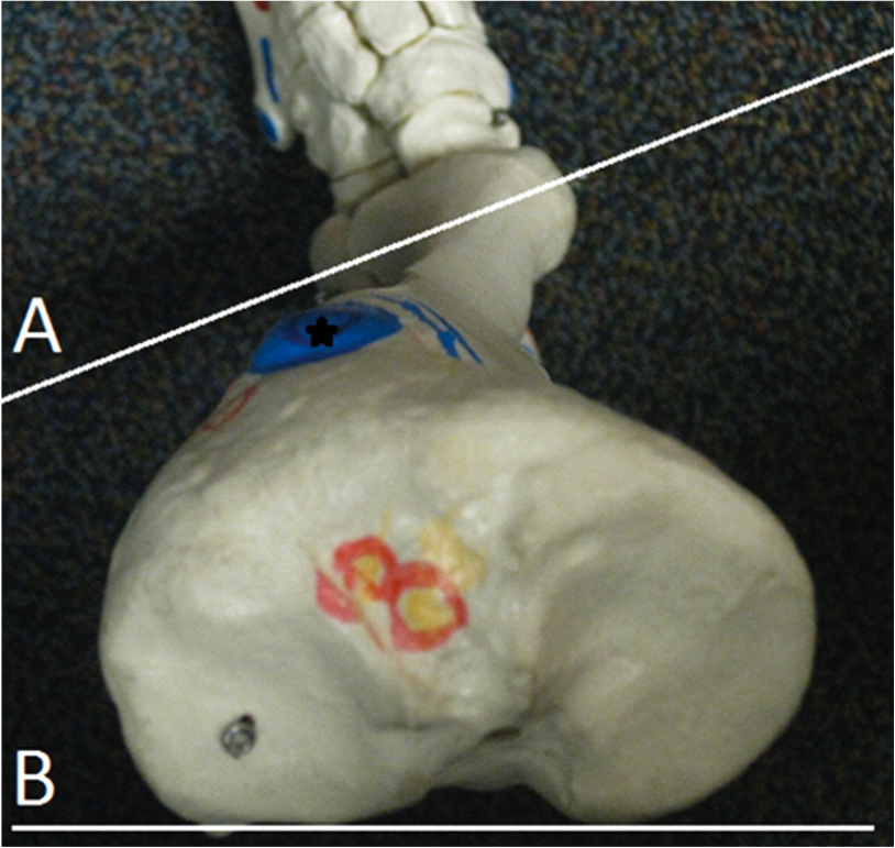
A torsion is the same condition with the amount of twist 1 to 2 standard deviations greater. An example is when the angle of the femoral neck and greater than 15 degrees, the condition of femoral ante torsion exists (see photo above).

There are at least 3 reasons you need to understand about developmental torsions and versions that occur with growth:

  1. Since they occur in the transverse (horizontal) plane, they affect the progression angle of the foot and thus gait
  2. They affect available ranges of motion of a limb (ex the femur needs to internally rotate 4-6 degrees for normal gait) and can cause pain and/or gait alterations
  3. They can affect the coronal (frontal) plane orientation of the lower limb, which can affect gait and shoe choices. A Rothbart foot type with an elevated 1st metatarsal head will often result in a varus (or inverted) position of the forefoot with respect to the rear foot.

In this series, we will explore these 3 major versional changes, one at a time.

The Gait Guys. Bald? Yes! Good looking? You bet! Yes, we are a little more twisted than most folks : )

All material copyright 2013 The Gait Guys/ The Homunculus Group. All rights reserved.  Please ask before recycling our stuff!