tumblr_lm2pqrc0fP1qhko2so1_1280.jpg
tumblr_lm2pqrc0fP1qhko2so2_1280.jpg

There are 2 photos here (move your cursor over to the little triangle on the right to highlight the bar to toggle between photos)

Can you believe that an experienced runner would let a pair of shoes get this far on both sides.  This person does a few marathons a year and has been a patient of our for years…..he tends to milk out every last step in a pair of shoes but we had to hog-tie him and flog him repeatedly for going overboard this time. I bet there had to be 2000 miles on these puppies !  And get this…..he had no foot or knee pain ! (his response was, i saw the wear but i was not having any issues).

Can you imagine how far out side the contact line the knees had to be ? This has the old Nike Cesium beat by a long shot (they were rear foot posted varus by about 3degrees). Heck, this could be 20-25degrees varus !  There is no question that he is avoiding and rear or mid foot pronation……the dude is fixed in supination. 

OF clinical note, he has a fixed right hallux limtus (turf toe) so this likely helps him to avoid medial toe off and forcing dorsiflexion through the big toe joint……however, there are better strategies than this to avoid a hearty toe off !

It is amazing what the body can endure !  Take this as a lesson of what NOT TO DO !  keep the miles between 400-500 miles gang……there are only so many compression cycles in EVA foam before deformation occurs.  In this case there was both deformation and just pure and simple friction wear !

PS: this was an easy pick up clinically……he sat in the waiting room with the 55 gallon barrel set beside him ….the one that he had been carrying around between his knees to force this much rear foot varus !

Good Form video from Newton

Great little video from Danny at Newton…….it doesnt matter what shoe you have if your technique is crummy. Here he demonstrates very well what good contact form is……..we like to say…..“land so subtle on the forefoot that it is a hair distance to kiss your heel to the ground before you push off…..if you remain high on your forefoot at contact you lose your shock absorption because the foot mechanics are more close to supination.”

Short discussion on the toe extensor muscles

We received a great question from a doctor active on our Facebook page (Thegaitguys PAGE, not our user portfolio, make sure you are on the “PAGE”)
Here was the comment:I do like the crouch gaits to help with proximal muscle activation. However I am still not sold on the long toe extensor activation. It would seem to me that the function of this muscle in close chain (ie gait) would be more to aid in pulling the body over the talus (while keeping the toes fully anchored and wide) as opposed to extending the distal phalanx in an open chain fashion. While open chain exercises may ‘strengthen’ this muscle the neurological processing would seem to be different than closed chain and therefore the transfer to more dynamic exercises would be difficult.? I would think that it would not necessarily change the gait but instead allow for better compensatory strength and durability. Although I still have yet to develop a great exercise for this closed chain control. Any ideas?See More

10 hours ago · LikeUnlike

The Gait Guys when you activate the toe extensors the arch is increased from the windlass mechanism across the metatarsophalangeal joints. Raising the arch will help bring it to neutral since the crouched gait is a pronation challenge. give it a try…..….try the crouch shuffles with toes down and toes up…..you will feel the increased demand on the anterior leg compartment, the greater awareness of the foot tripod esp the first metatarsal head anchor point and the improved ability to control the internal tibial spin (and pronation challenge( that occurs with shuffling with toes down. Remember, closed chain is not any more important that open chain activities……arm swing it gait is open chain but it is necessary…..leg swing is open chain but it is necessary for normal progression and pelvic/core use. also remember……we are a flexor dominant society…..look at how many of your clients toes have either a gentle flexion to them or significant…..the balance of the function across the metatarsophalangeal joint is necessary on balance of extensors and flexors……the shuffle gait with toes up is a huge challenge to the toe extensors……that feeling of the strain on the top of the foot and into the shin is confirmatory. OF course, you are right in what you said……but to get the toes optimally anchored you have to have enough long toe extensor strength to override the long flexor dominance…..otherwise you being hammering the toes and enter into the spiraling vortex of flexor dominance, lumbrical inhibition, short extensor overactivation, proximal fat pad drag etc.

tumblr_llu784oY6o1qhko2so1_400.gif
tumblr_llu784oY6o1qhko2so2_400.gif

Shoe Anatomy 101:

If you are truly to be a shoe geek, then you must be familiar with some “shoe anatomy.” Simply put, there is the sole ( the part that contacts the ground), the midsole (the part right between the sole and the last), the shank (the stiff rear part of the midsole), the last (the part on top of the midsole and where the insert or orthotic will sit), the insole (the removable insert), the upper (the part above the last that has the sides, laces, etc)the heel counter (the part that holds the heel in place), and the toe box.

Lets discuss each in turn


The Sole (also called the outsole)

This is the part of the shoe that comes in contact with the ground. It is usually made of rubber and provides for some degree of shock absorption and traction. For running shoes, it is usually cemented to the midsole.

Remember that the heel strikes the ground at approximately a 16° angle lateral from the center of the heel  (in a heel strike gait, no we aren’t condoning this, this is how shoes are designed).  The force is then transmitted from the sole of the shoe, up the lateral column of the foot and across to the first metatarsal for propulsion.  This can be assisted by a “rocker” which is a “drop” put into the front portion of some stiffer trail shoes, to ease walking and assist in toe off.  (This is good for people with Morton’s toes or hallux rigidus).

A flare to the sole of the shoe, particularly lateral can be important for stability on uneven surfaces.  A lateral flare provides extra stability upon heel strike, but it speeds up the rate of pronation.  This may be a bad thing, depending on your feet. This flare must extend the length of the sole, otherwise injury can occur at the mid tarsal joint as the foot comes through mid stance. A medial flare can help to prevent overpronation, as the foot comes through mid stance. Again, it must run the length of the shoe.

Look at the lugs on the sole. Are they beveled or straight? A straight lug or cleat will hold on to mud, whereas a beveled one will shed it. This is an important consideration if running off road. How about the cleat pattern (front vs. back and side to side)? Are they symmetrical or opposing? Opposing patterns will enable you to ascend and descend easier at the expense of a slight amount of speed.

The Midsole (sandwiched between the sole and upper)

Midsole material is very important, as it will accommodate to the load imposed on it from the person and their body weight. It serves as the intermediary for load transfer between the ground and the person.  Softer density material in the heel of the shoe softens the forces acting at heel strike and is good for impact and shock absorption. The stiffer the material, the more motion control.  Air is an excellent shock absorber, however it does not deform, it displaces. This creates and unstable surface for the foot, promoting ankle injuries. Foam and gel are much better as they transduce the force and dissipate it. Often midsoles are made with something called “dual density”.  This means that the midsole is softer on its lateral aspect, to absorb force and decrease the velocity of pronation during heel strike and midstance, with a firmer material medially that protects against overpronation as you come through mid stance and go through toe off.

The Shank (this can be within the midsole)

The shank is the stiff area of the shoe between the heel to the transverse tarsal joint. It corresponds to the medial longitudinal arch of the foot, provides torsional rigidity to this shoe and helps to limit the amount of pronation and motion at the subtalar and mid tarsal joints.

The Last (the part between the midsole and insole)

The last (look inside the shoe on top of the shank) is the surface that the insole of the shoe lays on, where the sole and upper are attached).    Shoes are board lasted, slip lasted or combination lasted. A board lasted shoe is very stiff and has a piece of cardboard or fiber overlying the shank and sole (sometimes the shank is incorporated into the midsole or last) .  It is very effective for motion control (pronation) but can be uncomfortable for somebody who does not have this problem.  A slip lasted shoe is made like a slipper and is sewn up the middle.  It allows great amounts of flexibility, which is better for people with more rigid feet.  A combination lasted shoe has a board lasted heel and slip lasted front portion, giving you the best of both worlds.

When evaluating a shoe, you want to look at the shape of the last (or sole).   Bisecting the heel and drawing an imaginary line along the sole of the shoe determines the last shape.  This line should pass between the second and third metatarsal.  Drawing this imaginary line, you are looking for equal amounts of shoe to be on either side of this line. Shoes have either a straight or curved last.  The original idea of a curved last (banana shaped shoe) was to help with pronation.  A curved last puts more motion into the foot and may force the foot through mechanics that is not accustomed to. Most people should have a straight last shoe. 

The Upper (the sides and top of the shoe)

This is the part above the midsole that holds your foot on the sole. It is usually made of nylon, Gore-Tex or some other man made material. Pick something light and breathable.

The Heel Counter (the back of the upper)

This is part of the upper. A strong, deep heel counter with medial and lateral support is also important for motion control; lateral support especially for people who invert a great deal or when you’re going to place an orthotic in the shoe which inverts the foot a great deal.  The lateral counter provides the foot something to give resistance against.  This needs to extend at least to the base of the fifth metatarsal, otherwise it can affect the foot during propulsion. A deep heel pocket helps to limit the motion of the calcaneus and will also allow space for an orthotic. The heel counter should grip right above the calcaneus, hugging the Achilles tendon.

The Toe Box

The toe box should be generous enough to prevent crowding and pressure on the metatarsal heads.  The widest portion of the shoe should parallel a line bisecting the metatarsal heads.  Excessive pressure can result in bunions and/or hammertoes.  The shoe may soften and break down laterally, but it will not increase in length.

When measuring feet and determining shoe sizes, do it both sitting and standing, because the laxity of ligaments can become very evident, especially when the foot is weight bearing or you have the weight of a pack on your back.  If the person has greater than one size of splaying in both length and width when going from one position to the other, go for the bigger size.  Always use ball length rather than sole length. People usually buy smaller shoes because when you pronate, there is less volume in the mid foot and a smaller size shoe will feel better.

The Insole (the removable inner footbed)

This is the part of the shoe that most people remove to put in an orthotic. They have come a long way in construction and make a big difference in shoe fit. They are usually made of some type of foam or EVA material. Some of the newer ones are even dual density foam.

Well, if you made it through this, you are officially as nerdy as us. We’ll see you in the shoe isle…..

 We remain, The Gait guys….

horse: walk, trot, canter, gallop
man: walk, jog, run, sprint….very similar
….amble…..well that is another story……horse “ambling” gaits, a collection of several other smooth footfall patterns that may appear naturally in some individuals but which usually occur only in certain breeds, and often require special training…….as for humans? hummm. time for some homework

" target="_blank">Why I run......amazing video

thanks Kyle and Jason and Newton…….this one is what it is really all about……. “in life we dont have it all figure out, there are key moments that change your life forever”.

So…..to close out the week…..we put aside anatomy, biomechanics, physics, neurology, orthopedics, research etc……we put it all aside……..and give you the big picture…..

“why i run”………don’t miss this one……it just might pull a tear from your eye……Thanks Kyle and Jason…….

tumblr_llt5akcND81qhko2so1_r1_400.gif
tumblr_llt5akcND81qhko2so2_1280.jpg

Today’s post is a very nice follow up to the one earlier in the week entitled “Materials: do soft soles improve running shoes?”……which also commented on client perceptions of the footwear.  We hope the shoe and R&D companies are paying close attention to the work were are making available here. 

__________________________________________________________

Today…..

Perceiving is believing

What you think matters! Perception of shoe cushioning seems congruent with what you believe.  If you THINK the cushioned shoe will be softer, you will probably perceive it as so, even though there may be little to no change in ground reaction forces. Most studies show cushioned shoes actually INCREASE impact forces.


McCAW, STEVEN T.; HEIL, MARK E.; HAMILL, JOSEPH: The effect of comments about shoe construction on impact forces during walking

Medicine & Science in Sports & Exercise: July 2000 - Volume 32 - Issue 7 - pp 1258-1264

journal link:

http://journals.lww.com/acsm-msse/Abstract/2000/07000/The_effect_of_comments_about_shoe_construction_on.12.aspx

conclusions as quoted in their article:

Footwear designers incorporate a variety of midsole materials and mechanical systems to cushion shock while controlling rearfoot motion. To market shoes, advertising copy and the claims of salespeople often extol the benefits of the materials and design incorporated in a shoe. This marketing strategy is common, as evident from a perusal of a fitness magazine, in spite of a paucity of unbiased biomechanical testing of the validity of many of these claims, and a lack of understanding of how such claims may influence the gait behavior of a purchaser of a pair of shoes.”

It is vital to understand their next research finding from this study,

“The results indicated the impact ground reaction force varied as a function of the advertising message. When subjects were informed that a particular surface provided additional cushioning, impact ground reaction force data were higher (121% of body weight) than when subjects were provided with a warning message (110% of body weight). The results were interpreted as suggestive of subjects moderating impact in accordance with the expected cushioning of the material. That is, subjects were less inclined to use a landing strategy that would reduce impact force if they had been told that cushioning would be provided by the surface material. The study raises the question of how subjects would respond if the cushioning characteristics of a shoe, rather than a landing surface, were altered, because a shoe represents a more personalized aspect of the foot/ground interface.”

From their study they referenced Robbins and Waked:

“Robbins and Waked (15) demonstrated that subjects may be deceived into accepting higher forces during landing if provided with misleading information regarding the energy absorbing characteristics of the landing. Because of research suggesting a possible cause of lower impact peaks in harder shoes to be an intrinsic avoidance mechanism (1), it may be theorized based on expectancy theory that influencing someone to believe that one shoe can absorb more energy than another may result in different ground reaction force values. Contrary and sometimes confusing findings in the literature emphasize the need for further study in the area of perception and energy absorption. The purpose of this study was, therefore, to determine the effect of investigator comments regarding shoe construction on the ground reaction force measured during walking.”

This is an excellent article that seems to suggest that there is far more to it than the engeneering that goes into the shoe. That marketing and consumer assumptions are a part of how a shoe is used and works. 

Very interesting !  We hightly recommend you purchase this article here…..

http://journals.lww.com/acsm-msse/Abstract/2000/07000/The_effect_of_comments_about_shoe_construction_on.12.aspx

Shuffle Gait DVD

Varations of this crouch style gait (we call them shuffles) are a major staple in our gait and core and lower limb rehab and exercise progressions……after dozens of emails already in just an hour of this posting……we can see that we need to do a DVD on our gait progressions and Shuffle Gait variants…..as there are multiple variants to correct various problems…..we will do our best to get it out soon……it is now in the hopper.

Muscle contributions to support and progression during single-limb stance in crouch gait.

Steele KM, Seth A, Hicks JL, Schwartz MS, Delp SL. J Biomech. 2010 Aug 10;43(11):2099-105. Epub 2010 May 20.

What the Gait Guys have to say about this article:

We have long advocated the use of the “shuffle gait” exercise for increased gluteal activation (see Youtube clip entitled “the shuffle gait exercise”) and this paper provides further support for our recommendation.

Try this simple experiment. Stand on 1 leg. Now close your eyes and try and stay that way for 30 seconds, trying not to lose your balance, touch the ground with your other foot or steady yourself with your hands. Once you have regained your composure, with your eyes closed, extend our head (ie. Look up). Tough to balance, eh? So what just transpired?

3 integrated sensory systems enable us to remain upright in the gravitational plane: vision, the vestibular system and the proprioceptive system (joint/muscle mechanoreceptors). When you 1st stood on one leg, you were utlilizing all 3 systems. When you closed your eyes you were eliminating vision from the equation. When you tilted your head back, you were taking out the lateral semicircular canals from the vestibular system, relying predominantly on the joint and muscle proprioceptors. Let’s guess that you probably need some work on your proprioceptors!

Placing someone in a crouched gait generally means you are at least partially flexed at the waist and need to extend your head somewhat to keep your eyes parallel with the horizon (the brain is wired to keep the visual axes upright and parallel). Extending your neck activates the local joint mechanoreceptors and more importantly, the muscle mechanoreceptors in the suboccipital muscles, which have the highest density of mechanoreceptors of any muscles in the body. These muscle mechanoreceptors feed information into the cerebellum and vestibular nuclei, which fire the antigravity extensor muscles that keep us upright in the gravitational plane. This includes muscles like the glutes, quads and lumbar erectors, just like in the study.

It was interesting that in crouched gait, the muscles which provided upward momentum (gastroc, soleus, quads) remained active throughout single leg stance. These are also stabilizers of the foot and placing the ankle in dorsiflexion (as in a crouch) increases the amount of pronation occurring (remember pronation is dorsiflexion, eversion and abduction); this requires additional stabilization (medial gastroc is an inverter, gluteus Maximus and posterior fibers of glute med control the speed of internal rotation of the lower extremity, along with vastus medialis).

Look to crouched positions as training tools when rehabbing gait, and don’t neglect the shuffle!

Invalid video embed.

Following the Flintstones….

In this next clip, Barneys barefoot technique has only improved marginally. He initiates with a much better forward lean. His ankle rocker is improved, but not enough for him to gain traction. His arm swing is better, but he has a tendency to hunch his shoulders. His leap into the car is a tribute to his gastroc soleus group…

Invalid video embed.

Hey

Check out Barney joing the barefoot craze! We slowed him down a bit to observe his technique a little closer

Not the best barefoot technique we have witnessed. Though he has a some forward lean, he still heel strikes with an extended knee in front of his body (ouch!) and has little to no ankle rocker with premature heel rise. This results in a “bouncing” gait with most likely sore calves!

We hope Betty was a massage therapist…

graphic above by Edward Muybridge
Leg length discrepancies and heel lifts. To lift or not to lift…
The Gait Guys
Leg length discrepancies (LLD’s) are encountered on a daily basis. They are the root of many ankle, knee, hip and spinal problems. The q…

graphic above by Edward Muybridge

Leg length discrepancies and heel lifts. To lift or not to lift…

The Gait Guys

Leg length discrepancies (LLD’s) are encountered on a daily basis. They are the root of many ankle, knee, hip and spinal problems. The questions the clinician must ask are “How much is significant?”, “How much do I add?” What are some of the signs and symptoms?” “What is the etiology?” and “How do I detect it?” A literature search (2003) provided the following information and answers.

How much is significant?

Most authorities claim that deficiencies of greater than ¼ inch (6mm) are clinically significant (1, 2) though some sources state that differences as little as 4 mm are significant (5). Subotnick (3) states that because of the threefold increase in ground reactive forces with running, lifts should be used with inequalities of greater than 1/8” inch (3mm).

How much do I add?

One of the easiest ways to determine the amount of lift needed is to examine the person in a weight bearing posture and add lifts under the short leg until the pelvis is even or until the lumbar spine is straight. If using off weight bearing measurements, you need to add 1/3 more height than measured because the talus is positioned 1/3 of the way between the calcaneus and metatarsal heads (4, 13). So, a heel lift placed under the calcaneus will only raise the talus 2/3 of that height. Lifts placed under the calcaneus can shorten the tricep surae muscles (4, 6) and apply increased pressure to the metatarsal heads (12); full length sole lifts are more physiological, though not always practical. Due to the supinatory moment of the short leg on heel strike, a lift may cause overcompensation and increased supination, with a tendency to overweight the lateral column and possibly injure the lateral ankle. Careful observation of gait post addition of a lift is in order and a valgus post running at least the length of the 5th metatarsal along with the lift should be considered (8, 9). Heel lifts also cause EMG changes of leg muscles, with decreased recruitment of gastrocnemius and tibialis anterior directly proportional to the height of the heel lift (18, 19). A lift or LLD changes the ground reactive forces associated with gait, increasing vertical force on the longer leg, along with increased joint stresses along the kinetic chain (14, 20).

Generally speaking, lifts greater than 3/8” (9mm) require extrinsic modifications to footwear (4, 6, 8). Find a competent individual to perform this work for you. Large discrepancies should be treated gradually, at a rate of ¼ inch every 4 weeks, less if symptoms do not permit.

What are signs and symptoms associated with LLD’s?

Compensation comes in many forms, depending whether it is acute (recent injury caused an LLD or compensation resulting in one, or long term. The deficiency can cause injury on the short or long legged side (or both).

The long leg moves through a greater arc during all portions of swing phase (7). The person may flex the knee to compensate and shorten the arc. The individual may also maximally pronate and evert the calcaneus an additional 3 degrees or greater on that side in an attempt to lower the navicular to the ground and shorten that leg. This causes an increased amount of internal rotation of the tibia and thigh causing muscular dysfunction (tightness of the hip flexors, strain of the intrinsic external rotators from eccentric deceleration of the thigh), along with medial knee strain (especially with concomitant genu valgus) (4, 6, 8, 9, 10, 11, 21, 22).

The short leg side will often supinate in an attempt to lengthen and cushion some of the shock of heel strike, since it has a greater vertical distance to travel (14); this often occurs with hyperextension of that knee. This lessens the dampening ability of the knee (since it flexes almost 20 degrees between heel strike and full forefoot load), and speeds the rate of subtalar pronation (since the rear foot is inverted and still must pronate the same amount (4). Many individuals will try and attenuate impact by contracting the contralateral hip abductor muscles and eccentrically lower the shorter extremity (4, 14). This can produce excessive strain of that musculature (trochanteric bursitis) as well as pathomechanical abnormalities of the L4 and L5 motion segments (due to increased body rotation toward the short side and attachments of the iliolumbar ligaments; this can cause degenerative changes if present long term (11, 12)).

What’s the etiology?

LLD’s can be structural (anatomical) or functional (pathomechanics, compensation). LLD’s can be due to foot problems (overpronation/supination, fractures), leg or thigh problems (congenital shortening, deformity, fracture), or pelvic compensation (rotation of ilia, fractures).

Text Box: Long leg adaptations "	Drooping of shoulder with elevation of iliac crest on long leg side "	Pirformis/external rotator tightness "	Tightness of hip flexors "	Increased lordosis "	Posterior rotation (flexion) of ilia (can shorten leg up to 6mm "	Medial knee degenerative changes/pain "	Increased pronationText Box: Short leg adaptations "	raised shoulder with depression of iliac crest on long leg side "	TFL tightness "	Decreased lordosis "	Anterior rotation (extension) of the ilia (can lengthen leg up to 6mm) "	Lateral knee degenerative changes "	Increased supination


So, what is the etiology? A lot can be gleaned from the history. Past trauma is the most obvious so pay close attention. This could result in flattening of the calcaneus or overpronation due to ligamentous laxity; tibial fractures can cause shortening as well as increased or decreased tibial torsion; similar findings can occur in the femur, along with anteversion or retroversion; pelvic trauma can be more subtle and x-ray can often provide the most information (1, 2, 4, 6).

How do you determine a leg length inequality?

There are a number of methods, each with their own merit. X –ray is most accurate, but exposes the patient to ionizing radiation. Weight bearing seems most appropriate, since symptomatology usually presents itself then. Supine measurements are said to be influenced by asymmetrical muscle tension, table pressure on the innominates and hip flexor length (15).

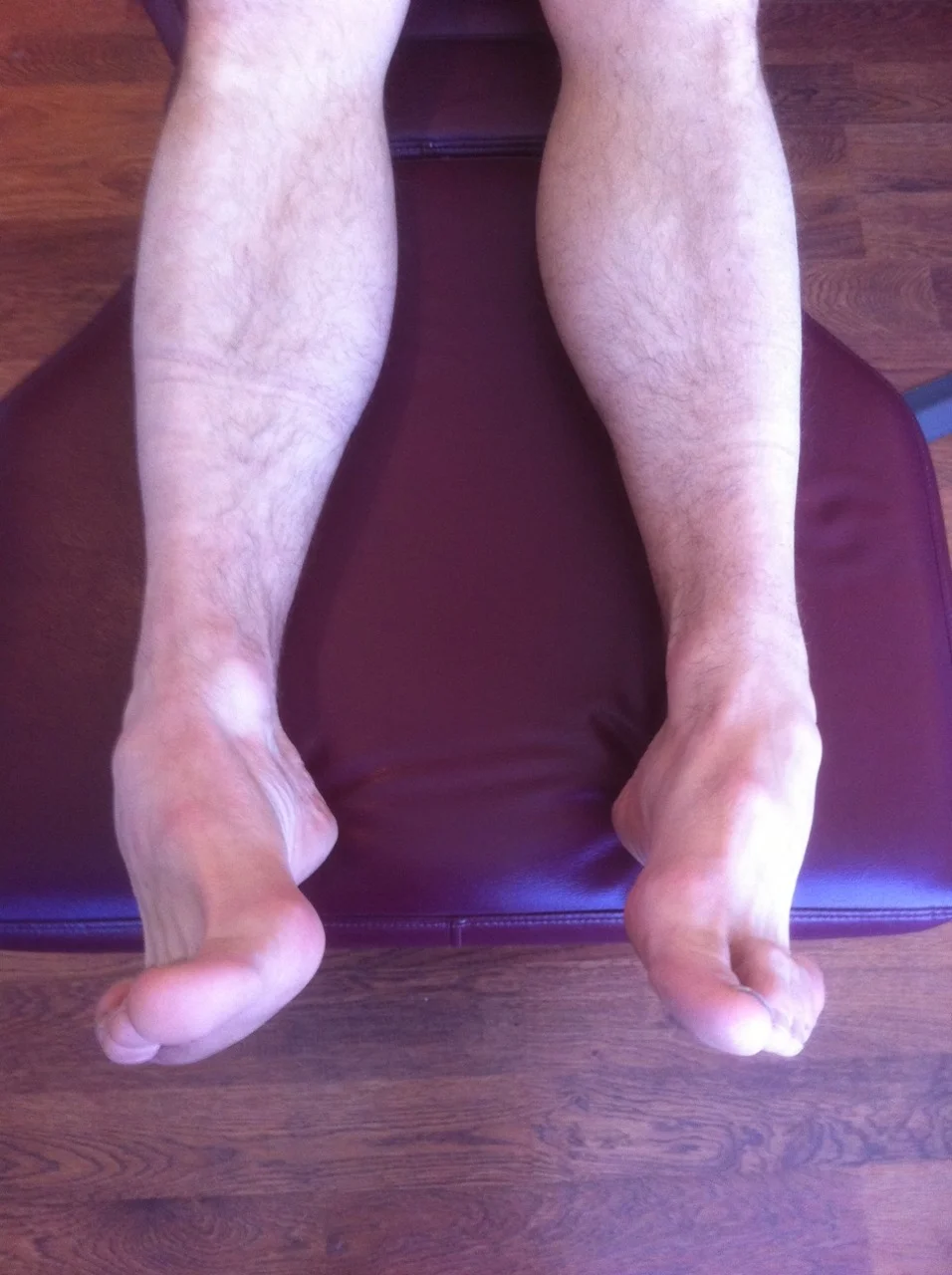
With the patient weight bearing and both feet placed below the trochanters, observe the level of the medial malleoli. Next, compare the heights of the tibial plateaus. Femoral length can be judged by the heights of the greater trochanters, and pelvic alignment judged by the heights of the iliac crests (4, 17).

Alternately, lay the person supine and observe the heels and medial malleoli. If there is noticeable discrepancy, they may have a short leg; if there isn’t, they still may have a discrepancy that they are compensating for. Check the range of motion of the foot and ankle in 6 general directions: plantar flexion (40-45 degrees), dorsiflexion (20-25 degrees, depending on whether the knee is flexed or extended), inversion of the forefoot (3-60 degrees, on average), and eversion of the forefoot (20-45 degrees on average), calcaneal inversion (4-20 degrees) and calcaneal eversion (4-10 degrees). Excessive calcaneal eversion usually means over pronation due to a longer leg on that side; excessive inversion can mean a long leg due to a cavus foot type (2, 4, 6, 8, 9, 12). Lack of flexibility in the posterior compartment of the calf usually causes a greater degree of pronation (16).

Now, perform Allis’s test. Bend both knees to 90 degrees and observe the height of the tibial plateaus. The lower one is usually the side of the discrepancy (which can be in tibial length or due to excessive pronation). Now walk superior to the knees and observe the femurs from more cephalad (4). Is there a discrepancy? If so, the problem may be in the femur length, femoral head angle or pelvis. Extend the knees so that the legs are lying flat on the exam table. Palpate the greater trochanters on both sides. Is one lower than the other? If so, they probably have coxa vara on the short side or coxa valga on the long side. If they are even, you need to look at the pelvis. Does one ASIS palpate more anterior or posterior than the other? This could represent compensation. A posterior or “flexed” ilia, usually causes a short leg on that side; an anterior or extended ilia usually causes a long leg on that side. Now stand the patient up and perform a Gillet Test. Have them stand erect and hold onto something for balance. Palpate the PSIS on one side along with the 2nd sacral tubercle. Have them raise their thigh to 90 degrees on the side you are palpating. The PSIS should nutate backward (flex) and drop .5-1.5 cm on the side of the raised leg. Now have them raise the opposite leg. The sacrum should nutate backward and down. If either of these movements does not occur, consider pelvic pathomechanics and treat accordingly. Recheck for motion as well as leg length when done.

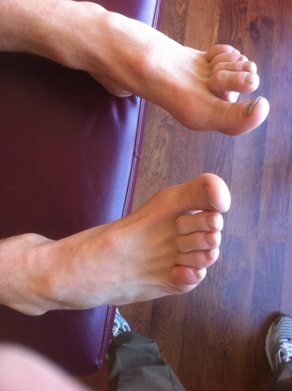
Standing observation often (but not always) reveals overpronation on the long leg side and relative supination on the short leg side. The shoulder is often higher on the short side and the waistline dips to the long side because of posterior rotation of the innominate. The shoulder will dip to the side of the short leg on heel strike during dynamic evaluation (4, 6, 8, 9, 10, 11). Gait observation usually reveals adduction of the pelvis toward the stance phase leg with a lateral sway in excess of 1” during stance phase. The person will seem like they are “stepping into a hole” on the short side.

Conclusion

Leg length inequalities occur due to a variety of anatomical and physiological conditions. Careful analysis and examination can often reveal its etiology. To lift or not to lift is a clinical decision that is left to the clinician and patient, with a careful balance between what is perceived as improved biomechanics and tolerance levels of the patient with regards to their presenting symptomatology.

References available by request

Materials: Do soft soles improve running shoes ?

BioMechanics
April 1998
Materials: 
Do soft soles improve running shoes?
Most athletic shoes advertise injury protectiong through “cushioning,” but real world studies have not shown impact moderation.
By Steven Robbins, MD, Edward Waked, PhD, and Gad Saad, PhD

here are their conclusions word for word:

Please honor the authors and purchase the article for your own use.

Conclusion
“Shoes with cushioning fail to absorb impact when humans run and jump, and amplify force under certain conditions, because soft materials used as interfaces between the foot and support surface elicit a predictable reduction in impact-moderating behavior.5-10,13-17 This behavior is not a response to sensations directly caused by impact because, whereas barefoot humans estimate impact precisely, humans judge it inaccurately when shod.14-17 This situation has recently been made clearer. Reduction of impact-moderating behavior is a response to loss of stability induced by soft-soled cushioned shoes: Humans reduce impact-moderating behavior in direct relation to increased instability.27 This is presumably an attempt to achieve equilibrium by obtaining a stable, rigid support base through compression of sole materials.27
After considering footwear advertising, additional factors appear to influence impact-moderating behavior. Recent reports also indicate that humans reduce impact-moderating behavior, thereby amplifying impact, when they are convinced that they are well protected by the footwear they are wearing. Advertising that suggests good protection results in higher impact, whereas advertising that suggests injury risk attenuates impact.19 Deceptive advertising, suggesting that expensive cushioned footwear offers advanced technology that protects against impact, accounts for the 123% greater frequency of injuries with the most expensive shoes found by Marti.18
Public health could be advanced through truth in advertising of footwear products with cushioned soles. Furthermore, footwear must be required to provide good balance. Current athletic footwear undoubtedly causes falls, since footwear with thick yielding soles destabilizes humans by as much as 300% compared with hard-soled shoes.
Now that the destabilizing nature of cushioned footwear is well established, continued manufacture of these hazardous items without explicit warning labels represents risk for liability claims from users who are injured from falls and ankle sprains while wearing them. In the context of this report, footwear that provides superior balance will probably be effective at attenuating vertical impact. Clearly, highly resilient materials must be removed from shoe soles for many reasons. This move will portend better health through improved stability and fewer injuries from excessive repetitive impact in sports. ”

Run, Carey, Run?

Lets look at this Hitchcock classic “North by Northwest” and check out Cary’s form.

1st of all, what an arm swing! Think of all that energy it is sapping from the rest of his muscular system. He must be hiding something, but what? We can only see him from the waist up, so we may never actually know. Did you notice how he initially only turns to the right? Did you pick up on the flexion at the waist? How about that torso bob from side to side? Not much to his hip abductors now are there?

The only thing he has going for him is he is wearing leather soled shoes, which have been shown to have one of the lowest impact loading on the body (yes, you read that right; increased cushioning INCREASES impact forces, but that’s not what we are here to talk about). Oh yea, he actually impacts the ground at the end of the sequence. I guess if his technique was better, he would have hit even HARDER.

Next sequence, we are off to a good start, look at that forward lean to start! This is essential to good technique. He loses that form pretty quickly; we can still see that forward flexion at the waist; certainly costing him energy by not using his core.

Finally, we get a posterior view at the end, but the uneven surface makes it difficult to make an analysis.

We think Cary would certainly give Lola a run for her money. Cary, next time, engage your core and watch your step…

We Remain….The Gait Guys

Chicago Lecture , "World Class Shoe Fit"

Yesterdays IRRA event (Independant Running Retailers Assoc) lecture in Chicago went 2.5 hours yesterday ! Great group, great feedback. We talked at length about shoe anatomy and the various anatomic foot types and how to evaluate a client or patient for a good shoe match and fit depending on their foot type.  Everyone, please welcome aboard another 60 foot/shoe/gait nerds ! The more the merrier !

Passive Arch Stability Anatomy Review

Anatomy review: No matter how good the shoe choice is for a client’s/patient’s foot type……if muscular weakness has persisted long enough to compromise one of the big 6 (plantar aponeurosis, long-short plantar ligaments, plantar calcaneonavicular ligament (spring ligament), medial talocalcaneal ligament, talocalcaneal interosseous ligament, and tibionavicular portion of the deltoid ligament) there is likely to be recurrent foot problems.

Foot Ankle Int. 1997 Oct;18(10):644-8.

Stability of the arch of the foot.

Kitaoka HB, Ahn TK, Luo ZP, An KN.

Source

Department of Orthopedics, Mayo Clinic, Rochester, Minnesota 55905, USA.

tumblr_ll7x9gTvYb1qhko2so1_1280.jpg
tumblr_ll7x9gTvYb1qhko2so2_1280.jpg

What did you notice? Cavus foot? Loss of the transverse arch? Prominence of extensor tendons?

The question is: Why?

It’s about reciprocal inhibition. The concept, though observed in the 19th century, was not fully understood and accepted until it earned a Nobel prize for its creditor, Sir Charles Sherrington, in 1932. Simply put, when a muscle contracts, its antagonist is neurologically inhibited, So when your bicep contracts, your tricep is inhibited. This holds true whether you actively contract the muscle or if the muscle is irritated (causing contraction).

So how does this apply to this foot?

We see prominence of the extensor tendons (particularly the extensor digitorum brevis EDB; the longus would have caused extension at the distal interphalangeal joint). The belly of the muscle is visible, telling us that it is active. It is neurologically linked to the flexor digitorum brevis (FDB). This muscle, in turn, has slips which attach it to the abductor hallucis brevis (AHB) medially and the abductor digiti minimi (ADM) laterally. These muscles together form 2 triangles (to be discussed in another post) on the bottom of the foot, which lend to the stability of the foot and the arches, especially the transverse.

When the EDB fires, it inhibits the FDB, (which, in addition to flexing the MTP’s, assists in maintaining the arch). The EDB has an effect which drops the distal heads of the metatarsals as well (Hmm, think about all the people with met head pain) Now, look at the course of the tendons of the EDB. In a cavus foot, there is also a mild abductory moment, which flattens the arch. Conversely, the FDB in a cavus foot would serve to actually increase the arch, and would have a ,mild adductory moment. Net result? A flattened transverse arch.

Now look at the Flexor digitorum longus, overactive in tbis foot (as evidenced by the flexion of the distal interphalangeal joints, mild adduction of the toes (due to the change of direction of pull in a cavus foot) and lowering of the met heads due to hyperextesnion at the MTP joints ). This mm is reciprocally linked with the extensor digitorum longus. The prominence of the extensor tendons is do to increased activity of the EDB (go ahead, extend all your fingers and look at the tendons in your hand. Now flex the  DIP and IP joints and extend the MTP; see how they become more prominent?).

Reciprocal inhibition. It’s not just for dinner anymore…

We are and remain; The Gait Guys

Part 2 of a case study from Northern Ireland. This video discusses the dynamic findings and how they correlate clinically with the history. Treatment recommendations are discussed as well.

Follow up question from a doctor…..

Thanks for the post. Interesting case study. Are most hernias at this point a result of overactive hip flexors? What would be your exercise dosage/prescription of the exercises mentioned in part 2?

The Gait Guys In our experience, most inguinal hernias are due to weakness of the lower abdominal wall, in this case, the external obliques, not being able to fire appropriately to guard against the load. Exercise would most likely progress along the lines of skill 1st (can he perform the exercise appropriately), endurance 2nd (increased reps to increase capillarization, myoglobin content, mitochondrial content; beginning with 8-12 reps and increasing to 5-10 sets daily) and strength last (low reps, high weight; dependent on progress)