tumblr_oa5thqROml1qhko2so1_1280.jpg
tumblr_oa5thqROml1qhko2so2_1280.jpg
tumblr_oa5thqROml1qhko2so3_1280.jpg
tumblr_oa5thqROml1qhko2so4_1280.jpg

 Why does this gal have so much limited external rotation of her legs? 

 We have discussed torsions and versions here on the blog many times before. We rarely see femoral antetorsion. She came in to see us with the pain following a total hip replacement on the right.

 Note that she has fairly good internal rotation of the hips bilaterally but limited external rotation. This is usually not the case, as most folks lose internal rotation. We need 4 to 6° internal and external rotation to walk normally. This poor gal has very little external rotation available to her.

Have you figured out what’s going on with hips yet? She has a condition called femoral ante torsion.   This means that the angle of the femoral neck is in excess of 12°. This will allow her to have a lot of internal rotation but very little external rotation.  She will need to either “create” or “borrow” her requisite external rotation from somewhere. In this case she decreases her progression of gait (intoed), and borrows the remainder from her lumbar spine.

 So what do we do? We attempt to create more external rotation. We are accomplishing this with exercises that emphasize external rotation, acupuncture/needling of the hip capsule and musculature which would promote external rotation (posterior fibers of gluteus medius,  gluteus maximus, vastus medialis, biceps femoris). A few degrees can go a very long way as they have in this patient. 

confused? Did you miss our awesome post on femoral torsions: click here to learn more.

tumblr_n9y3v5nRcH1qhko2so1_1280.jpg
tumblr_n9y3v5nRcH1qhko2so2_1280.jpg
tumblr_n9y3v5nRcH1qhko2so3_1280.jpg
tumblr_n9y3v5nRcH1qhko2so4_1280.jpg
tumblr_n9y3v5nRcH1qhko2so5_1280.jpg

More on the “little guy”

We have been following this little guy for some time now. If you have not been keeping up, perhaps you should read herehere and here 1st. 

So, what do we see in these latest pictures?

Top left: neutral view.

  • He enjoys flip flops; probably not the best thing for a developing kiddo, in light of the excessive engagement of the posterior compartment (and reciprocal inhibition of the anterior compartment)
  • he has some tibial varum (ie bowleggedness) L > R
  • he has some developmental genu valgum whnich appears to be improving (need a Q angle review? click here)
  • no tibial torsion present on L: for a review on torsions, click here
  • still some external tibial torsion present on R (see section below on middle shots)

Top right and bottom: full internal rotation of R thigh: compare with bottom: full internal rotation of L thigh

  • he has adequate internal rotation (4 degrees needed) but not as great as left side (see bottom shot); this represents some improvement since we started
  • he has generous internal rotation of the left thigh

Middle Left: full external rotation of right thigh

  • note the position of the knee and the position of the foot; external tibial torsion is present. for a review of torsions, click here.
  • he has limited external rotation of the right thigh (compared with the left. The knee should fall more outside the saggital plane

Middle right: full external rotation of the left thigh

  • note the position of the knee and the position of the foot; internal tibial torsion is present. 
  • he has generous external rotation of the left thigh (compared with the left)


of other significant note: most of his calcaneal valgus has resolved; longitudinal arches are improved.

What now?

  • He continues to develop normally and continues to improve since his original presentation to the office
  • Having the child continue to walk barefoot
  • Continue to wear shoes with little torsional rigidity, to encourage additional additional intrinsic strength to the feet
  • He should continue to limit “W” sitting, as this will tend to increase the genu valgus present
  • We reviewed 1 leg balancing “games” and encouraged continuing agility activities, like balance beam, hopping, skipping and jumping on each leg individually
  • added in using a push and pedal bike
  • added in heel walking exercises

Ivo and Shawn. Bald. Good looking. Extraordinary Gait Geeks. Taking the world of gait literacy by storm with each and every post.

tumblr_n9jt5dtWlc1qhko2so1_1280.jpg
tumblr_n9jt5dtWlc1qhko2so2_1280.jpg
tumblr_n9jt5dtWlc1qhko2so3_1280.jpg
tumblr_n9jt5dtWlc1qhko2so4_r1_1280.gif

Subtle clues. Helping someone around their anatomy

This patient comes in with low back pain of years duration, helped temporarily with manipulation and activity. Her exam is relatively benign, save for increased lumbar discomfort with axial compression in extension and extension combined with lateral bending. Believe it or not, her abdominal and gluteal muscles (yes, all of them) test strong (no, we couldn’t believe it either; she is extremely regular with her exercises). She has bilateral internal tibial torsion (ITT) and bilateral femoral retro torsion (FRT). She has a decreased progression angle of the feet during walking and the knees do not progress past midlilne. There is a loss of active ankle rocker with gait, but not on the exam table; same with hip extension. 

We know she has a sweater on which obscures things a bit, but this is what you have to work with. Look carefully at her posture from the side. The gravitational line should pass from the earlobe, through the shoulder, greater trochanter and through or just anterior to the lateral malleolus.

In the top picture, can you see how her pelvis is anterior to this line? Do you see how it gets worse when she lifts her hands over her head (yes, they are directly over head)? This can signify many things, but often indicates a lack of flexibility in the lumbar lordosis; in this case, she cannot extend her lumbar spine further so she translates her pelvis forward. Most folks should have enough range of motion from a neutral pelvis and enough stability to allow the movement to occur without a significant change. Go ahead, we know you are curious, go watch yourself do this in a mirror and see if YOU change.

Looking at the bottom left picture, can you pick out that she has a genu valgus? Look at the hips and look at the tibial angle.

In the bottom left picture, did you note the progression angle (or lack of) in her feet? This is a common finding (but NOT pathognomonic) in patients with internal tibial torsion. Notice the forefoot adductus on the right foot?

So what do we think is going on?

  • ITT and FRT both limit the amount of internal rotation of the thigh and lower leg. Remember you NEED 4 degrees of each to walk normally. Most folks have significantly more
  • if you don’t have enough internal rotation of the lower extremity, you will need to “create” it. You can do this by extending the lumbar spine (bottom picture, right) or externally rotating the lower extremity
  • Since her ITT and FRT are bilateral, she flexes the pelvis and nutates the pelvis anteriorly.
  • the lumbar facet joints should only carry 20% of load
  • she is increasing the load and causing facet imbercation resulting in LBP.

What did we do?

  • taught her about neutral pelvic positioning, creating more ROM in the lumbar spine
  • had her consciously alter her progression angle of her foot on strike, to create more available ROM in internal rotation
  • encouraged her to wear neutral shoes
  • worked on helping her to create more ankle rocker and hip extension with active drills and exercise (ie gait rehabilitation); shuffle walks, Texas walk, toes up walking, etc

why didn’t we put her in an orthotic to externally rotate her lower extremity? Because with internal tibial torsion, this would move her knee outside the saggital plane and create a biomechanical conflict at the knee and possibly compromising her meniscus.

Cool case, eh? We thought so. Keep on learning so your brain keeps expanding. If you are not growing your brain, you are shrinking it!

The Gait Guys

tumblr_n7yjctusWm1qhko2so1_1280.jpg
tumblr_n7yjctusWm1qhko2so2_1280.jpg
tumblr_n7yjctusWm1qhko2so3_1280.jpg
tumblr_n7yjctusWm1qhko2so4_1280.jpg
tumblr_n7yjctusWm1qhko2so7_1280.jpg
tumblr_n7yjctusWm1qhko2so9_1280.jpg

You can only “borrow” so much before you need to “pay it back”

How can feet relate to golf swing?

This 52 year old right handed gentleman presented with pain at the thoracolumbar junction after playing golf. He noticed he had a limited amount of “back swing” and pain at the end of his “follow through”.

Take a look a these pix and think about why.

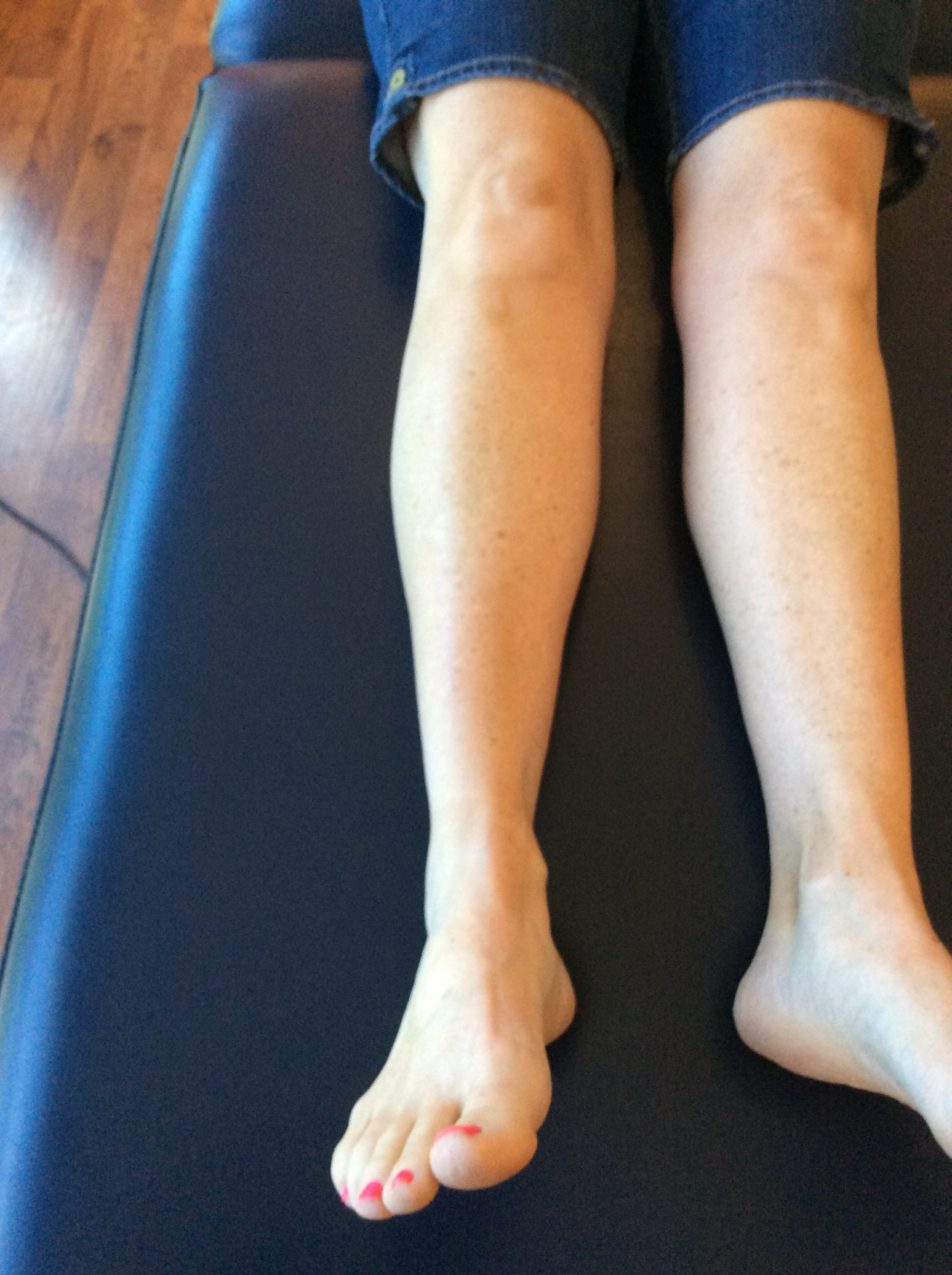
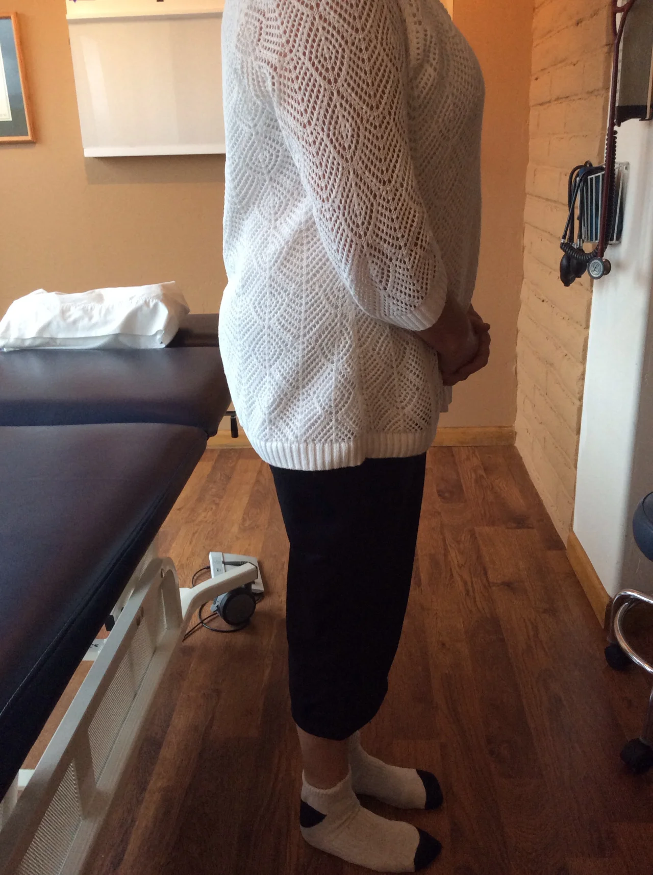
Hopefully, in addition to he having hairy and scarred legs (he is a contractor by trade), you noted the following

  • Top left: note the normal internal rotation of the right hip; You need 4 degrees to walk normally and most folks have close to 40 degrees. He also has internal tibial torsion.
  • Top right: loss of external rotation of the right hip. Again, you need 4 degrees (from neutral) of external rotation of the hip to supinate and walk normally.
  • Top center:normal internal rotation of the left hip; internal tibial torsion
  • 3rd photo down: limited external rotation of the left hip, especially with respect ti the amount of internal rotation present; this is to a greater degree than the right
  • 4th and 5th photos down: note the amount of tibial varum and tibial torsion. Yes, with this much varum, he has a forefoot varus.

The brain is wired so that it will (generally) not allow you to walk with your toes pointing in (pigeon toed), so you rotate them out to somewhat of a normal progression angle (for more on progression angles, click here). If you have internal tibial torsion, this places the knees outside the saggital plane. (For more on tibial torsion, click here.) If you rotate your extremity outward, and already have a limited amount of range of motion available, you will take up some of that range of motion, making less available for normal physiological function. If the motion cannot occur at the knee or hip, it will usually occur at the next available joint cephalad, in this case the spine.

The lumbar spine has a limited amount of rotation available, ranging from 1.2-1.7 degrees per segment in a normal spine (1). This is generally less in degenerative conditions (2).

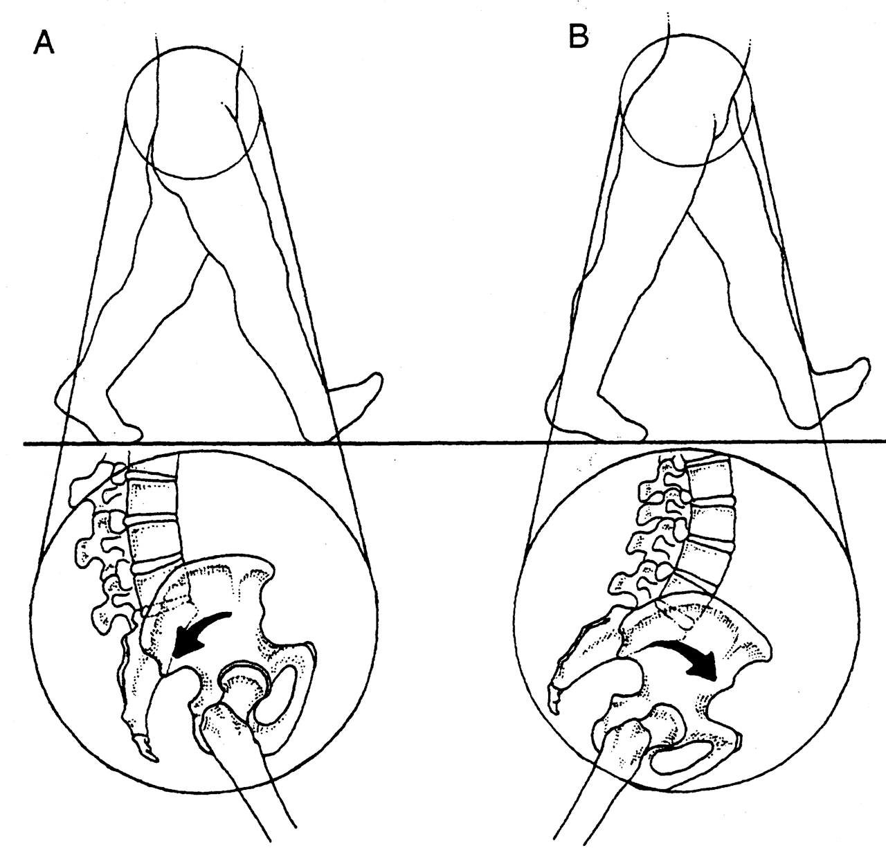
Place your feet on the ground with your feet pointing straight ahead. Now simulate a right handed golf swing, bending slightly at the waist and  rotating your body backward to the right. Now slowly swing and follow through from right to left. Note what happens to your hips: as you wind back to the right, the left hip is externally rotating and the right hip is internally rotating. As you follow through to the left, your right, your hip must externally rotate and your left hip must externally rotate. Can you see how his left hip is inhibiting his back swing and his right hip is limiting  his follow through? Can you see that because of his internal tibial torsion, he has already “used up” some of his external rotation range of motion?

If he does not have enough range of motion in the hip, where will it come from?

he will “borrow it” from a joint more north of the hip, in this case, his spine. More motion will occur at the thoracolumbar junction, since most likely (because of degenerative change) the most is available there; but you can only “borrow” so much before you need to “Pay it back”. In this case, he over rotated and injured the joint.

What did we do?

  • we treated the injured joint locally, with manipulation of the pathomechanical segments
  • we reduced inflammation and muscle spasm with acupuncture
  • we gave him some lumbar and throacolumbar stabilization exercises: founders exercise, extension holds, non tripod, cross crawl, pull ups
  • we gave him foot exercises to reduce his forefoot varus: tripod standing, EHB, lift-spread-reach
  • we had him externally rotate both feet (duck) when playing golf

The Gait Guys. Helping you to store up lots “in your bank” of foot and gait literacy, so you can help people when they need to “pay it back”, one case at a time.

(1) http://www.ncbi.nlm.nih.gov/pmc/articles/PMC2223353/

(2) http://www.ncbi.nlm.nih.gov/pmc/articles/PMC3705911/

tumblr_mr3b80hz4Y1qhko2so1_1280.jpg
tumblr_mr3b80hz4Y1qhko2so2_1280.jpg
tumblr_mr3b80hz4Y1qhko2so3_1280.jpg
tumblr_mr3b80hz4Y1qhko2so4_1280.jpg
tumblr_mr3b80hz4Y1qhko2so5_1280.jpg

Holy Twisted Femurs, Batman. What is going on here?

So, this is what femoral antetorsion looks like!

Remember that ante torsion occurs during development and is when the neck of the femur makes greater than a 12 degree angle with the shaft. We did a great post on this a while ago, click here to read it.

If you remember that the femur heads point anteriorly in a standing position, this would accentuate that, so they stand with an increased progression angle (ie feet toed out; see 1st picture).

With the increased femoral neck angle, these folks have a greater range of internal rotation of the femur, and decreased external rotation. Can you see this in the pictures above? We have rotated her legs fully internally and externally.

A few questions for you:

if you look carefully at the 1st picture, you will note she has external tibial torsion. Why?

  • this condition can develop in utero, but more commonly occurs postnatally with"W" sitting (sitting with knees together and legs abducted, with buttocks between the legs or feet. Think about that constant internal force on the femurs and external rotatory force on the lower tibia! Have your kids sit differently!

What type of shoe should this person be in?

  • The condition itself does not dictate the type of shoe thay should be in. This individual has a rigid, cavus foot BUT has an uncompensated forefoot varus with a great deal of forefoot pronation. In addition to exercises to strengthen the external rotators of the thigh, and inverters of the foot, a shoe with some motion control features is indicated in this instance

The Gait Guys…..Twisted? Yes! And still bald, middle aged and geeky as well.

A bit confused? Dig into our blog more, or watch our youtube channel. Maybe it’s time to push your knowledge base to the next level and take the National Shoe Fit Program. email us at thegaitguys@gmail.com

Photo: Where is your knee joint hinge point ?  Say that 4 times fast. 
Here is a photo of 4 elite runners. We suspect it is an 800m race  because #100 is Ahmed Bile who is the son of Olympian and world champion Abdi Bile.
In this photo you can see t…

Photo: Where is your knee joint hinge point ?  Say that 4 times fast.

Here is a photo of 4 elite runners. We suspect it is an 800m race  because #100 is Ahmed Bile who is the son of Olympian and world champion Abdi Bile.

In this photo you can see that Ahmed #100 has a significantly large foot progression angle (large foot turn out) and this likely represents external tibial torsion or femoral antetorsion while #454 has a neutral foot turn out as does #232.  #46 has a modest foot progression angle. Grossly, #46 also has the patella right over the foot and so tibial torsion is not likely. Now, move up to observe their knee progression. All of them have a forward (sagittally) oriented knee progression. How can that be? Well, it is simple if you know your torsional issues. After all, the knee is a hinge and if you are running forward your knee pretty much should hinge forward as well.  Now, there is much room for conversation here and debate but we are just trying to make and observation and a point. To a large extent the knee rules the roost in the lower limb in terms of sagittal progression because it is the joint with the least number of tolerances. The knee only hinges in flexion and extension where as the hip and ankle/foot have frontal and axial planes they can notably tap into when the sagittal is challenged.  Again, look at #100 and our point is made.

Look at the 2 fellas in the middle (454 and 232). they have a internally (medially) postured knee/thigh yet their foot progression angle is mostly neutral and the knees are hinging forward.  Does #454 have internal tibial torsion? It could be (hint, look at his right trailing leg, specifically the patella and foot postures) but the left limb looks cleaner although adducted suggesting he might like the cross-over gait or it is more external tibial torsioned. Where as the 2 outer fellas, 100 and 46, are more neutrally oriented knees/thighs (one could make the case that #100 has a more externally oriented femur) yet increased progression foot progression angle in an environment of a forward hinging knee.

So what gives ? Torsions. Yes, we are soapboxing on torsions again. Torsions in the tibia, torsions in the femur. Versions are normal expressed angles, tibial torsions are abnormal.

Now, as life would have it, look over the right shoulder of #100. See the fella in the red headband? Ya, that guy losing.  He has the cleanest lines of the bunch. How is that for cruel irony ?  Sometimes it ain’t what you got, it is what you do with what you got.  Unless of course he is actually wincing in pain and trailing behind because he got spiked by #100 and that hideously frontal plane splayed foot !

Lastly, this wouldn’t be an official Gait Guys post if we did not preach to remember that “what you see is not the problem, what you see in a gait analysis is the person’s compensatory strategy around their deficits”. And here we see deficits. Our observations today are merely just that, observations. Now someone has to get them on a table and examine them and confirm our observations, prove them wrong and/or discover the joint, muscular and motor pattern deficits that created these observations.  Or, someone has to confirm that all parts are working and that they were at the end of the line when the straight long bones were first handed out.  

Today’s Lesson:  Get in line, and get in line early. (just kidding of course)

The Gait Guys.  Calling it they way we see it, but reserving the right to plead the 5th or change our minds after an examination.  We would suggest to everyone, when it counts and when your reputation is on the line, plead the 5th, until you have completed your hands on clinical examination.  "Seeing may be believing" but that still doesn’t always make it so.

Want to learn more about these kinds of things, foot beds, foot types, shoe anatomy and shoe function, proper shoe prescription etc ?  Our National Shoe Fit program will help you get smarter about this stuff. email us at : thegaitguys@gmail.com 

Gait Guys online /download store:http://store.payloadz.com/results/results.aspx?m=80204

*Photo courtesy of BIG EAST Conference