tumblr_oa5t63IlXj1qhko2so1_1280.jpg
tumblr_oa5t63IlXj1qhko2so2_1280.jpg
tumblr_oa5t63IlXj1qhko2so3_1280.jpg
tumblr_oa5t63IlXj1qhko2so4_1280.jpg

A visual example of the consequences of a leg length discrepancy.

This patient has an anatomical (femoral) discrepancy between three and 5 mm. She has occasional lower back discomfort and also describes being very “aware” of her second and third metatarsals on the left foot during running.

You can clearly see the difference in where patterns on her flip-flops. Note how much more in varus wear on the left side compared to the right. This is most likely in compensation for an increased supination moment on that side. She is constantly trying to lengthen her left side by anteriorly rotated pelvis on that side and supinating her foot  and trying to “short” the right side by rotating the pelvis posteriorly and pronating the foot.

With the pelvic rotation present described above (which is what we found in the exam) you can see how she has intermittent low back pain. Combine this with the fact that she runs a daycare and is extremely right-handed and you can see part of the problem.

Leg length discrepancies become clinically important when they resulting in a compensation pattern that no longer works for the patient. Be on the lookout for differences and wear patterns from side to side.

Leg length discrepancies and total joint replacments.

5mm cut off ?  MaybeYou are likely to come across hip and knee arthroplasty clients (total joint replacements). When they take a joint out and replace it with a new one, it can be a true challenge to restore leg lengths to equality side to side. Problems often arise down the road once gait is resumed and rehabilitation is completed. It can take time for the leg length discrepancy (LLD) to begin to create compensatory problems. This article seems to suggest that 5mm is the tipping point where gait changes becoming a problem are founded. Other sources will render different numbers, this article found 5mm. The authors found that both over- and underrestoration of leg length/offset showed similar effects on gait and that Gait analysis was able to assess restoration of biomechanics after hip replacement.  I would chose to use the word “change” over restore, since the gait analysis is merely showing the deployed strategies and compensations, never the problem.  But it is a tool, and gait analysis can be a decent tool to show “change”.*Remember, it is not always a product of true length, it can come from the pelvis posturing and/or from the acetabular orrientation, which can be a postoperative sequella. One cannot over look  acetabular inclination, anteversion and femoral component anteversion/retroversion issues.Just remember, before you start making LLD changes with inserts, cork, orthotics etc be sure that you have restored as best as possible, pelvis-hip-spine mechanics because changes here can reflect as a mere leg length discrepancy. And it goes the other way as well, a LLD can cause those changes above.

* Just use your brain and don’t just lift the heel, give them a full sole lift. Heel lifts for this problem are newbie mistakes. Don’t be a newbie.


- Dr. Shawn Allen


Leg length and offset differences above 5 mm after total hip arthroplasty are associated with altered gait kinematicsTobias Renkawitz, Tim Weber, Silvia Dullien, Michael Woerner, Sebastian Dendorfer, Joachim Grifka,Markus Weber
http://www.gaitposture.com/article/S0966-6362(16)30148-5/abstract?platform=hootsuite

tumblr_o92viq6Er51qhko2so1_1280.jpg
tumblr_o92viq6Er51qhko2so2_1280.jpg
tumblr_o92viq6Er51qhko2so3_1280.jpg

Holy Leg Length discrepancy!

These pix come to us from one of our brethren, Dr Scott Tesoro in Carbondale of a 73 yr old golfer with mild LBP and a  L knee replacement three yrs ago. He has a VERY short R leg (close to an inch).

What you are seeing is he ultimate compensation for a short leg. Note how he takes the shorter side and supinates it (to the max!). You can see the external rotation of the lower leg and thigh to go along with it. If you look carefully and extrapolate how his left leg would look “neutral”, you can see he has internal tibial torsion on this (right) side as well. He has some increased midfoot pronation on the right compared to the left, but not an excessive amount.

A full length sole lift would probably be in order, as well as potentially addressing some of his compensations. Wow, what a great set of pictures !

Using a boot to heal a bone, tendon, post-op ?  Think deeper please.Please please, please ! If you are going to put your client in a CAM rocker boot/shoe for a fracture, or post-op can you please try to level out the leg length discrepancy caused by…

Using a boot to heal a bone, tendon, post-op ?  Think deeper please.

Please please, please ! If you are going to put your client in a CAM rocker boot/shoe for a fracture, or post-op can you please try to level out the leg length discrepancy caused by the thickness of the boot’s sole ? Please ? Pretty please with sugar on top?

Some boot brands have a huge midsole thickness. This leads to a functionally longer leg length. If they are barefoot much of the day, there will be a huge leg length discrepancy. If in shoes all day, you can offset this with a sole lift in the healthy foot’s shoe or you can add something like this to the outsole. Use common sense. IF someone is in a CAM boot for 6 weeks and thus a longer leg, this is going to promote a knee flexed posture on the boot side (ie. shortens the leg) and/or hyperextension of the healthy leg’s knee, supination of the foot, more forefoot habitus (sustained calf loads) and even frontal plane lurch pelvis gait mechanics (this is why many folks will get opposite hip pain). These embedded gait flaws must be addressed and remedied after they are out of the boot to reset normal gait. We have seen enough problems come to our offices that are suspect as a result of prolonged boot use and failure to reteach normal gait patterns, meaning, to reduce the learned gait behaviors of being in a boot for prolonged periods. Gait retraining is just as important as the rehab post-boot removal.  Of course, this is rarely done.  Using logic is never a bad thing.   

Dr. Shawn Allen, one of the gait guys

Here is a neat device we found to help.http://www.braceshop.com/procare-evenup-shoe-balancer-walker-system.htm?gdftrk=gdfV28018_a_7c2568_a_7c10961_a_7c32290&gclid=Cj0KEQiA37CnBRChp7e-pM2Mzp0BEiQAlSxQCCeL74AvCkYXbQX_jV1jEP27mfocB87f8pSfbo2PZMIaAsOV8P8HAQ

Falling hard; Using supination to stop the drop.

“One thing, affects all things. One change necessitates global change. The more you know, the more you will see (and understand).  The more you know, see and understand, the more responsible you will and should feel to get it right and the more global your approach should become. If your head does not spin at times with all the issues that need to be juggled, you are likely not seeing all the issues you should be seeing.” -Dr. Allen (from an upcoming CME course)

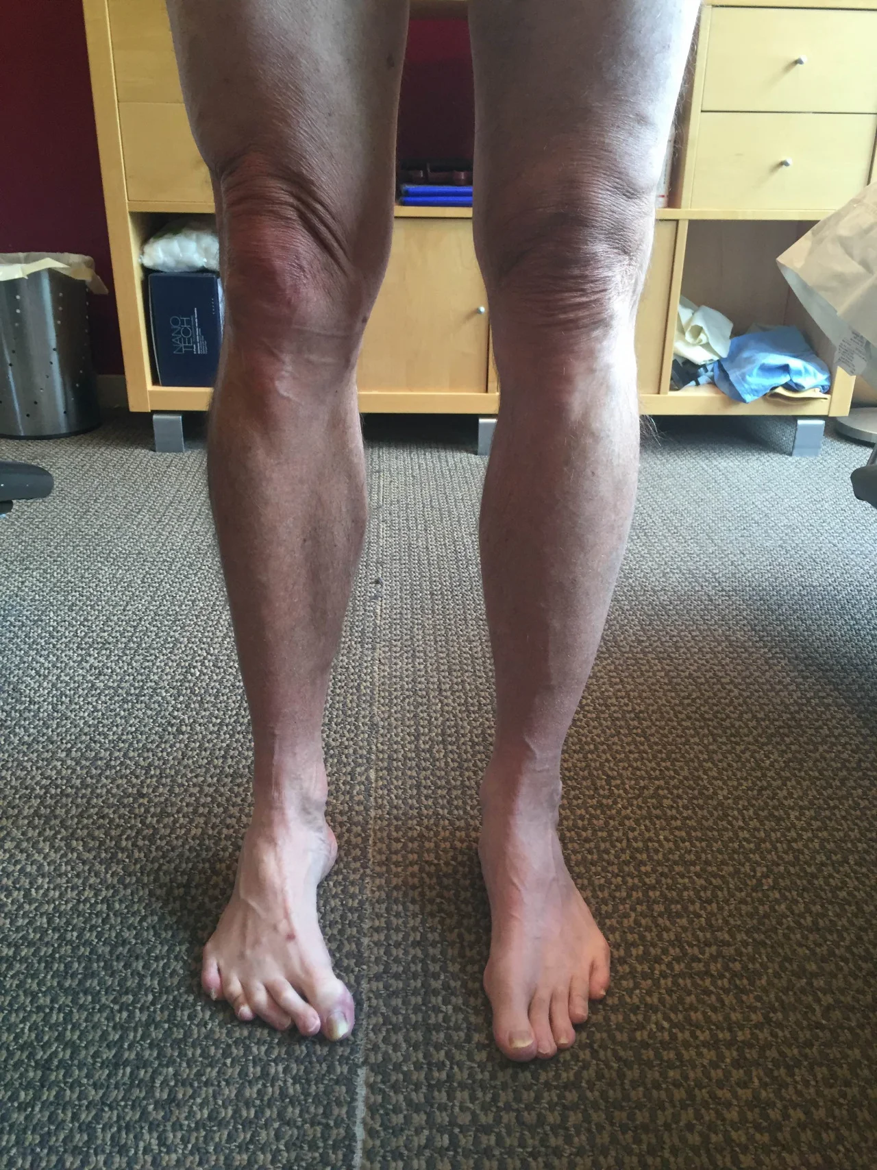
This is a case that has been looked at before but today with new video. This is a client with a known anatomic short leg on the right (sock-less foot) from a diseased right hip joint.  

In this video, it is clear to see the subconscious brain attempting to lengthen the right leg by right foot strike laterally (in supination) in an attempt to keep the arch and talus as high as possible.  Supination should raise the arch and thus the resting height of the talus, which will functionally lengthen the leg.  This is great for the early stance phase of gait and help to normalize pelvis symmetry, however, it will certainly result in (as seen in this video) a sudden late stance phase pronation event as they move over to the medial foot for toe off. Pronation will occur abruptly and excessively, which can have its own set of biomechanical compensations all the way up the chain, from metatarsal stress responses and plantar fasciitis to hip rotational pathologies.  It will also result in a sudden plummet downwards back into the anatomic short leg as the functional lengthening strategy is aborted out of necessity to move forward.  

This is a case where use of a full length sole lift is imperative at all times. The closer you get to normalizing the functional length, the less you need to worry about controlling pronation with a controlling orthotic (controlling rate and extent of arch drop in many cases). Do not use a heel lift only in these cases, you can see this client is already rushing quickly into forefoot loading from the issues at hand, the last thing you should be doing is plantarflexing the foot-ankle and helping them get to the forefoot even faster !  This will cause toe hammering and gripping and set the client up for further risk to fat pat displacement, abnormal metatarsal loading, challenges to the lumbricals as well as imbalances in the harmony of the long and short flexors and extensors (ie. hammer toes). 

How much do you lift ?  Be patient, go little by little. Give time for adaptation. Gauge the amount on improved function, not trying to match the right and the left precisely, after all the two hips are not the same to begin with. So go with cleaner function over choosing matching equal leg lengths.  Give time for compensatory adaptation, it is going to take time.  

Finally, do not forget that these types of clients will always need therapy and retraining of normal ankle rocker and hip extension mechanics as well as lumbopelvic stability (because they will be most likely be dumping into anterior pelvic tilt and knee flexion during the sudden forefoot loading in the late midstance phase of gait). So ramp up those lower abdominals (especially on the right) !  

Oh, and do not forget that left arm swing will be all distorted since it pairs with this right limp challenge. Leave those therapeutic issues to the end, they will not change until they see more equal functional leg lengths. This is why we say never (ok, almost never) retrain arm swing until you know you have two closely symmetrical lower limbs. Otherwise you will be teaching them to compensate on an already faulty motor compensation. Remember, to get proper anti-phasic gait, or better put, to slow the tendency towards spinal protective phasic gait, you need the pelvic and shoulder “girdles” to cooperate. When you get it right, opposite arm and leg will swing together in same pendulum direction, and this will be matched and set up by an antiphasic gait.

One last thing, rushing to the right forefoot will force an early departure off that right limb during gait, which will have to be caught by the left quad to dampen the premature load on the left. They will also likely have a left frontal plane pelvis drift which will also have to be addressed at some point or concurrently. This could set up a cross over gait in some folks, so watch for that as well.

“One thing, affects all things. One change necessitates global change. The more you know, the more you will see (and understand).  The more you know, see and understand, the more responsible you will and should feel to get it right and the more global your approach should become. If your head does not spin at times with all the issues that need to be juggled, you are likely not seeing all the issues you should be seeing.” -Dr. Allen (from an upcoming CME course)

Shawn Allen, one of the gait guys.

Difference between adult and infant gait compensation.

We highly doubt the infants compensated to the point of “recovering symmetrical gait”. It just isn’t possible seeing as there was frank asymmetry in leg length. However, it is quite possible they accomodated quicker with a more reasonable compensation, that MAY have appeared to have less limp. We did not do the study, but over a beer we might guess that the investigators might agree that our verbiage is closer to accurate. None the less, cool stuff to cogitate. We are very appreciative of this study, there is something to take from this study.

“The stability of a system affects how it will handle a perturbation: The system may compensate for the perturbation or not. This study examined how 14-month-old infants-notoriously unstable walkers-and adults cope with a perturbation to walking. We attached a platform to one of participants’ shoes, forcing them to walk with one elongated leg. At first, the platform shoe caused both age groups to slow down and limp, and caused infants to misstep and fall. But after a few trials, infants altered their gait to compensate for the platform shoe whereas adults did not; infants recovered symmetrical gait whereas adults continued to limp. Apparently, adult walking was stable enough to cope with the perturbation, but infants risked falling if they did not compensate. Compensation depends on the interplay of multiple factors: The availability of a compensatory response, the cost of compensation, and the stability of the system being perturbed.”- From the Cole et all study (reference below)

- thoughts by Shawn Allen

references:

Infant Behav Dev. 2014 Aug;37(3):305-14. doi: 10.1016/j.infbeh.2014.04.006. Epub 2014 May 20.Coping with asymmetry: how infants and adults walk with one elongated leg.Cole WG1, Gill SV2, Vereijken B3, Adolph KE4.

http://www.ncbi.nlm.nih.gov/pubmed/24857934

Eating up a cardinal plane.Simple post, simple principle today.  We found this case on the web, somewhere. Wish we could remember so we could give credit. Looks like simple right leg length discrepancy but the point we wanted to make is that any tim…

Eating up a cardinal plane.

Simple post, simple principle today.  We found this case on the web, somewhere. Wish we could remember so we could give credit. 

Looks like simple right leg length discrepancy but the point we wanted to make is that any time you deviate into a plane, you eat up length. In this case, the right knee is severely valgus and that has at least in part contributed to a shorter limb and unleveling of the pelvis. And, it is not uncommon that rotation axis are changed when frontal or sagittal planes are compromised. It is easy to see this on the x-rays, if the foot posturing isn’t at least noted, look at the spacing between the tibia and fibula .  .  .  . rotational planes have changed as well. Is it from the femur or tibia? That is the topic of another day. 

In the larger photo you will notice that even with the right foot lifted there is still a pelvis unleveling. How can that be, unless it was further unleveled that what we are seeing ?  Well, just because you lift to fix doesn’t mean the lift will not enable further collapsing into the weakness and deformity.  We have described this principle on the topic of EVA shoe foam deformation.  When the foot presses the foam into the deformation, it leaves more room for possibly further and faster deformation loads (perhaps more so than had a new shoe been prescribed). So in some cases, more lift can allow more deformation.  How far, well as in this photo, at least until the right knee slams into the left knee and stops further deformation.

So, seeing a plane deficit clues you into possible unleveling of the pelvis and abnormal joint loading responses. It should clue you into looking for another cardinal plane compromise as well. But make no mistake, just adding a lift doesn’t mean the deformation is remedied and not enabling further deformation. It is possible that you can make your client worse if you do not teach them how to find the appropriate motor patterns with the lift so they can learn to protect the parts. Often teaching these types of clients how to control their deformities (when and if possible) is where the gold lies, not in just leveling out the foundation. 

One more “beating of the dead  horse”, lift the whole foot, heel and forefoot with a sole lift when you are “lifting and leveling”. Lifting only the heel puts them into ankle plantar flexion and can often facilitated earlier and faster forefoot loading and even earlier knee flexion.  Save the heel lift as a possible consideration when there are posterior compartment contractures or inflammation.  Certainly we could have gone into functional and structural leg length discrepancies, but we have blogged excessively on that topic in the past. Go ahead and search our blog if you want more on those topics.

Take home point, “just because you lift, doesn’t mean you are truly lifting, you may enable the opposite”.

Shawn Allen, one of the gait guys

tumblr_nglhhhvhOP1qhko2so1_1280.png
tumblr_nglhhhvhOP1qhko2so2_r1_1280.png

Test your Mental Clinical Thinking Skills with this pedograph case. 

A few months ago, we discussed this case in great detail. There is likely little chance you will see our thinking progression with these final conclusions without sitting down with a warm cup of coffee and going over these 2 prior blog posts on this case (part 1 and part 2).  Besides, it will be a good review for you and it is great mental gymnastics.  This kind of analysis gets easier each time you do it but we have to through out our standard warning. This is the kind of stuff one needs to be able to go through on the fly in one’s practice, it is something to aspire to.

First of all, caveats:

  • Our discussions on this case were all theoretical.  What we went through was an exercise in static assessment and clinical thinking
  • One cannot, and must not, make clinical decisions from a static assessment. 
  • As in all assessments, information is taken in, digested and then MUST be confirmed, denied and/or at the very least, folded into a functional and clinically relevant assessment of the client before the findings are accepted, dismissed and acted upon. 
  • Gait analysis or pedograph-type assessment are helpful tools, but not the final answer.

Our static exam proposal on this case came up with the following theories (please stand up and mimic as we discuss, trust us, it will help you). *Remember: the foot on YOUR LEFT is the RIGHT foot for the purposes of this discussion. And remember, this is all theoretical, this is an exercise in biomechanical and clinical thinking, nothing more.

  • Suspect Counter-clockwise pelvis distortion pattern (causes relative internal rotation on LEFT and external rotation on RIGHT), this will drive Left knee hyperextension and Right knee flexion (hence foot plantar pressures as we discussed in previous 2 blog posts linked above). This of course cannot be seen, but we are extrapolating from our clinical experiences.
  • poor pronation and internal limb spin control on the left (hence longer foot and toe hammering). Obviously, we would see a dramatic shift of the pressures to the medial foot if this were truly the case.  Perhaps this is because of the greater lateral left pelvis drift forcing the glute and foot pronatory controls to have to work harder and longer, and maybe even quicker, to control the internal spin and pronation. Over time, they fatigue and fail rendering a flatter, more pronated and longer heel:toe ball length ratio. This would also give credence to the left toe hammering/gripping response.
  • static increased left limb weight bearing (left hip drift)
  • abrupt right foot loading pattern (more mid-forefoot strike), perhaps as reflected by the static forefoot loading. Again, supposition.
  • with all of the above, it is suspect that this client will appear to have a subtle limp, coming off the left quickly or prematurely as they speed through uncontrolled pronation and resulting in an abrupt right limb loading response that mostly skips through heel strike and results in a more aggressive mid-forefoot loading response.  This, sort of, creates a catching of the loading response by the quadriceps more than the gluteals. This can cause medial knee drift (valgus loading) if the medial knee stabilizers are not up to task, this also creates a sudden patellofemoral compresson event and unappreciated sudden tension on the extensor mechanism (the quad-patella-patellar tendon complex).  Can you say generic anterior knee pain ?

Just some thoughts. Please go back to the prior 2 blog posts to delve deeper into the conclusions we have brought about here, we have other good reasoning to suspect the above as the scenario. But remember, what you see is not the problem, we see people’s compensations, their strategies. This was just an exercise in “what ifs”, nothing more. But you will see it in your clinic, just substantiate it with an exam, not what you necessarily see in your clients gait or static assessment. Static assessments are for fools, don’t be a fooled fool.  What  you see is not the problem.

Remember this critical fact.  After an injury or a long standing problem, the job of muscles and motor patterns is to stabilize and manage loads (stability and mobility) for adequate and necessary movement. Injuries often leave a mark on the system as a whole because adaptation was necessary during the initial healing phase. This usually spills over during the early movement re-introduction phase, particularly if movement is reintroduced too early or too aggressively.  Plasticity is often a culprit. Just because the injury has come and gone does not mean that new patterns of skill, endurance, strength (S.E.S -our favorite mnemonic), stability and mobility were not subsequently built onto the apparently trivial remnants of the injury. There is nothing trivial if it is abnormal. The forces must, and will, play out somewhere in the body and this is often where pain or injury occurs but it is rarely where the underlying problem lives. Is the compensation top down, bottom up, or both ?

Don;t be a fooled fool. Get the facts.

Shawn and Ivo, the gait guys

The “Standing on Glass” Static Foot/Pedograph Assessment: Part 1
* note: This is a static assessment dialogue. One cannot, and must not, make clinical decisions from a static assessment. As in all assessments, information is taken in, di…

The “Standing on Glass” Static Foot/Pedograph Assessment: Part 1

* note: This is a static assessment dialogue. One cannot, and must not, make clinical decisions from a static assessment. As in all assessments, information is taken in, digested and them MUST be confirmed, denied and/or at the very least, folded into a functional and clinically relevant assessment of the client before the findings are accepted, dismissed and acted upon. As we always say, a gait analysis or pedograph-type assessment is never enough to make decisions on treatment to resolve problems and injuries. What is seen and represented on either are the client’s strategies around clinical problems or compensations.  Today’s photo and blog post are an exercise in critical clinical thinking to get the juices flowing and to get the observer thinking about the client’s presentation and to help open up the field to questions the observer should be entertaining.  The big questions should be, “why do i see this, what could be causing these observances ?”

* note the right and left sides by the R and L circled in pink.

ORANGE lines: The right foot appears to be shorter, or is it that the left is longer (see the lines and arrows drawing your attention to these differences)? A shorter foot could be represented by a supinated foot (if you raise the arch via the windlass mechanism you will shorten the foot distance between the rear and forefoot). A longer foot could be represented by a more pronated foot.  Is that what we have here ? There is no way to know, this is a static presentation of a client standing on glass. What we should remember is that the goal is always to get the pelvis square and level.  If an anatomically or functionally short leg is present, the short leg side MAY supinate to raise the mortise and somewhat lengthen the leg.  In that same client, they may try to meet the process part way by pronating the other foot to functionally “shorten” that leg.  Is that what is happening here ? So, does this client have a shorter right leg ? Longer left ?  Do you see a plunking down heavily onto the right foot in gait ? Remember, what you see is their compensation.  Perhaps the right foot is supinating, and thus working harder at the bottom end of the limb (via more supination), to make up for a weak right glute failing to eccentrically control the internal spin of the leg during stance phase ? OR, perhaps the left foot is pronating more to drive more internal rotation on the left limb because there is a restricted left internal hip rotation from the top ? Is the compensation top-down or bottom up ? These are all viable possibilities and you must have these things flowing freely through your head during the clinical examination as you rule in/rule out your hands-on findings.  Remember, just going by a FMS-type screen to drive prescription exercises from what you see on a movement screen is not going to necessarily fix the problem, it could in fact lead one to drive a deeper compensation pattern. You can be sure that Gray Cook’s turbo charged brain is juggling all of these issues (and more !) when he sees a screen impairment, although we are not speaking for him here.

Remember this critical fact.  After an injury or a long standing problem, muscles and motor patterns jobs are to stabilize and manage loads (stability and mobility) for adequate and necessary movement. Injuries leave a mark on the system as a whole because adaptation was necessary during the initial healing phase. This usually spills over during the early movement re-introduction phase, particularly if movement is reintroduced too early or too aggressively.  Plasticity is the culprit. Just because the injury has come and gone does not mean that new patterns of skill, endurance, strength (S.E.S -our favorite mnemonic), stability and mobility were not subsequently built onto the apparently trivial remnants of the injury.  There is nothing trivial if it is abnormal. The forces must, and will, play out somewhere in the body and this is often where pain or injury occurs but it is rarely where the underlying problem lives.

Come back tomorrow, where we will open your mind into the yellow, pink, blue and lime markings on the photo. Are the hammering toes (lime) on the left a clue ? How about the width of the feet (yellow) ? The posturing differences of the 5th toe to the lateral foot border ?  What about the static plantar pressure differences from side to side (blue)? Maybe, just maybe, we can bring a logical clinical assumption together and then a few clinical exam methods to confirm or dis-confirm our working diagnostic assumption.  See you tomorrow friends !

Shawn and ivo, the gait guys

tumblr_n7yjq09EgU1qhko2so1_1280.jpg
tumblr_n7yjq09EgU1qhko2so2_1280.jpg
tumblr_n7yjq09EgU1qhko2so3_1280.jpg
tumblr_n7yjq09EgU1qhko2so4_1280.jpg
tumblr_n7yjq09EgU1qhko2so5_1280.jpg
tumblr_n7yjq09EgU1qhko2so7_1280.jpg
tumblr_n7yjq09EgU1qhko2so9_1280.jpg
tumblr_n7yjq09EgU1qhko2so6_r1_500.jpg

So a patient presents to your office with a recent history of a L total knee replacement 8 weeks ago AND a recent history of a resurgence of low back pain, supra iliac area on the L side. Hmmmm. Hope the flags went up for you too!

His global lumbar ROM’s were 70/90 flexion with low back discomfort at the lumbo sacral junction, 20/30 extension with lumbosacral discomfort, left lateral bending 10 degrees with increased pain (reproduction); right lateral bending 20 degrees with a pulling sensation on the right. Extension and axial compression of the lumbar spine in left lateral bending reproduced his pain.

Neurologically he had an absent patellar reflex on the left, with diminished sensation over the knee medially and laterally. Muscle strength 5/5 in LE; sl impaired balance in Left single leg standing. There was incomplete extension of the left knee, being at 5 degrees flexion (right side was zero).

He has a right sided leg length deficiency (or a left sided excess!) of 5 mm. Take a look at the tibial lengths in the 1st 3 pictures. See how the left is longer? In the next shot, do you see how the knee cannot completely extend? Can you imagine that the discrepancy would probably be larger if it did?

Now look at the x rays. We drew a line across from the non surgical leg to make things clearer.

Now, think about the mechanics of a longer leg. That leg will usually pronate more in an attempt to shorten the leg, and the opposite side will supinate to attempt to lengthen. Can you see how this would cause clockwise pelvic rotation (in addition to anterior pelvic rotation)? Can you see this patients in the view of the knees from the top? Do you understand that the lumbar spine has very limited rotation (about 5-10 degrees, with more movement superiorly (1)  ). Does it make sense that the increased range of motion could effect the disc and facet joints and increase the patients low back pain?

So, how do we fix it? Have you seen the movie “Gattica”? Hmmm….A bit extreme. How about a full length 3mm sole lift to start, along with specific joint manipulation to restore normal motion and some acupuncture to reduce inflammation? We say that is a good start.

The Gait Guys. Increasing your gait literacy with each and every post. If you liked this post, please send it to someone else for them to enjoy and learn. 

(1) Three-Dimensional In Vivo Measurement of Lumbar Spine Segmental Motion Ruth S. Ochia, PhD, Nozomu Inoue, MD, PhD, Susan M. Renner, MS, Eric P. Lorenz, MS, Tae-Hong Lim, PhD, Gunnar B. Andersson, J. MD, PhD, Howard S. An, MD Spine. 2006;31(15):2073-2078.

Podcast 36: Heel lift lies, the Exercise Drug & Malcolm Gladwell's 10,000 hours.

http://thegaitguys.libsyn.com/podcast-36-heel-lift-lies-and-the-exercise-drug

iTunes link:

https://itunes.apple.com/us/podcast/the-gait-guys-podcast/id559864138

Gait Guys online /download store:

http://store.payloadz.com/results/results.aspx?m=80204

other web based Gait Guys lectures:

www.onlinece.com   type in Dr. Waerlop or Dr. Allen  Biomechanics

Today’s show notes:

Neuroscience piece:

Description
1. Health Scanner Scanadu Scout: the future of medical info gathering ?

2. The Exercise Drug:

www.gizmag.com/scripps-drug-sr9009-exercise-mimic/28651

3. FB reader sent us a message:

- am posting this to see if I can get a little bit of help from the best professionals in the area (you). 
I have read some of the information on your site and I think I have quite a problem on my right foot. It happens that the medial part of the foot tripod does not touches the floor at all and I have lack of support in that zone. So it seems like my forefoot is varus. I have also noticed that when I am standing it looks like my rear foot is valgus. So, I can’t really compensate this problem because if the forefoot is varus and I try to put it neutral, the rear foot gets even more valgus, and if it I try to put the rear foot neutral, the forefoot gets even more varus and my big toe does not touch any part of the floor. Can you please help me? I do not know what to do and I am a little bit desperate because nobody I went to could help me. You are probably my last hope. I know I can correct this and I have the will and dedication to pull it off. I bet there are some exercises I can do but I do not really know which at all. 
Thanks in advance. -Jorge
4. Another TUMBLR reader asks question about
Guys what are the possible muscular causes of genu varum during initial swing?
5. Another off tumblr:  Anything  you can talk about on this topic ?
How does running in low-to-no light conditions effect your gait/running/injuries/etc?
6. Topic: step  width

Changing step width alters lower extremity biomechanics during running

7.  heel lift vs. sole lift
why and when would you use only a heel lift…..unilaterally ?
8. National Shoe Fit program: 
Link: http://store.payloadz.com/results/results.aspx?m=80204
9. Questions from a field doc:
Hey guys,
 I have heard you guys say many times that many people who choose to venture into minimalistic footwear have not “earned their right" to do such without increasing their risk for problems.  I was wondering if you could explain what parameters you use to determine if and when they are ready.  
Thanks,
Ryan 
10. Shoes: does pronation matter
11. Shoes #2:
12. Malcolm Gladwell debate, 10,000 hours
tumblr_msj34fqgpW1qhko2so1_1280.jpg
tumblr_msj34fqgpW1qhko2so2_1280.jpg

What does sustained rearfoot and forefoot inversion look like in terms of shoe wear ?

This is a client who came to us with a history of several months of big toe pain (specifically 1st metatarsophalangeal joint pain). The pain was dorsally located (ie. top of the big toe joint).  It was clear that on examination the pain was being caused by osseous and soft tissue dorsal impingement due to a progressing hallux rigidus/limitus. 

This painful status obviously is creating both a conscious medial toe off pattern during the propulsive phase of late stance phases.  The client is more than obviously steering push off laterally as can be seen in this photo. The grey lateral pods are almost obliterated. This means the foot is perpetually supinated during the entire stance phase of gait and this means that pronation shock absorption is not present.

Remember, a perpetually supinated foot means the talus and arch never descend as part of the pronation/shock absorption cycle and so the same side limb will always remain longer than the other limb which is seeing the internal rotation/pronation effects which functionally shorten the leg during stance phase.  So in this case, we have a pelvic unlevelling and a frontal plane shift to the functionally shorter leg during its stance phase.  It should not surprise you that this client has hip pain contralateral to this abnormal shoe wear/hallux limitus side. 

There are plenty of other issues here to be discussed, like eccentric weakness of the same side g.maximus, patellar tracking issues, lack of hip extension and thus weakening of the glutes and thus resultant shortness of the quadriceps group which will all often be found in this clinical picture. But we will save that all for another time.

Remember, the longer this client stays in this shoe, the easier it is mechanically on them because the eVA foam and the shoe are broken down into their compensatory avoidance behaviour.  But, this is where the pattern becomes subconsciously embedded and thus when the pattern drives many of the other compensatory patterns off of this one since it is the new norm. The faster you address this problem, the sooner you stop the compensatory cascade.  And on that note, if you read our blog post re-run of the Arm Swing last week you will understand why  these folks will begin altering the opposite arm swing phase.

Shawn and Ivo, The Gait Guys

tumblr_mrrb0hsxBX1qhko2so1_1280.jpg
tumblr_mrrb0hsxBX1qhko2so2_1280.jpg
tumblr_mrrb0hsxBX1qhko2so3_1280.jpg
tumblr_mrrb0hsxBX1qhko2so4_1280.jpg
tumblr_mrrb0hsxBX1qhko2so5_1280.jpg
tumblr_mrrb0hsxBX1qhko2so6_1280.jpg

Subtle clues often tell the story. A case 

A young athlete that wants to be faster (he is a 4:17 miler).

In the 1st shot we see he has an anatomically short R tibia (and the gait to match it). You will note the right tibial plateau is lower than the left. The 2nd shot backs this up; look at the malleoli.

The next shot shows a FABER test f the L hip. Compare the range of motion with the right one. Hmmm.  Limited external rotation and abduction on the right.  It should be noted he had normal and relatively symmetrical internal rotation of both hips.

Now come the feet. 1st the left. A relatively neutral foot. Next the right. What’s different? Note how much more pronounced the right 1st ray (ie 1st metatarsal phalangeal joint).

Think about his short side. Most likely, he will be trying to lengthen it, right? How would he accomplish that? By supinating the foot (making it more rigid) and attempting to lengthen that leg, by anterior rotation of the pelvis. If you anteriorly rotate the pelvis (ie the innominate rotates forward, bringing the ASIS forward), what happens to external rotation of the hip? Stand up, edtend your hip on your pelvis and find out. It limits it.

How else might he try to lengthen that leg? If he supinates the foot (ie planytarflexion, adducion and inversion), the foot will be more inverted. He will be trying to get that medial tripod down to the ground. How might he accomplish that? By plantarflexing the 1st ray!

So how can we make him faster?

  • Place sole lift under r foot
  • Correct pelvic pathomechanics with manipulation
  • Support coorection with appropriate exercise (he had weak R lower external oblique’s)
  • Foot mobilization
  • R Foot intrinsic exercises to promote rasing of the 1st ray (extensor hallicus longus  and flexor hallicus brevis exercises)  and lowering of the lesser metatarsal heads (extensor digitorum brevis exercises ).

The answers are often in the details. Be detail oriented. That’s one of the things that makes us foot geeks.

Ivo and Shawn

* remember: by clicking on the YOUTUBE logo in the lower right you will be immediately linked to a larger viewing screen on youtube.

This is a video case of a triathlete who presented with left calf pain and right quadriceps leg pain after months of training. In the video we discuss altered ankle rocker (dorsiflexion), lower crossed syndrome, altered arm swing patterning, unilateral quadriceps tightness and several other functional gait pathologies with this case.