The Cheetah man: A new perspective on Cross Crawl and neurologic patterning.

A few months ago we wrote a piece about Uner Tan Syndrome.  Here was a key point from that blog post (blog post link) and it links beautifully to our most recent controversial blog post on the “Bird Dog” rehab exercise (link here):

[In the video presented in that blog post, and in our “Bird Dog” post photo] there is ipsilateral interference between the foot and hand in this quadrupedal gait. In this diagonal quadrupedal locomotion (QL) the forward moving lower limb is impaired from further forward progression by the posting up (contact) hand of the same side. This would not occur if the QL gait was non-diagonal (ie. unilateral), the forward progression of the lower limb would be met with same time forward progression of the upper limb, allowing a larger striding out of both limbs.  This would enable faster locomotion without increasing cadence (which would be the only way of speeding up in the diagonal QL), at the possible limitation of necessitating greater unilateral truncal postural control (which is a typical problem in some of these Uner Tan Syndrome individuals who typically have profound truncal ataxia).  

So, why are we showing you the video above today ?  If you do not know, go read those 2 blog posts again and look more closely at the video above. At exactly 0:21 seconds into the video, at the slow motion section, you can see what we were talking about in the Bird Dog post last week, that being that the quadrupedal pattern that is neurologically substantiated is that when the right lower limb is in flexion, so is the left upper limb. (we will show these 2 photos in tomorrows post).  Where as, “Bird Dog” shows the opposite, that the contralateral upper limb will be in the opposite phase of the contralateral lower limb.

Who cares right ?  Well, it matters.  In the video above, this could be a problem because if the right leg is in flexion that means that the right arm will be moving into extension. This means that the knee and the hand will be running into each other (look at the baby photo here). As we discussed in the Uner Tan article this will impair faster quadrupedal locomotion. It is also one of the theories as to what may have pushed us to become bipedal and allow faster ambulation (there are many theories of course).  So, how then does this guy in the video move like a cheetah ? How is he going so fast with the quadrupedal pattern we have clearly outlined here ?

Within days a blog reader (Micheal L, thanks Michael) messaged us and said this:

  • As a person who likes what’s going on at MoveNat, this type of quadrupedal movement is referred to by them as contralateral movement and is how they teach people to crawl at their seminars. In CrossFit workouts, we also do bear crawls as an exercise, and I always try to maintain a contralateral gait. i.e. Right arm moves forward as left foot comes forwards and vice versa. 
    So, in other words, in the Uner Tan Syndrome (UTS) the gait is cumbersome and inefficient. In the video above and at MoveNat seminars, it’s a technique/skill.
    Did you guys intend for this comparison, or am I out in the cornfield on this?
    Here was Dr. Uner Tan  himself chiming in on the dialogue:
  • Üner Tan It is not the same type of locomotion, i.e., not “the diagonal-sequence quadrupedal locomotion”, which is also used by non-human primates.. .
    Michael: The guy runs so fast it’s hard for me to see it well. Okay, so with UTS the lower limb runs into the upper limb. In this video, his upper limb quickly gets out of the way, giving room for the lower limb (to further flex forward increasing swing phase forward step length). It’s just really hard to see it without slow motion. Thank you for clarifying.

As we said in last weeks post on all of this:
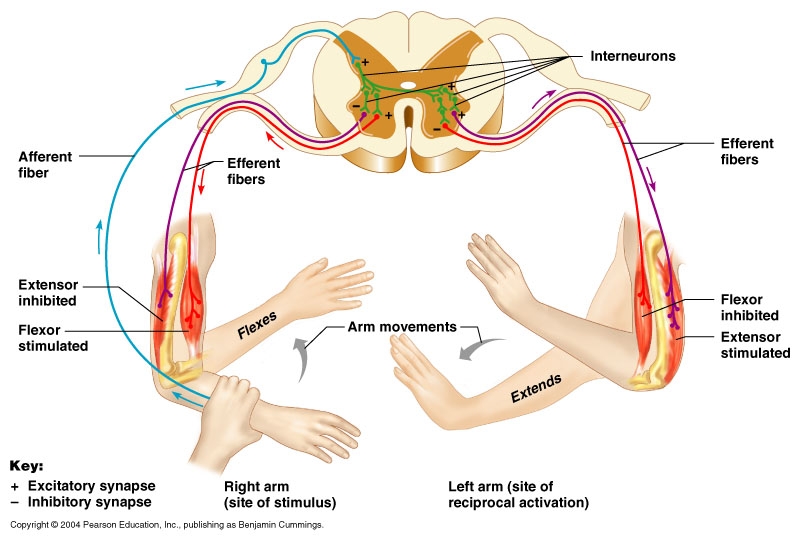
“Think about gait. Your right leg and left arm flex until about midstance, when they start to extend; the left leg and right arm are doing the opposite. At no point are the arm and opposite leg opposing one another. 
If you look at it neurologically, it is a crossed extensor reflex.  It is very similar to a protective reflex called the “flexor reflex” or “flexor reflex afferent”. 

In this video case today, it appeared on the surface because of the speed of this fella, that all that we have been talking about had been left in the dust. But, after looking at things closer and more slowly, the principles remain intact.  For now.
Just a little open thinking digging today. Hope you enjoyed.
Shawn and Ivo,
The gait guys
tumblr_n4swdbdmHS1qhko2so2_1280.jpg
tumblr_n4swdbdmHS1qhko2so1_1280.jpg

The case of the missing toes.

OK, a bit dramatic but as you can see in the plantar view above, all you can see is the toe pads, the rest of the digit shafts are hidden.  

This is a classic example of a foot imbalance. We have talked about this many times before but the attached video link here  ( http://youtu.be/IIyg7ejYNOg ) shows it very well.  Read on.

There is shortness and increased resting tone in the short toe extensors (EDB, extensor digitorum brevis) and long toe flexors (FDL=flexor dig. longus) with insufficiency in the short flexors and long extensors. This pairing creates a hammer toe effect.  In the video, you can see that these toes are showing early hammering characteristics, but not yet rigid ones. The key word there is, “yet” so this is still a correctable phenomenon at this point.  You can also clearly see the distal migration of the metatarsal fat pad. The fat pad has migrated forward of the MET heads and is being pulled forward by the excess tension in the long toe flexors. As this imbalance in the toe flexors and extensors develops, the forefoot mechanics get impaired and the lumbricals (which anchor off off the FDL) become challenged. Their contributory biomechanics, amongst other things, help to keep the fat pad in place under the metatarsal heads. You can see in this video link above that by proximally migrating (towards the heel) just the fat pad back under the MET heads the resting mechanics of the toes changes, for the better.  

Remember the other functions of the lumbricals ?  their other major functions, namely: thinking from a distal to proximal orientation (a closed chain mode of thinking), they actually plantarflex the metatarsal on the fixed phalynx, assist in dorsiflexion of the ankle, and help to keep the toes from clawing from over recruitment of the flexor digitorum longus.

Here is another blog post we did on a similar presentation.http://thegaitguys.tumblr.com/post/14766494068/a-case-of-plantar-foot-pain-during-gait-this

Proper balance of the toe flexors and extensors, and their harmony with lumbricals and fat pad amongst other things will give healthy long flat toes that can help the proximal biomechanics of the foot.  If you have neuromas, metatarsalgia, hammer toes, claw toes, migrating toes, bunions or hallux valgus amongst many other things, this might be a good place to start.   

There are exercises that can help this presentation, but understanding “the why” is the first step.

Shawn and Ivo

The Gait Guys

tumblr_n4p3qkoqT81qhko2so1_250.jpg
tumblr_n4p3qkoqT81qhko2so2_1280.jpg

This simple screening test becomes a form of exercise.

Last week we explored the “Lean” test to see how your QL and gluteus medius were paired. Today we look at a simple CNS screen for your “central pattern generators” or “CPG’s”. If you do not pass, then the exercise becomes the rehab exercise. If you (or your client) does not have good coordination between the upper and lower extremity, then they will not be that efficient, physiologically or metabolically.

The “cross crawl” or “step test” looks at upper and lower extremity coordination, rather than muscular strength. If performed for a few minutes, it becomes a test that can look at endurance as well.

It is based on the “crossed extensor” response, we looked at last week. That is, when one lower limb flexes, the other extends; the contralateral upper limb also flexes and the ipsilateral upper limb extends. It mimics the way things should move when walking or running.

  • Stand (or have your client stand) in a place where you will not run into anything.
  • Begin marching in place.
  • Observe for a few seconds. When you (or your client) are flexing the right thigh, the left arm should flex as well; then the left thigh and right arm. Are your (their) arms moving? Are they coordinated with the lower extremity?
  • What happens after a few minutes? Is motion good at 1st and then breaks down?
  • Now speed up. What happens? Is the movement smooth and coordinated? Choppy? Discoordinated?
  • now slow back down and try it with your (their) eyes closed


If  movement is smooth and coordinated, you (they) pass

If movement is choppy or discoordinated, there can be many causes, from simple (muscle not firing, injury) to complex (physical or physiological lesion in the CNS).

  • If movement is not smooth and coordinated, try doing the exercise for a few minutes a day. You can even start sitting down, if you (they) cannot perform it standing. If it improves, great; you were able to help “reprogram” the system. If not, then you (they) should seek out a qualified individual for some assistance and to get to the root of the problem.


The Gait Guys. Giving you information you can use and taking you a little deeper down the rabbit hole with each post.

Rewind double feature! Part 2

(for part 1, click here)

In conjunction with the latest PODcast talking about efferent copy, we thought it appropriate to talk about the cerebellum here. In this capsule we talk about the efferent pathways

Enjoy! and have a nice weekend (not that we are telling you what to do…)

Ivo and Shawn

Rewind double feature! Part 1

A first ever her on TGG. With the latest PODcast talking about efferent copy, we thought it appropriate to talk about the cerebellum here.

Enjoy! and have a nice weekend (not that we are telling you what to do…)

Ivo and Shawn

Podcast 60: Speeding up at the finish line & Efferent Copy

A. Link to our server:

Direct Download: 

http://traffic.libsyn.com/thegaitguys/pod_60final.mp3

Permalink: 

http://thegaitguys.libsyn.com/podcast-60-speeding-up-at-the-finish-line-efferent-copy

B. iTunes link:

https://itunes.apple.com/us/podcast/the-gait-guys-podcast/id559864138

C. Gait Guys online /download store (National Shoe Fit Certification and more !) :

http://store.payloadz.com/results/results.aspx?m=80204

D. other web based Gait Guys lectures:

www.onlinece.com   type in Dr. Waerlop or Dr. Allen,  ”Biomechanics”

______________

Today’s Show notes:

1.  Move Your Body, Grow Your Brain

6. Blog reader: Hey again, I personally compete in this uphill/ downhill running race in 2 months. The slops are really steep(incline/ decline).I’m just looking for some useful tips on running biomechanics, cadence etc. Any hints on what things I should be doing and of course avoid doings that will lead to injury? An international podcast fun!

7. Blog reader:

How does subtalar supination/pronation affect plane deviations farther up the chain, specifically at the pelvis? I was going over some notes and found something saying that increased pronation leads to increased frontal plane motion at the pelvis during gait, and increased sup leads to increased transverse plane motion. However, in this video I was watching, the clinician states that increased sup will lead to increased frontal plane motion. Is this not a contradiction? Can both be true? Thanks
Invalid video embed.

Can you see something strange in this gait ? You gotta look closely, there are several things that should bother you.

Chronic Inflammatory Demyelinating Polyradiculoneuropathy (CIDP)

Not every gait problem is related to faulty shoes, bad feet or impaired arm swing.  

This is a case of CIDP that walked (terribly) into our clinic.  We did not initially know what this was but as you can see from this gait, if you look closely enough, something is just missing and wrong.

Arm swing is very problematic and there is no anti-phasic shoulder/pelvic girdle motions.  There are subtle demonstrations of both wide based gait steps and cross-overs. The gait is syncopated, sort of.  We had to shorten the clip for patient ID preservation, we wish we could have shown you more, it is really quite obvious that there is a systemic neurologic coordination problem in this patient.  With demyelinating polyneuropathies as the peripheral receptors become more and more impaired the brain eventually starts to lose more and more signals from the peripheral joints as to where the body parts are in space. IF the brain cannot find the parts, it cannot control them. If the brain just gets some of the signal the cerebellum cannot use its EFFERENT copy of the motor program, which is send out into the limbs, to orchestrate smooth coordinated tasks.  Go watch the video again, look at the global feel of the video, it is ratchety and syncopated.

CIDP is an acquired immune-mediated inflammatory disorder of the peripheral nervous system. The myelin sheath is slowly destroyed in this disease so nerve conduction is gradually lost. Some sources compare this disease to Guillian Barre disease. 

From wikipedia: 

Chronic inflammatory demyelinating polyneuropathy[3] is believed to be due to immune cells, cells which normally protect the body from foreign infection, but here begin incorrectly attacking the nerves in the body instead. As a result, the affected nerves fail to respond, or respond only weakly, to stimuli, causing numbing, tingling, pain, progressive muscle weakness, loss of deep tendon reflexes (areflexia), fatigue, and abnormal sensations. The likelihood of progression of the disease is high.

After seeing the client a few times, it was clear that there were several fixed neurologic parameters which could not be mediated and the client was sent an EMG/NCV and the specific diagnosis was made. The client was put on a monthly immunogloblulin IV drip and has remarkably stabilized.

Not all gaits are from a bad ankle, a slumbering cerebellum or cruddy arm swing.  Trust your clinical judgement, if it doesn’t feel right, refer up or laterally.  

Shawn and Ivo

tumblr_n4b8plBI1q1qhko2so1_400.png
tumblr_n4b8plBI1q1qhko2so2_1280.jpg
tumblr_n4b8plBI1q1qhko2so3_1280.jpg
tumblr_n4b8plBI1q1qhko2so4_400.png
tumblr_n4b8plBI1q1qhko2so5_500.jpg

A new twist on an old exercise

Do you know the the “Bird Dog” exercise? It looks like the picture above. The upper and contralateral lower extremities are extended, the the opposite ones are flexed. Seems to make make sense, unless you think about gait and neurology (yes, as you can see, those things seem to always be intertwined).

Think about gait. Your right leg and left arm flex until about midstance, when they start to extend; the left leg and right arm are doing the opposite. At no point are the arm and opposite leg opposing one another. Hmmm.

If you look at it neurologically, it is a crossed extensor reflex (see above); again, flexion of the lower extremity is paired with flexion of the opposite upper extremity. It is very similar to a protective reflex called the “flexor reflex” or “flexor reflex afferent”.

Wouldn’t it make more sense to do a cross crawl pattern? Or maybe like the babies shown above? Seems like if that’s the way the system was programmed, maybe we should try and emulate that. Don’t we want to send the appropriate messages to our nervous system for neurological re patterning? If you are doing the classic “opposite” pattern, what is your reasoning? Can you provide a sound neurological or physiological reason?

Think before you act. Know what you are doing.

The Gait Guys. Bridging the gap between neurology and gait, so you can do a better job.

Welcome to rewind (Late) Friday. Sorry about the late entry, folks.

Along the vein of bike fit, to go with our lecture on onlinece.com this week, here is gentleman with right sided low back pain ONLY when ascending hills on his mountain bike. Can you figure out why?

*Stop, watch the video and think about it before we give you the answer… .

____________________________

This gentleman presented with low back pain, only on his mountain bike, only on long ascents.

He measures out with an 83 cm inseam which should put him on a 44 to 45.5 cm frame (measured via our method). His frame has a dropped top tube and measures 55 cm.

He has a knee bend angle of 20 degrees at bottom dead center. Knee is centered well over pedal axis.

His stem falls far in front of his line of sight with respect to his hub. Stem is a 100 mm stem with a 6 degree rise.

There is a 2" drop from the seat to the top of the handlebars.

He has an anatomically short Left leg (tibial)

Look at the tissue folds at the waist and amount of reach with each leg during the downstroke.

The frame, though he is a big dude (6’+), is too big and his stem is too long. He is stretched out too far over the top tube, causing him to have an even more rounded back (and less access to his glutes; glutes should rule the downstroke and abs the upstroke). This gets worse when he pushes back (on his seat) and settles in for a long uphill. Now throw in a leg length discrepancy and asymmetrical biomechanics.

Our recommendations: smaller frame (not going to happen) lower seat 5-7mm shorter stem (60-75mm) with greater than 15 degree rise lift in Left shoe

We ARE the Gait Guys, and we do bikes too!

Go ahead and try this at home.remember last mondays post? (if not, click here). Here is one way of telling whether your (or someone else’s) vestibular system is working. It will also give you an idea of how some people compensate. Ready?
Stand up (b…

Go ahead and try this at home.

remember last mondays post? (if not, click here). Here is one way of telling whether your (or someone else’s) vestibular system is working. It will also give you an idea of how some people compensate.

Ready?

  • Stand up (barefoot or shoes does not matter).
  • place your hands resting on the top of your hips with your thumbs to the back (like your Mom used to, when you were in trouble). Your thumbs should be resting on your quadratus lumborum (QL) muscle.
  • tilt your HEAD to the LEFT
  • you should feel the muscle (ie the QL) under your RIGHT thumb contract
  • come back upright


repeat, but this time lean your BODY to the LEFT

  • same thing right? Now check the other side.


Everything OK? Everything fire as it should?

Now lets add another dimension.

  • slide your fingers down so they are just below the crest of the hip, resting above the greater trochanter (the bump on the side of your upper thigh). This should place your fingers on the middle fibers of the gluteus medius.
  • tilt your head (or body ) to the LEFT.
  • You should feel the LEFT gluteus medius and the RIGHT QL contract. These muscles should be paired neurologically. When walking, during stance phase on the LEFT: the LEFT gluteus medius helps to maintain the pelvis level, while the RIGHT QL, assists in hiking the RIGHT side.


If everything works OK, then your vestibulospinal spinal system is intact and your QL and gluteus medius seem to be firing and appropriately paired. If not? That is the subject for another post.

The Gait Guys. Helping you to understand the concepts of WHY compensations occur.

tumblr_n3vbw7hW5t1qhko2so1_1280.jpg
tumblr_n3vbw7hW5t1qhko2so2_400.jpg

Welcome to Rewind Friday, Folks. Today we review the importance of the great toe extensor. Enjoy!

Gait Topic: The Mighty EHB (The Short extensor of the big toe, do not dismiss it !)

Look at this beautiful muscle in a foot that has not yet been exposed to hard planar surfaces and shoes that limit or alter motion! (2 pics above, toggle back and forth)

The Extensor Hallicus Brevis, or EHB as we fondly call it (beautifully pictured above causing the  extension (dorsiflexion) of the child’s proximal big toe) is an important muscle for descending the distal aspect of the 1st ray complex (1st metatarsal and medial cunieform) as well as extending the 1st metatarsophalangeal joint. It is in part responsible for affixing the medial tripod of the foot to the ground.  Its motion is generally triplanar, with the position being 45 degrees from the saggital (midline) plane and 45 degrees from the frontal (coronal) plane, angled medially, which places it almost parallel with the transverse plane. With pronation, it is believed to favor adduction (reference). Did you ever watch our video from 2 years ago ? If not, here it is, you will see good EHB demo and function in this video. click here

It arises from the anterior calcaneus and inserts on the dorsal aspect of the proximal phalynx. It is that quarter dollar sized fleshy protruding, mass on the lateral aspect of the dorsal foot.  The EHB is the upper part of that mass. It is innervated by the lateral portion of one of the terminal branches of the deep peronel nerve (S1, S2), which happens to be the same as the extensor digitorum brevis (EDB), which is why some sources believe it is actually the medial part of that muscle. It appears to fire from loading response to nearly toe off, just like the EDB; another reason it may phylogenetically represent an extension of the same muscle.

*The EDB and EHB are quite frequently damaged during inversion sprains but few seem to ever look to assess it, largely out of ignorance. We had a young runner this past year who had clearly torn just the EHB and could not engage it at all. He was being treated for lateral ankle ligament injury when clearly the problem was the EHB, the lateral ligamentous system had healed fine and this residual was his chief problem.  Thankfully we got the case on film so we will present this one soon for you !  In chronic cases we have been known to take xrays on a non-standard tangential view (local radiographic clinics hate us, but learn alot from our creativity) to demonstrate small bony avulsion fragments proving its damage in unresolving chronic ankle sprains not to mention small myositis ossificans deposits within the muscle mass proper.

Because the tendon travels behind the axis of rotation of the 1st metatarsal phalangeal joint, in addition to providing extension of the proximal phalynx of the hallux (as seen in the child above), it can also provide a downward moment on the distal 1st metatarsal (when properly coupled to and temporally sequenced with the flexor hallicus brevis and longus), assisting in formation of the foot tripod we have all come to love (the head of the 1st met, the head of the 5th met and the calcaneus).

Wow, all that from a little muscle on the dorsum of the foot.

The Gait Guys. Definitive Foot Geeks. We are the kind of people your podiatrist warned you about…

tumblr_n2n3xcTGG81qhko2so1_250.jpg
tumblr_n2n3xcTGG81qhko2so2_r1_250.jpg

Things may not always be how they appear.

What can you notice about all these kids that you may not have noticed before?

Look north for a moment. What do you notice about all the kids with a head tilt? We are talking about girl in pink on viewers left, gentleman in red 2nd from left, blue shirt all the way on viewers right. Notice how the posture of the 2 on the left are very similar and the one on the right is the mirror image?

What can be said about the rest of their body posture? Can you see how the body is trying to move so that the eyes can be parallel with the horizon? This is part of a vestibulo cerebellar reflex. The system is designed to try and keep the eyes parallel with the horizon. The semicircular canals (see above), located medial to your ears, sense linear and angular acceleration. These structures feed head position information to the cerebellum which then forwards it to the vestibular nucleii, which sends messages down the vestibulo spinal tract and up the medial longitudinal fasiculus to adjust the body position and eye position accordingly. 

Can you see how when we add another parameter to the postural position (in this case, running; yes, it may be staged, but the reflex persists despite that. Neurology does not lie), that there can be a compensation that you may not have expected?

What if one of these 3 (or all three) kids had neck pain. Can you see how it may not be coming from the neck. What do you think happens with cortical (re)mapping over many years of a compensation like this? Hmmm. Makes you think, eh?

Ivo and Shawn. The Gait Guys. Taking you a little further down the rabbit hole, each and every post.

The turned out foot. How far ahead (and how fast) can you think ? 
There are many causes of the turned out foot. The above slide is just one of many logical and possible chain of events.  
There are also reasons above the neck that cannot be ignored…

The turned out foot. How far ahead (and how fast) can you think ? 

There are many causes of the turned out foot. The above slide is just one of many logical and possible chain of events.  

There are also reasons above the neck that cannot be ignored in creating the externally rotated foot (and in resolving it). Things are not always biomechanical in origin so remember this when you are continually doing activation and rehab interventions to get more glute or drive more internal limb spin and your results are met with a non-response.  

Most of us like a biomechanical line of thinking when it comes to apparent biomechanical aberrancies from the norm.  However, more often than you probably think (go back and listen to podcast 58 on Cortical Brain Mapping of injuries), several more purely neurologic reasons are plausible.  For example, changes in input/output in unilateral activity within the pontomedullary reticular formation (PMRF) of the brain can lead to inhibition of the posterior chain muscles below the T6 spinal level (And anterior muscles above T6. And what is awesome is that there are ways to test this kinda stuff on a physical exam !  However, this blog post is not the place to teach these neurologic examination procedures.  But, if this sounds like Janda’s Upper and Lower Crossed Syndromes you are thinking soundly. Just remember though, if you are fixing what you see, you may not be fixing the problem, fix the cause that drove what you are seeing.  If you know your functional neurology you will know where these things come from, they are a cortical phenomenon).  

Of the posterior compartment muscles below T6, the gluteus maximus is probably the largest of this group and when it is inhibited there is loss of control of its ability to stabilize single leg stance.  One strategy around a stability challenge would be to turn the foot/leg into the frontal plane (toe out) via external limb rotation.  Now we can use the remaining muscles in both the sagittal and frontal planes ! We are always more stable when we can engage two or more cardinal planes at the same time.

There are  many more reasons for the externally rotated limb/foot, for example vestibular dysfunction, cerebellar dysfunction, core dysfunction, impaired normal arm swing and the list goes on. We have talked about many of these reasons on many of our blog posts and podcasts.

Mental gymnastics when it comes to the brain are important, Keep your gait and human movement game sharp, work through scenarios in your head regularly because it is what is necessary when you are working up a client.  

Shawn and Ivo

the gait guys

Podcast 58: Brain Mapping Injuries, Muscle Activation & Sleep

The intricacies of how the brain maps a compensation pattern.

A. Link to our server:

Direct Download: 

http://traffic.libsyn.com/thegaitguys/pod_58f.mp3

Permalink: 

http://thegaitguys.libsyn.com/podcast-58-brain-mapping-injuries-muscle-activation-sleep

B. iTunes link:

https://itunes.apple.com/us/podcast/the-gait-guys-podcast/id559864138

C. Gait Guys online /download store (National Shoe Fit Certification and more !) :

http://store.payloadz.com/results/results.aspx?m=80204

D. other web based Gait Guys lectures:

www.onlinece.com   type in Dr. Waerlop or Dr. Allen,  ”Biomechanics”

_______________
Today’s Show notes:
Neuroscience pieces:
1. Gait Posture. 2013 Jul;38(3):549-51. doi: 10.1016/j.gaitpost.2013.02.008. Epub 2013 Mar 11.

Altered gait termination strategies following a concussion.

2. lack of sleep, brain damage……… honor  your recovery days and as importantly, honor the things that make you a better runner, that includes sleep !!!!!

4. New minimalist casual shoes:

Zed’s