Tibial torsion and the effect on progression angle

more tibial torsion = a change in progression angle.

How does tibial torsion impact the development of the foot progression angle?

Stratifying the data by Foot Progression Angle (FPA) revealed there were significant differences in tibial torsion among the groups and provided evidence that tibial torsion influences the direction and magnitude of the FPA. Offsetting torsions between the tibia and femur were more common in people with higher and lower FPA and had clearer patterns where the tibia tended to follow the direction of the FPA.

So, got that? The foot progression angle follows the tibial torsion...

Why do we care?

the greater the “kickstand” angle to the foot, the more we progress through the mid foot (rather than from the lateral aspect of the heel, up the lateral column, across the transverse metatarsal arch and through the 1st ray). This causes more mid foot pronation and more medial knee fall, resulting in gait inefficiency and often times in our experiences, increased knee pain.

Gait Posture. 2016 Sep;49:426-30. doi: 10.1016/j.gaitpost.2016.08.004. Epub 2016 Aug 3.
The rotational profile: A study of lower limb axial torsion, hip rotation, and the foot progression angle in healthy adults.
Hudson D1.

A primer on tibal torsions and versions....

We get tired of reading posts on squats, lifting, lunges and the whole “have your toes in”, “Have your tires pointing out”, “keep your feet straight” sort of advice for best performance. The truth of the matter is, when the knee is in the saggital plane, you will have the best results and cause the least amount of damage to the knee and menisci. In our opinion, if you are not paying attention to femoral and tibial torsions and versions, you are missing the boat.

This is not a post for the faint of heart, but hopefully will help clear up some questions you may (or may not) have had. Grab a cup of your favorite beverage and enjoy...

The tibia and femur are more prone to torsional defects, as they are longer lamellar (layered) bones as opposed to the cancellous bone that makes up the talus. These often present as an “in toeing” or “out toeing” of the foot with respect to the leg; changing the progression angle of gait.

Tibial versions and torsions can be measured by the “thigh foot angle” (the angulation of the foot to the thigh with the leg bent 90 degrees: above right) or the “transmalleolar angle” (the angle that a line drawn between the medial and lateral malleoli of the ankle makes with the tibial plateau).

At a gestational age of 5 months, the fetus has approximately 20° of internal tibial torsion. As the fetus matures, The tibia then rotates externally, and most newborns have an average of 0- 4° of internal tibial torsion. At birth, there should be little to no torsion of the tibia; the proximal and distal portions of the bone have little angular difference (see above: top). Postnatally, the tibia should twist outward (externally) a total of 1.5 degrees per year until adult values are reached between ages 8 and 10 years of 23° of external tibial torsion (range, 0° to 40°).

Sometimes the rotation at birth is excessive. This is called a torsion. Five in 10,000 children born will have rotational deformities of the legs. The most common cause is position and pressure (on the lower legs) in the uterus (an unstretched uterus in a first pregnancy causes greater pressuremaking the first-born child more prone to rotational deformities. Growth of the unborn child accelerates during the last 10 weeks and the compression from the uterus thus increases. As you would guess, premature infants have less rotational deformities than full-term infants. This is probably due to decreased pressure in the uterus. Twins take up more space in the uterus and are more likely to have rotational deformities.

Of interesting note, there is a 2:1 preponderance of left sided deformities believed to be due to most babies being carried on their backs on the left side of the mother in utero, causing the left leg to overlie the right in an externally rotated and abducted position.

Normal ranges of versions and torsions are highly variable. Ranges less than 2 standard deviations are considered internal tibial torsion and greater external tibial torsion.

Internal tibial torsion (ITT) usually corrects 1 to 2 years after physiological bowing of the tibia (ie tibial varum) resolves. External tibial torsion (TT) is less common in infancy than ITT but is more likely to persist in later childhood and NOT resolve with growth because the natural progression of development is toward increasing external torsion.

Males and females seem to be affected equally, with about two thirds of patients are affected bilaterally and the differences in normal tibial version values are often expected to be cultural, lifestyle and posture related.

The ability to compensate for a tibial torsion depends on the amount of inversion and eversion present in the foot and on the amount of rotation possible at the hip. Internal torsion causes the foot to adduct, and the patient tries to compensate by everting the foot and/or by externally rotating at the hip. Similarly, persons with external tibial torsion invert at the foot and internally rotate at the hip. Both can decrease walking agility and speed if severe. With an external tibial torsion deformity of 30 degrees , the capacities of soleus, posterior gluteus medius, and gluteus maximus to extend both the hip and knee were all reduced by over 10%.

So, there you have it. Ina nutshell, the basics that will take you far and wide on your journey to better performance and biomechanics for yourself and your patients/clients.


Childhood Long Bone Torsions: Neurodevelopmental Considerations

Key Tag words:  torsions, gait, long bone torsions, femoral torsion, tibial torsion, neuromotor, neuroscience, locomotion, DNS, ambulation, walking, running, gait analysis, infant gait, childhood gait, jiu jitsu, crossover gait, cross over, vestibular, Parkinson's disease

We hit some good topics today, from childhood torsional issues, fix or leave alone ? What to look for when first observing and examining your client's gait plus Balance and vestibular function in gait and bike riding, exercise and neurodegenerative disorders and diseases and even developing proper neuromotor patterns, and inhibiting improper ones.
Plus we hit a favorite topic, the cross over gait and Ivo hits some highlights on gating inhibitory pathways.


Links to find the podcast:

iTunes page: https://itunes.apple.com/us/podcast/the-gait-guys-podcast/id559864138?mt=2

Direct Download: http://traffic.libsyn.com/thegaitguys/pod_134_Final.mp3

Permalink URL: http://thegaitguys.libsyn.com/childhood-long-bone-torsions-neurodevelopmental-considerations

Libsyn URL: http://directory.libsyn.com/episode/index/id/6247612


Our Websites:
www.thegaitguys.com

summitchiroandrehab.com doctorallen.co shawnallen.net

Our website is all you need to remember. Everything you want, need and wish for is right there on the site.
Interested in our stuff ? Want to buy some of our lectures or our National Shoe Fit program? Click here (thegaitguys.com or thegaitguys.tumblr.com) and you will come to our websites. In the tabs, you will find tabs for STORE, SEMINARS, BOOK etc. We also lecture every 3rd Wednesday of the month on onlineCE.com. We have an extensive catalogued library of our courses there, you can take them any time for a nominal fee (~$20).

Our podcast is on iTunes and just about every other podcast harbor site, just google "the gait guys podcast", you will find us.

Show Notes:

When the wrong shoe, meets the right foot

Is it any wonder that this gentleman has pain on the dorsum of this his feet?

1st of all, how about his internal tibial torsion? It is bilateral, L > R. This places the majority of his weight on the outside of his feet, keeping him somewhat supinated most of the time.

2nd: he has an anatomical leg length discrepancy on the right which is tibial (see pictures 2 and 3). This will place EVEN MORE weight on the outside of the right foot, as it will often remain in supination in an attempt to "lengthen" itself.

3rd, take a look at his shoes. Is this particular model supposed to be rear foot posted in varus? Talk about adding insult to injury! This will place this guys feet into EVEN MORE supination and EVEN MORE on the outside of his feet. maybe the right shoe is worn into more supination because of his right sided LLD?

And if that wasn't enough, this particular shoe has increased torsional rigidity through the midfoot, slowing or arresting any hope of shock absorption that he may have. 

Yikes! We sure wish more folks knew more about feet and shoes! Maybe they should think about taking the National Shoe Fit Program? Email us for more info.

What internal tibial torsion and tibial varum looks like in a world champ.

You can be an effective athlete with internal tibial torsion and tibial varum.

The video of world champ Mirinda Carfrae shows it beautifully on the right side during loading. The question is always, "how durable is your given anatomy ?". 

How durable is your compensation ? And is there a cost when your endurance runs outor when the load gets too high ?  Those are the big questions you have to ask.

In this video, stop the play at 34-37 seconds, keep playing that loop over and over again until you can clearly lock this in your head -- internal tibial torsion and tibial varum.  See how far laterally she appears in initial weight bearing ? See the appearance of her apparent "in-toe"?  You cannot correct this. You would be a fool to tell them to toe out more -- this would take her knee outside the sagittal plane. You leave this one alone. You make your athlete durable, giving them frontal plane and rotational-axial plane work to control those torsional forces during loading and unloading.  The difference been you and her, is she has done this loading a trillion times, safely and built durability on the entire chain from foot to spine so the tissues can tolerate it. The question is, will there be a limit for her ? Will there be a point where the bone and soft tissues say they have had enough ?  This is the golden question. 

Some folks with this can be assisted by more step width separation, getting away from a Cross Over gait but Mirinda has a beautiful running form.  However, in this particular video so does show some cross over gait, very narrow foot separation, and this magnified what you are seeing during her foot strike.  In many other videos she does not cross over if you have studied her running form elsewhere.

As she says in her video, being a world champ is all about the details, details like we pointed out here today.

Know your anatomical variances. Know how they play out, and how they fail.

Shawn and Ivo, the gait guys

Do you have enough in the anterior tank ? Dr. Allen’s quiz question and lesson of the week.One of my favorite sayings to my clients, “Do you have enough anterior strength to achieve and maintain posterior length?”  Translation, do you have enough an…

Do you have enough in the anterior tank ? Dr. Allen’s quiz question and lesson of the week.

One of my favorite sayings to my clients, “Do you have enough anterior strength to achieve and maintain posterior length?”  

Translation, do you have enough anterior lower leg compartment strength (tibialis anterior, long toe extensor muscle group, peroneus tertius) to achieve sufficient ankle dorsiflexion in order to achieve posterior compartment length (gastric, soleus, tibialis posterior, long toe flexor muscle) ?  You see, you can either regularly stretch the calf-achilles complex or you can achieve great anterior compartment strength, to drive sufficient ankle dorsiflexion, in effect EARNING the posterior compartment length. This is a grounded principle in our offices. It is the premise of the Shuffle Walk exercise (link) and many others we implement in restoring someones biomechanics.

Now on to today’s quiz question.

In this photo, both people are just mere moments before heel strike. 

1. Who is gonna need to have more eccentric strength in the anterior compartment ? And what if they don’t have it ? Repercussions ?  

2. Who is toeing off the lateral forefoot ? 

3. Who is crossing over more and thus could have more gluteus medius weakness ?

A picture is worth a thousand words. Answers and dialogue below.

.

.

.

.

.

.

1. The lady in the high heeled shoes. If she heel strikes first, the larger longer heel on her shoe will mean she will need more of a prolonged eccentric loading of the anterior compartment to lower the forefoot to the ground. I hope she shortens her strike so she can get close to mid foot strike, it will negate most of this issue.  Repercussions? Forefoot pain, clenching/hammering of her toes from use of the long flexors to dampen loading of the metatarsal heads, and even possibly anterior shin splint like pain.

2. The lady is clearly in more lateral toe off, this is from the intoe’ing we see. This is low gear toe off. She may have limb torsion, internal tibial torsion to be specific, or insufficent external hip rotation control as a possibility. There are several possibilities here.

3. Hard to say, but the man seems to be crossing over more.

There is also no arm swing, hands are in the pockets, this is a big hit to gait economy. We have discussed these numbers in previous blog posts, the numbers are significant and real.  Step width is also a real factor, reduced step width leads to joint stacking challenges and is found with weaker hip abductors and changes in the iliotibial band length.

A picture can be worth a thousand words. I am a few short of the mark today, but I wanted to keep it short.

Dr. Shawn Allen, one of the gait guys

tumblr_o471xcV05d1qhko2so1_1280.jpg
tumblr_o471xcV05d1qhko2so3_1280.jpg
tumblr_o471xcV05d1qhko2so2_1280.jpg
tumblr_o471xcV05d1qhko2so4_1280.jpg

Holy twisted tibias Batman! What is going here in this R sided knee pain patient?

In the 1st picture note this patient is in a neutral posture. Note how far externally rotated her right foot is compared to the left. Note that when you drop a plumbline down from the tibial tuberosity it does not pass-through or between the second and third metatarsals. Also note the incident left short leg
In the next picture both of the patients legs are fully externally rotated. Note the large disparity from right to left. Because of the limited extra rotation of the right hip this patient most likely has femoral retro torsion. This means that the angle of her femoral head is at a greater than 12° angle. We would normally expect approximately 40° of external Rotation. 4 to 6° is requisite for normal gait and supination.

In the next picture the patients knees are fully internally rotated you can see that she has an excessive amount of internal rotation on the right compare to left, confirming her femoral antetorsion.

When this patient puts her feet straight (last picture), her knees point to the inside causing the patello femoral dysfunction right greater than left. No wonder she has right-sided knee pain!

Because of the degree of external tibial torsion (14 to 21° considered normal), activity modification is imperative. A foot leveling orthotic with a modified UCB, also inverting the orthotic is helpful to bring her foot somewhat more to the midline (the orthotic pushes the knee further outside the sagittal plane and the patient internally rotate the need to compensate, thus giving a better alignment).

a note on tibial torsion. As the fetus matures, The tibia then rotates externally, and most newborns have an average of 0- 4° of internal tibial torsion. At birth, there should be little to no torsion of the tibia; the proximal and distal portions of the bone have little angular difference (see above: top). Postnatally, the tibia should twist outward (externally) a total of 15 degrees until adult values are reached between ages 8 and 10 years of 23° of external tibial torsion (range, 0° to 40°). more cool stuff on torsions here

Wow, cool stuff, eh?

tumblr_nxo5gvpjDh1qhko2so1_1280.jpg
tumblr_nxo5gvpjDh1qhko2so2_1280.jpg

Now THERE”S some internal tibial torsion!

So, this gent came in to see us with L sided knee pain after it collapsed with an audible “pop” during a baseball game. He has +1/+2 laxity in his ACL on that side. He has subpatellar and joint line pain on full flexion, which is limited slightly to 130 (compared to 145 right)

 We know he has internal torsion because a line drawn from the tibial tuberosity dropped inferiorly does not pass through or near the plane of the 2nd metatarsal (more on tibial torsions here)

What would you do? Here’s what we did:

  • acupuncture to reduce swelling
  • took him out of his motion control shoes (which pitch him further outside the saggital plane)
  • gave him propriosensory exercises (1 leg balance: eyes open/ eyes closed; 1 legged mini squats, BOSU ball standing: eyes open/eyes closed)
  • potty squats in a pain free range
  • ice prn
  • asked him to avoid full flexion

Is it any wonder he injured his knee? Imagine placing the FOOT in the saggital plane, which places the knee FAR outside it; now load the joint an twist, OUCH!

Rearfoot to Hip Pathomechanical considerations.

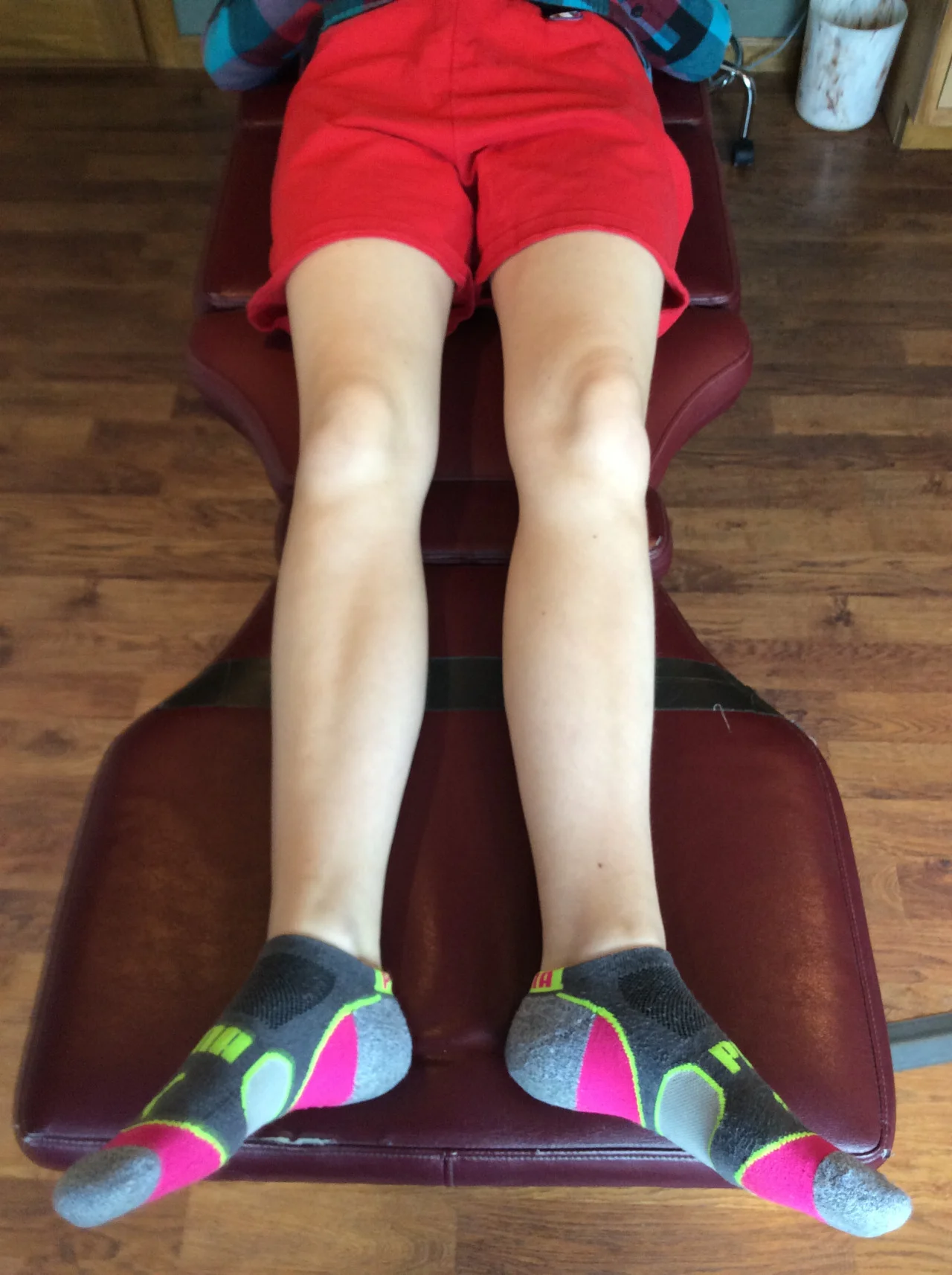
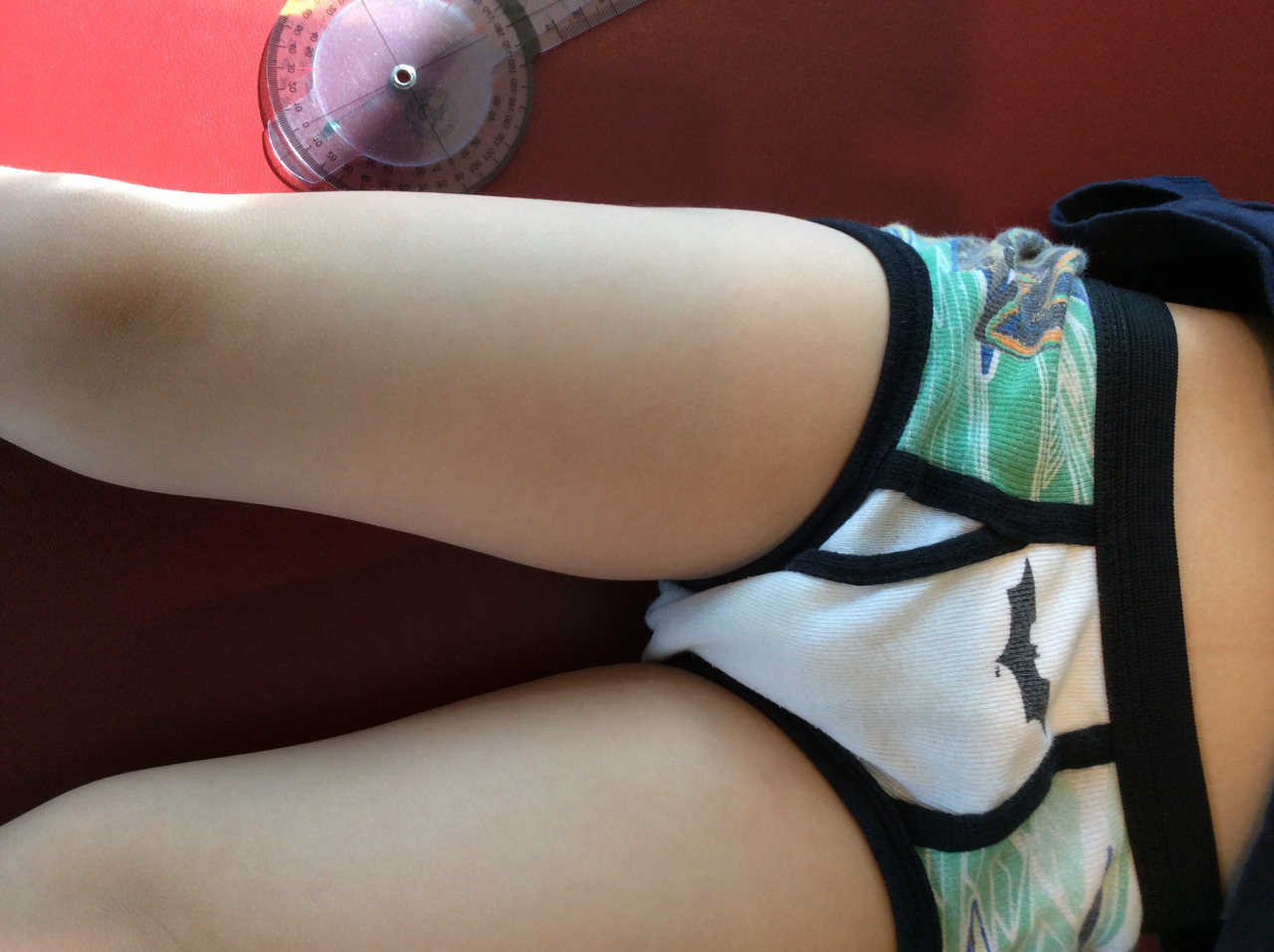
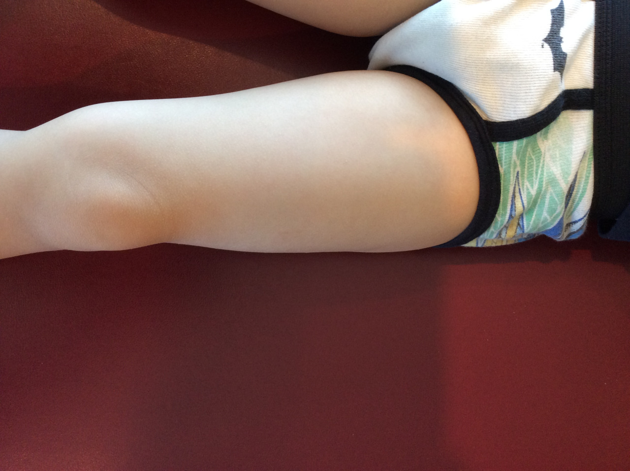
In normal gait, the rearfoot strikes in slight inversion and then quickly moves through eversion in the frontal plane to help with the midfoot through forefoot pronation phases of gait. Some sources would refer this rearfoot eversion as the rearfoot pronatory phase, after all. pronation can occur at the rear, mid or forefoot. As with all pronation in all areas, when it occurs too fast, too soon or too much, it can be a problem and rearfoot eversion is no different.  If uncontrolled via muscles such as through tibialis posterior eccentric capabilities (Skill, endurance, strength) or from a structural presentation of Rearfoot Valgus pain can arise. 

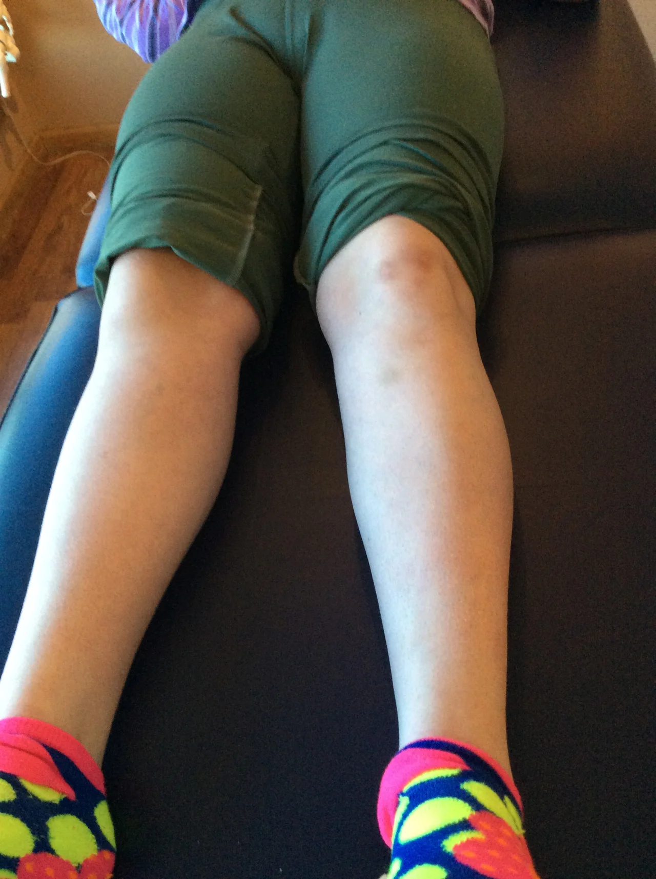
From a scenario like in the video above, where a more rearfoot varus presentation is observed,  where the lateral to medial pronation progression is excessive and extreme in terms of speed, duration and magnitude this can also create too much lateral to medial foot, ankle and knee movement.  This will often accompany unchecked movements of internal spin through the hip. So one should see that these pronation and spin issues can occur and be controlled from the bottom or from the top, and hopefully adequately from both in a normal scenario.  It is when there is a biomechanical limitation or insufficiency somewhere in the chain that problems can arise. And remember, pain does not have to occur where the failure occurs, in fact it usually does not. So when you have knee pain from an apparent valgus posturing knee, make sure you look above and below that knee.  Also, keep in mind that as discussed last week in the blog post on ischiofemoral impingment syndrome (link), these spin scenarios can be quite frequently found with ipsilateral frontal plane lateral deviations (bumping of the hip-pelvis outside the vertical stacking of the foot-knee-hip stacking line). This stacking failure can also be the source of many of the issues discussed above, so be sure you are looking locally and globally. And remember, what you see is not the problem, it is their compensation around their deeper problem quite often.

If you have not read the blog post from last week on ischiofemoral impingement syndrome you might not know where the components of the cross over gait come in to play here nor how a rearfoot problem can present with a hip impingement scenario, so I can recommend that article one more time.

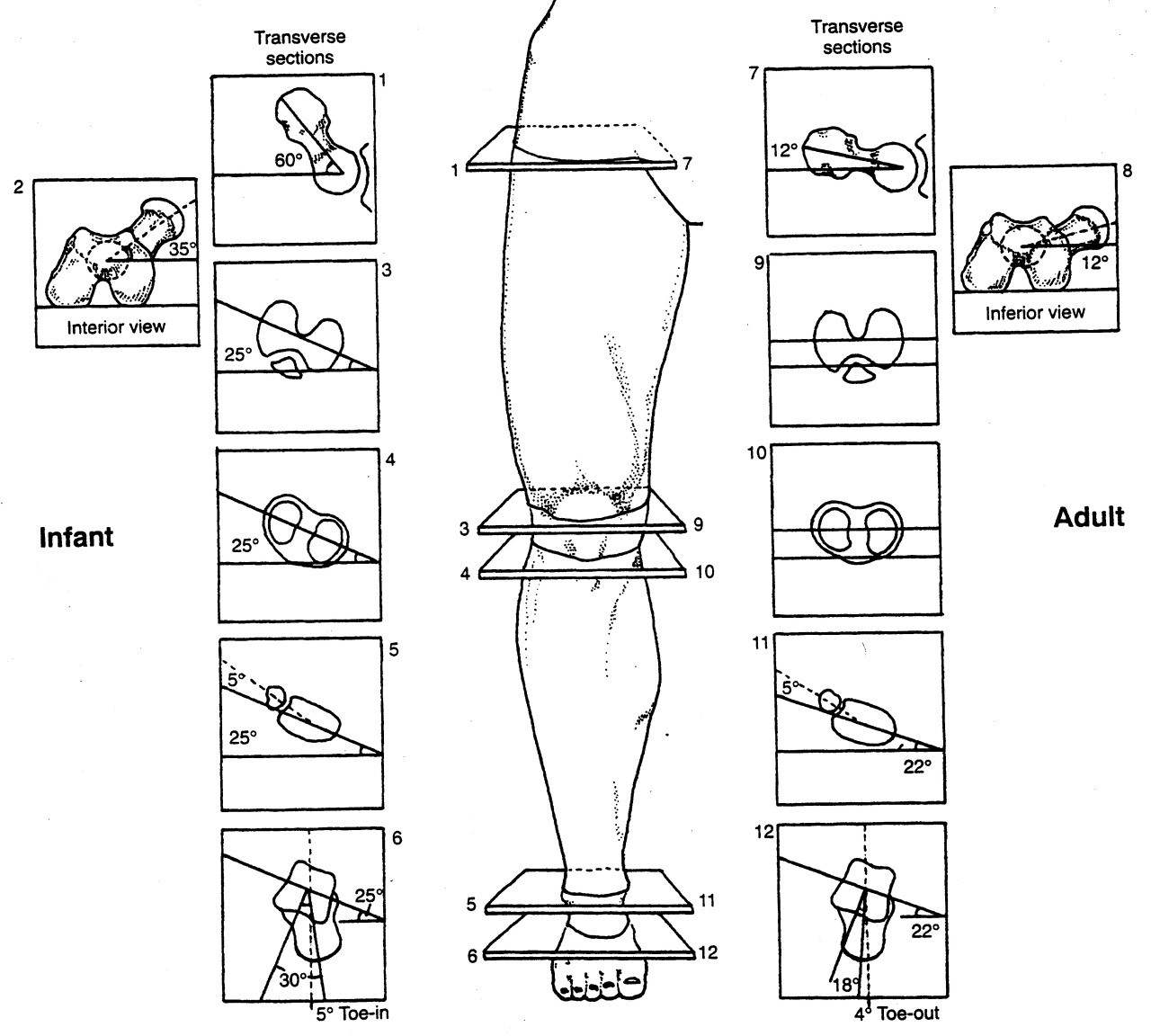
One last thing, just in case you think this stuff is easy to work through, remember that these rearfoot varus and valgus problems, and pronation rates. and limb spin rates are all highly variable when someone has varying degrees of femoral torsion, tibial torsion or talar torsion. Each case is different, and each will be unique in their presentation and in the uniqueness of the treatment recipe. I just thought I would throw that in to make your head spin a little in case it wasn’t already.

For example, a case where the rearfoot is a semi rigid varus, with tibial varum, and frontal plane lateral pelvic drift with components of cross over gait (ie. the video case above) will require a different treatment plan and strategy than the same rearfoot varus in a presentation of femoral torsion challenges and genu valgum. Same body parts, different orientations, different mechanics, different treatment recipe.  

So, you can fiddle with a dozen pair of shoes to find one that helps minimize your pains, you can go for massages and hope for the best, you can go and get activated over and over, you can try yet another new orthotic, you can go to a running clinic and try some form changes, throw in some yoga or pilates, compression wear, voodoo bands and gosh who knows what else. Sometimes they are the answer or stumble across it … or you can find someone who understands the pieces of the puzzle and how to piece a reasonable recipe together to bake the cake just right. We do not always get there, but we try.  

Want more ? Try our National Shoe Fit certification program for a starter or try our online teleseminars at www.onlinece.com (we did a one hour course on the RearFoot just the other night, and it was recorded over at onlineCE.com).

Dr. Shawn Allen,  of the gait guys


Reference:

Man Ther.  2014 Oct;19(5):379-85. doi: 10.1016/j.math.2013.10.003. Epub 2013 Oct 29.Clinical measures of hip and foot-ankle mechanics as predictors of rearfoot motion and posture.  Souza TR et al.

Health professionals are frequently interested in predicting rearfoot pronation during weight-bearing activities. Previous inconsistent results regarding the ability of clinical measures to predict rearfoot kinematics may have been influenced by the neglect of possible combined effects of alignment and mobility at the foot-ankle complex and by the disregard of possible influences of hip mobility on foot kinematics. The present study tested whether using a measure that combines frontal-plane bone alignment and mobility at the foot-ankle complex and a measure of hip internal rotation mobility predicts rearfoot kinematics, in walking and upright stance. Twenty-three healthy subjects underwent assessment of forefoot-shank angle (which combines varus bone alignments at the foot-ankle complex with inversion mobility at the midfoot joints), with a goniometer, and hip internal rotation mobility, with an inclinometer. Frontal-plane kinematics of the rearfoot was assessed with a three-dimensional system, during treadmill walking and upright stance. Multivariate linear regressions tested the predictive strength of these measures to inform about rearfoot kinematics. The measures significantly predicted (p ≤ 0.041) mean eversion-inversion position, during walking (r(2) = 0.40) and standing (r(2) = 0.31), and eversion peak in walking (r(2) = 0.27). Greater values of varus alignment at the foot-ankle complex combined with inversion mobility at the midfoot joints and greater hip internal rotation mobility are related to greater weight-bearing rearfoot eversion. Each measure (forefoot-shank angle and hip internal rotation mobility) alone and their combination partially predicted rearfoot kinematics. These measures may help detecting foot-ankle and hip mechanical variables possibly involved in an observed rearfoot motion or posture.

tumblr_nfzg0nM4pC1qhko2so1_1280.jpg
tumblr_nfzg0nM4pC1qhko2so2_1280.jpg
tumblr_nfzg0nM4pC1qhko2so3_1280.jpg
tumblr_nfzg0nM4pC1qhko2so4_1280.jpg
tumblr_nfzg0nM4pC1qhko2so5_1280.jpg
tumblr_nfzg0nM4pC1qhko2so6_1280.jpg

What would you do? This is what we did.

History:

This 7 year old girl is brought in by her mother because of knee misalignment while skiing, L > R. No history of trauma; normal term birth with no complications. No knee pain. Of incidental note, she is deaf in the left ear.

Exam findings:

She has bi-lat. external tibial torsion, left much worse than right (40 degrees transmallolear angle vs 22 degrees. for info on measuring torsions, click here). remember, you should be able to draw a line from the tibial tuberosity down through the 2nd metatarsal head. 

She has a 5mm anatomical leg length deficiency on the right (see top above left).

She has femoral antetorsion right side with very little external rotation, approximately 10 degrees,  internal rotation is in excess of 50.  Left side has normal femoral versions (for a review of femoral versions and torsions, click here).  See last 2 pictures which are full internal and external rotation respectively.

She has a mild uncompensated forefoot varus (cannot really see from the pictures, you will need to take our word for it) with a relatively cavus arch to her foot(see center and last picture on right.

Neurologically, she appeared to have integrity with respect to sensation, motor strength and deep tendon reflexes in the lower extremities.

Assessment:

Pathomechanical alignment as described.  Severe left external tibial torsion. MIld to moderate right. Femoral antetorsion right.

Plan:

We are going to build her a medium heel cup full length modified UCB orthotic inverting the cast bi-lat. left greater than right.  We gave her  balance and coordination exercises, heel walking, lift/spread/reach and one leg balancing. She will follow up for a dispense.  Her mother will try to get a better fitting ski boot as the one she has currently is two sizes too big. She will return for a dispense. She should consider wearing the orthotics in everyday footwear as well. We will do a follow up post in a few weeks. 

The Gait Guys. Teaching you something new in each and every post. Like this post? Tell and share it with a friend. Don’t like this post? Let us know!

tumblr_nc9grebaIQ1qhko2so1_1280.jpg
tumblr_nc9grebaIQ1qhko2so2_1280.jpg
tumblr_nc9grebaIQ1qhko2so3_1280.jpg

Wow! What would you do?

This is part 1 of a 2 part post. Look for the other one a few minutes after this one with a video up top for the conclusion

PRESENTING PROBLEM: This 54 YO female patient presents with with left sided knee pain.  She had a total knee replacement (TKR) done in 2011.  She’s had a significant amount of discomfort on the medial aspect of the knee since then. She had an MRI of the hip done thinking the problem was there, and found nothing.   She is walking with a bad limp, left leg is half inch shorter than the right.  Pain is worse at night, changes with weather. 

She has knee pain on the lateral aspect (points to tibial plateau and joint line) with swelling that goes down to the ankle left side.  She has been wearing a “Good Feet” OTC orthotic on the left side which she states helps quite a bit.

Generally speaking, stretching and analgesics make the discomfort better.    Ibuprofen 400 mg. b.i.d. can take the edge off  Soft sided brace (neoprene sleeve) makes a difference as well. The hard sided brace gives her difficulty.

WORK HISTORY: She works for a preschool.  Her job involves standing and getting up and down a lot.  

FAMILY HISTORY:  She has left sided lid ptosis, this evidently is familial.  

PHYSICAL EXAM:  She stood 5’ 1” and weighed approx. 150 pounds.

Viewing the knees bi-lat., the left knee is markedly externally rotated.

She does have a left short leg; tibial and femoral.  She has bilateral tibial torsion (look at the tibial tuberosities and drop a line straight down; it should pass through the 2nd metatarsal head) and marked internal tibial torsion on the left side (>60 degrees) with femoral retrotorsion (less than 8 degree angle of femoral head with the shaft) on this side.  There is no rotation of the thigh or leg past zero degrees midline. .  She had 10 degrees of tibial varum on the left hand side.  Her Q-angle is 10 degrees on that side.  There is plantar flexion inversion of the foot.  Left lower extremity has less sensation secondary to the her TKR  surgery.

Gait evaluation reveals a fair amount of midfoot pronation noted on the left hand side in addition to an intoed gait.  She has to lean her body over to the left to get the right leg to clear.

Some mild weakness noted of hip abduction musculature left hand side gluteus medius, middle and anterior fibers. Knee stability tests were negative.

Neurologically, otherwise, she had full integrity with respect to sensation, motor strength and deep tendon reflexes in the upper and lower extremities.

Please see part 2 of this post for additional info including our assessment and what WE did.

 The Gait Guys. Making it real, each and every post here on the blog.

special thanks to SZ for allowing us to publish her case, so others can learn

tumblr_n9y3v5nRcH1qhko2so1_1280.jpg
tumblr_n9y3v5nRcH1qhko2so2_1280.jpg
tumblr_n9y3v5nRcH1qhko2so3_1280.jpg
tumblr_n9y3v5nRcH1qhko2so4_1280.jpg
tumblr_n9y3v5nRcH1qhko2so5_1280.jpg

More on the “little guy”

We have been following this little guy for some time now. If you have not been keeping up, perhaps you should read herehere and here 1st. 

So, what do we see in these latest pictures?

Top left: neutral view.

  • He enjoys flip flops; probably not the best thing for a developing kiddo, in light of the excessive engagement of the posterior compartment (and reciprocal inhibition of the anterior compartment)
  • he has some tibial varum (ie bowleggedness) L > R
  • he has some developmental genu valgum whnich appears to be improving (need a Q angle review? click here)
  • no tibial torsion present on L: for a review on torsions, click here
  • still some external tibial torsion present on R (see section below on middle shots)

Top right and bottom: full internal rotation of R thigh: compare with bottom: full internal rotation of L thigh

  • he has adequate internal rotation (4 degrees needed) but not as great as left side (see bottom shot); this represents some improvement since we started
  • he has generous internal rotation of the left thigh

Middle Left: full external rotation of right thigh

  • note the position of the knee and the position of the foot; external tibial torsion is present. for a review of torsions, click here.
  • he has limited external rotation of the right thigh (compared with the left. The knee should fall more outside the saggital plane

Middle right: full external rotation of the left thigh

  • note the position of the knee and the position of the foot; internal tibial torsion is present. 
  • he has generous external rotation of the left thigh (compared with the left)


of other significant note: most of his calcaneal valgus has resolved; longitudinal arches are improved.

What now?

  • He continues to develop normally and continues to improve since his original presentation to the office
  • Having the child continue to walk barefoot
  • Continue to wear shoes with little torsional rigidity, to encourage additional additional intrinsic strength to the feet
  • He should continue to limit “W” sitting, as this will tend to increase the genu valgus present
  • We reviewed 1 leg balancing “games” and encouraged continuing agility activities, like balance beam, hopping, skipping and jumping on each leg individually
  • added in using a push and pedal bike
  • added in heel walking exercises

Ivo and Shawn. Bald. Good looking. Extraordinary Gait Geeks. Taking the world of gait literacy by storm with each and every post.

tumblr_nb9a9pr6O71qhko2so1_1280.gif
tumblr_nb9a9pr6O71qhko2so2_1280.jpg

All that is twisted is not tibial

Last week we posted on measuring tibial torsions (click here to read that post). This week we are posting on measuring the other, often over looked torsion: “femoral torsion”.

Perhaps you have read some of our posts on femoral torsion, particularly this one.

We remember that as hip (thigh) flexion increases, the amount of internal rotation of the femur decreases. This is due largely to the direction of the hip capsule ligaments (ishiofemoral, iliofemoral and pubeofemoral ligaments) “spiraling” from their attachment from the femur to the innominate. This may seem like a subtle detail until you thing about how much hip flexion occurs when we do a squat, and what exactly, is the position of our feet.

We start life with the hips anteverted (ie, the angle of the neck of the femur with the shaft of the femur is > 12 degrees; in fact at birth it is around 35 degrees) and this angle should decrease as we age to about 8-12 degrees). When we stand, the heads of our femurs point anteriorly; it is just a matter of how much (ante version or ante torsion) or how little  (retro version or retro torsion) that is. If you are a precise person and would really like to geek out on the difference between versions or torsions, check out this post here

Measurement is important, because the more retro torsion you have (ie, the smaller the angle is), the less internal rotation of the femur you will have available to you. An important fact if you are planning on squatting. 

An easy way to do this is by approximating the angle of the femoral neck by performing “Craig’s Test”. Have your patient/client/athlete lie prone with their knee flexed 90 degrees. Palpate the greater trochanter (the bump on the side of the hip that the gluteus medius muscles attach to) with one hand while using the other hand to grasp around the ankle and internally and externally rotate the femur (we like to use the right hand on the right trochanter for the patient/client/athletes right leg). Note the position of the tibia when the greater trochanter is parallel to the table (see diagram above from Tom Michaud’s most excellent text: Human Locomotion: the conservative management of gait related disorders, available by clicking here). The smaller the angle, the more retro version/torsion present). 

This is also a convenient way to estimate the amount of internal and external rotation of the femur available. One source states that internal rotation of greater than 70 degrees and external rotation of less than 25 degrees means that there is excessive femoral ante torsion present (1).

Craig’s Test: a convenient way to measure torsions of the femur. Important if you squat! Brought to you by The Gait Guys: Uber Gait Geeks Extrodinaire. 

(1) Staheli LT. Rotational problems in the lower extremity. Orthop Clin North Am, 1987; 18:503-512

tumblr_n9y3ea9yyg1qhko2so4_r1_1280.jpg
tumblr_n9y3ea9yyg1qhko2so3_1280.png
tumblr_n9y3ea9yyg1qhko2so2_1280.jpg
tumblr_n9y3ea9yyg1qhko2so1_1280.jpg

How do you measure tibial torsion anyway?

With all the talk on the Crossfit blog about the knees out debate, we though we would shed some light on measuring torsions, beginning with tibial torsion, since this does not seem to have been taken account of in the discussion and we feel it is germane. 

Yo may have seen some of our other posts in tibial torsion here or here; this post will serve to help you measure it. 

Looking at the top left picture: we can see that the axis of the tibial plateau and the transmalleolar axis (an imaginary line drawn through the medial and lateral malleolus) are parallel at birth (net angle zero) and progress to 22 degrees at skeletal maturity, resulting from the outward rotation of the tibia of about 1-1.5 degrees per year. This results in a normal external tibial version of about 17-18 degrees (you subtract 5 degrees for the talar neck angle, talked about in the link above). Note that this is the normal or ideal angle we would expect (hope?) to see. Go 2 standard deviations in either direction and we have external and internal tibial torsions.

You can go about taking this measurement in may ways; we will outline 2 of them. 

  1. In the upper left picture, we see an individual who has their knee flexed to 90 degrees over the side of a table while seated. This represents the tibial plateau angle. You the use a protractor to measure the angle between the tibial plateau and an imaginary line drawn through the medial and lateral malleoli. This is the transmalleolar angle. You then subtract 5 degrees from this number (remember the talar neck angle?) to get the angle of tibial version (or torsion).
  2. In the lower left and right pictures, we have the patient supine with the knees pointed upward and tibial plateau flat on the table. Then, working from inferiorly, use a goniometer to measure the angle of the transmalleolar axis. Again, we subtract 5 degrees for the talar neck.

We would encourage you to read up on torsions. This post, which we wrote over a year ago, is probably one of the most important ones on tibial torsions. 

Torsions. Important stuff, especially when you are talking about the axis of the knees in activities like a squat. Remember, the knee is a hinge between 2 multiaxial joints (hip and ankle) and will often take the brunt of the (patho)mechanics, as it has fewer degrees of freedom of movement. If you have external tibial torsion and you push your knees (angle your feet) out further, you are moving the knees outside the saggital plane. You have better have a very competent medial tripod! If you have internal tibial torsion, angling the feet out may be a good idea. Know your (or your patients/clients/athletes) anatomy!

The Gait Guys. Bald, Good Looking and Twisted. Here to help you navigate your way through better biomechanics. 

tumblr_n9ql3rlqGi1qhko2so2_r1_1280.jpg
tumblr_n9ql3rlqGi1qhko2so1_1280.gif
tumblr_n9ql3rlqGi1qhko2so3_r1_1280.gif
tumblr_n9ql3rlqGi1qhko2so4_r1_1280.gif

ETT and Hip Extension

Not Extra Terrestrial Tricks, but rather External Tibial Torsion. How it effects hip extension.

We received this question from Matthew P on our Facebook post from 8/1 (original post from here) which was based on this article, and thought it would make an excellent opportunity to teach. 

I looked at this yesterday and had actually first come across it a year or more ago when I was trying to find some resources for femoral torsion. You guys are about the only ones discussing at length the impact and implication of adult femoral torsion.

Re: tibial torsion and your post yesterday saying that > 30deg external torsion can affect both knee and hip extension, what I still don’t understand is the mechanism behind the hip limitation. How would that torsion translate through the leg to the hip?

There are a few things we need to remember to make sense of this:

  • tibial torsion is the angular difference between the tibial plateau and distal tibial malleoli and refers only to the tibia, not the entire lower extremity (see top photo)
  • pronation can occur in the rear foot, mid foot, and fore foot
  • pronation causes internal spin of the leg and thigh, due to plantar flexion, eversion and abduction of the talus (see middle photo)
  • internal spin of the hip causes posterior translation of the femoral head via the “glide and roll” phenomenon
  • these are appropriate mechanics during the 1st ½ of the gait cycle (initial contact to mid stance)
  • pronation is one of the 4 shock absorbing mechanisms (pronation, ankle dorsiflexion, knee flexion, hip flexion) of the lower extremity

now try this (yes, at home!)

  • flex your knee
  • internally rotate you leg
  • allow your arch to flatten
  • try and extend your hip

Remember these facts about supination

  • supination is initiated by the swing phase leg as it starts in early swing and continues to terminal swing (see third picture)
  • supination (from full pronation), should occur from midstance to pre swing
  • supination makes the foot into a “rigid lever” to transpose forces from above the foot into the foot and allow for propulsion (see third picture)
  • supination involves external rotation of the lower leg and thigh (see pictures 3 and 4)
  • external rotation of the hip is accompanied by anterior glide of the femoral head via the “glide and roll” phenomenon
  • this position puts the gluteal muscles (max and posterior fibers of the gluteus medius) at a mechanical advantage

now try this:

  • extend your knee
  • externally rotate your leg
  • you should have a full arch at this point
  • try and extend your hip

What did you (hopefully) learn?

  1. When the hip is in an externally rotated position it is easier to extend; the femur head moves anteriorly, the femoral joint capsule becomes tighter and stability is created
  2. when the hip is internally rotated, it is more difficult to extend
  • the femur head glides posteriorly, changing the axis of rotation of the joint
  • the gluteus maximus and posterior fibers of the gluteus medius are at a mechanical disadvantage

OK. Got it? We sure hope so! Excellent question, Matthew. Thanks for the opportunity to teach this concept.

The Gait Guys. Taking you closer to mastering the gait cycle with each post. 

Foot Progression Angle Exaggeration: External Tibial Torsion

Take a look at the tibial tuberosity and then where you think the 2nd metatarsal head would be. What do you see? The 2nd metatarsal is lateral to the tibial tuberosity. You are looking at external tibial torsion. 

Lets see how this external tibail torsion behaves during a knee bending. Observe the medial drift of the knee during weight bearing knee flexion. Many folks would say that the problem here is the increased foot prontation, but that is not where the problem lies, that is where many of the forces are funneling though. The client is pronating more because the external tibial torsion that is creating this appearance has put the knee inside the foot tripods region of stability.

In external tibial torsion there is an external torsion or a “twist” along the length of the tibia (diaphysis or long section). This occurs in this example to the degree that the ankle joint (mortise joint) can no longer cooperate with sagittal knee joint.  When taking a client with external tibial torsion and pre-postioning their foot in a relatively acceptable/normal foot progression angle there is a conflict at the knee, meaning that the knee cannot hinge forward in its usual sagittal plane. In this case with the foot progression angle smaller than what this client would posture the foot, the knee the knee will be forced to drift medially.

Are you looking for torsions of the lower limb in your clients ?

Are you forcing them into foot postures that look better to  you but that which are conflicting to your clients given body mechanics ?  Would you correct this client’s foot turn out (increased progression angle) ? IF you did you would likely cause them knee pain in time.  Would you put them into a stability shoe to try and control the pronation ? Again, you are likely to draw their knee outside the saggital knee hinge that is presently pain free. There is more to shoe fit that just looking at the foot. First do no harm is our mantra ! 

Remember, telling someone to turn their foot in or out because it doesn’t appear correct to your eyes can significantly impair either local or global joints , and often both. Torsions can occur in the talus, the tibia and the femur.

Furthermore, torsions can have an impact on foot posturing at foot strike and affect the limbs loading response, from foot to core and even arm swing can be altered.  Letting your foot fall naturally beneath your body does not mean that you have the clean anatomy to do so without a short term or long term cost. 

This is some of the toughest stuff you will deal with clinically. The fence is narrow, if you do to little correction you fall off the fence into the wrong yard and create problems. If you do to much correction you get the same result. These torsional issues are a delicate balancing act many times. You first have to know what you have, then you have to know where the fix is, and then how much is safe.  Tricky stuff. This is exactly why in some folks a stability shoe can be magic or tragic and in others dropping into zero drop minimalism can be magic or tragic.  

Want more on torsion and versions ?  Type the words into the search box on our blog. We have plenty of good info for you.

Shawn and Ivo, The Gait Guys

tumblr_n7yjctusWm1qhko2so1_1280.jpg
tumblr_n7yjctusWm1qhko2so2_1280.jpg
tumblr_n7yjctusWm1qhko2so3_1280.jpg
tumblr_n7yjctusWm1qhko2so4_1280.jpg
tumblr_n7yjctusWm1qhko2so7_1280.jpg
tumblr_n7yjctusWm1qhko2so9_1280.jpg

You can only “borrow” so much before you need to “pay it back”

How can feet relate to golf swing?

This 52 year old right handed gentleman presented with pain at the thoracolumbar junction after playing golf. He noticed he had a limited amount of “back swing” and pain at the end of his “follow through”.

Take a look a these pix and think about why.

Hopefully, in addition to he having hairy and scarred legs (he is a contractor by trade), you noted the following

  • Top left: note the normal internal rotation of the right hip; You need 4 degrees to walk normally and most folks have close to 40 degrees. He also has internal tibial torsion.
  • Top right: loss of external rotation of the right hip. Again, you need 4 degrees (from neutral) of external rotation of the hip to supinate and walk normally.
  • Top center:normal internal rotation of the left hip; internal tibial torsion
  • 3rd photo down: limited external rotation of the left hip, especially with respect ti the amount of internal rotation present; this is to a greater degree than the right
  • 4th and 5th photos down: note the amount of tibial varum and tibial torsion. Yes, with this much varum, he has a forefoot varus.

The brain is wired so that it will (generally) not allow you to walk with your toes pointing in (pigeon toed), so you rotate them out to somewhat of a normal progression angle (for more on progression angles, click here). If you have internal tibial torsion, this places the knees outside the saggital plane. (For more on tibial torsion, click here.) If you rotate your extremity outward, and already have a limited amount of range of motion available, you will take up some of that range of motion, making less available for normal physiological function. If the motion cannot occur at the knee or hip, it will usually occur at the next available joint cephalad, in this case the spine.

The lumbar spine has a limited amount of rotation available, ranging from 1.2-1.7 degrees per segment in a normal spine (1). This is generally less in degenerative conditions (2).

Place your feet on the ground with your feet pointing straight ahead. Now simulate a right handed golf swing, bending slightly at the waist and  rotating your body backward to the right. Now slowly swing and follow through from right to left. Note what happens to your hips: as you wind back to the right, the left hip is externally rotating and the right hip is internally rotating. As you follow through to the left, your right, your hip must externally rotate and your left hip must externally rotate. Can you see how his left hip is inhibiting his back swing and his right hip is limiting  his follow through? Can you see that because of his internal tibial torsion, he has already “used up” some of his external rotation range of motion?

If he does not have enough range of motion in the hip, where will it come from?

he will “borrow it” from a joint more north of the hip, in this case, his spine. More motion will occur at the thoracolumbar junction, since most likely (because of degenerative change) the most is available there; but you can only “borrow” so much before you need to “Pay it back”. In this case, he over rotated and injured the joint.

What did we do?

  • we treated the injured joint locally, with manipulation of the pathomechanical segments
  • we reduced inflammation and muscle spasm with acupuncture
  • we gave him some lumbar and throacolumbar stabilization exercises: founders exercise, extension holds, non tripod, cross crawl, pull ups
  • we gave him foot exercises to reduce his forefoot varus: tripod standing, EHB, lift-spread-reach
  • we had him externally rotate both feet (duck) when playing golf

The Gait Guys. Helping you to store up lots “in your bank” of foot and gait literacy, so you can help people when they need to “pay it back”, one case at a time.

(1) http://www.ncbi.nlm.nih.gov/pmc/articles/PMC2223353/

(2) http://www.ncbi.nlm.nih.gov/pmc/articles/PMC3705911/

tumblr_n2ac0gX4j01qhko2so1_r1_1280.jpg
tumblr_n2ac0gX4j01qhko2so2_r1_1280.jpg
tumblr_n2ac0gX4j01qhko2so3_r1_1280.jpg
tumblr_n2ac0gX4j01qhko2so4_r1_1280.jpg
tumblr_n2ac0gX4j01qhko2so5_r1_1280.jpg
tumblr_n2ac0gX4j01qhko2so6_r1_1280.jpg

Remember this kiddo?

We have been following the natural development of this little guy for some time now. For a review, please see here (1 year ago) and here (2 years ago) for our previous posts on him.

In the top 2 shots, the legs are neutral. The 3rd and 4th shots are full internal rotation of the left and right hips respectively. The last 2 shots are full external rotation of the hips.

Well, what do you think now?

We remember that this child has external tibial torsion and pes planus. As seen in the supine photo, when the knees face forward, the feet have an increased progression angle (they turn out). We are born with some degree / or little to none, tibial torsion and the in-toeing of infants is due to the angle of the talar neck (30 degrees) and femoral anteversion (the angle of the neck of the femur and the distal end is 35 degrees).  The lower limbs rotate outward at a rate of approximately 1.5 degrees per year to reach a final angle of 22 degrees….. that is of course if the normal de rotation that a child’s lower limbs go through occurs timely and completely.

He still has a pronounced valgus angle at the the knees (need a review on Q angles? click here). We remember that the Q angle is negative at birth (ie genu varum) progresses to a maximal angulation of 10-15 degrees at about 3.5 years, then settles down to 5-7 degrees by the time they have stopped growing. He is almost 4 and it ihas lessend since the last check to 15 degrees.

His internal rotation of the hips should be about 40 degrees, which it appears to be. External rotation should match; his is a little more limited than internal rotation, L > R. Remember that the femoral neck angle will be reducing at the rate of about 1.5 degrees per year from 35 degrees to about 12 in the adult (ie, they are becoming less anteverted).

At the same time, the tibia is externally rotating (normal tibial version) from 0 to about 22 degrees. He has fairly normal external tibial version on the right and still has some persistent internal tibial version on the left. Picture the hips rotating in and the lower leg rotating out. In this little fellow, his tibia is outpacing the hips. Nothing to worry about, but we do need to keep and eye on it.

What do we tell his folks?

  • He is developing normally and has improved significantly since his original presentation to the office
  • Having the child walk barefoot has been a good thing and has provided some intrinsic strength to the feet
  • He needs to continue to walk barefoot and when not, wear shoes with little torsional rigidity, to encourage additional intrinsic strength to the feet
  • He should limit “W” sitting, as this will tend to increase the genu valgus present
  • We gave him 1 leg balancing “games” and encouraged agility activities, like balance beam, hopping, skipping and jumping on each leg individually

We are the Gait Guys, promoting gait and foot literacy, each and every post.

tumblr_n2aypmQpun1qhko2so1_1280.jpg
tumblr_n2aypmQpun1qhko2so2_1280.jpg
tumblr_n2aypmQpun1qhko2so3_1280.jpg
tumblr_n2aypmQpun1qhko2so4_1280.jpg

Holy Hand Grenades! What kind of shoe do I put these feet in?

Take a look at these feet. (* click on each of the photos to see the full photo, they get cropped in the viewer) Pretty bad, eh? How about a motion control shoe to help things along? NOT! OK. but WHY NOT? Let’s take a look and talk about it.

To orient you:

  • top photo: full internal rotation of the Left leg
  • 2nd photo: full internal rotation of the Right leg
  • 3rd photo: full external rotation of the Left leg
  • last photo: full external rotation of the Right leg

Yes, this gal has internal tibial torsion (yikes! what’s that? click here for a review).

Yes, it is worse on the Left side

Yes, she has a moderate genu valgus, bilaterally.

If someone has internal tibial torsion, the foot points inward when the knee is in the saggital plane (it is like a hinge). The brain will not allow us to walk this way, as we would trip, so we rotate the feet out. This moves the knee out of the saggital plane (ie. now it points outward).

What happens when we place a motion control shoe (with a generous arch and midfoot and rearfoot control) under the foot? It lifts the arch (ie it creates supination and it PREVENTS pronation). This creates EXTERNAL rotation of the leg and thigh, moving the knee EVEN FURTHER outside the saggital plane. No bueno for walking forward and bad news for the menisci.

Another point worth mentioning is the genu valgus. What happens when you pick up the arch? It forces the knee laterally, correct? It does this by externally rotating the leg. This places more pressure/compression on the medial aspect of the knee joint (particularly the medial condyle of the femur). Not a good idea if there is any degeneration present, as it will increase pain. And this is no way to let younger clients start out their life either.

So, what type of shoe would be best?

  • a shoe with little to no torsional rigidity (the shoe needs to have some “give”)
  • a shoe with no motion control features
  • a shoe with less of a ramp delta (ie; less drop, because more drop = more supination of the foot (supination is plantarflexion, inversion and adduction)
  • a shoe that matches her sox, so as not to interfere with the harmonic radiation of the colors (OK, maybe not so much…)

Sometimes giving the foot what it appears to need can wreak  havoc elsewhere. One needs to understand the whole system and understand what interventions will do to each part. Sometimes one has to compromise to a partial remedy in one area so as not to create a problem elsewhere. (Kind of like your eye-glass doctor. Rarely do they give you the full prescription you need, because the full prescription might be too much for the brain all at once.  Better to see decent and not fall over, than to see perfectly while face down in the dirt.) 

Want to know more? Consider taking the National Shoe Fit Certification Program. Email us for details: thegaitguys@gmail.com.

We are the Gait Guys, and yes, we like her sox : )