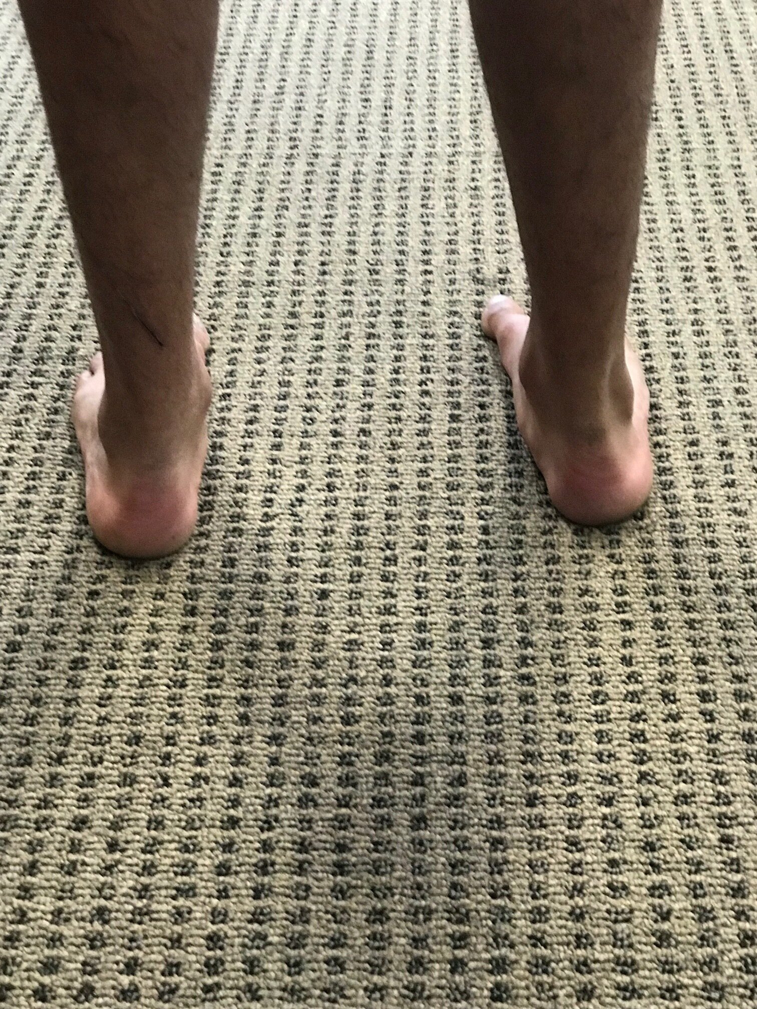
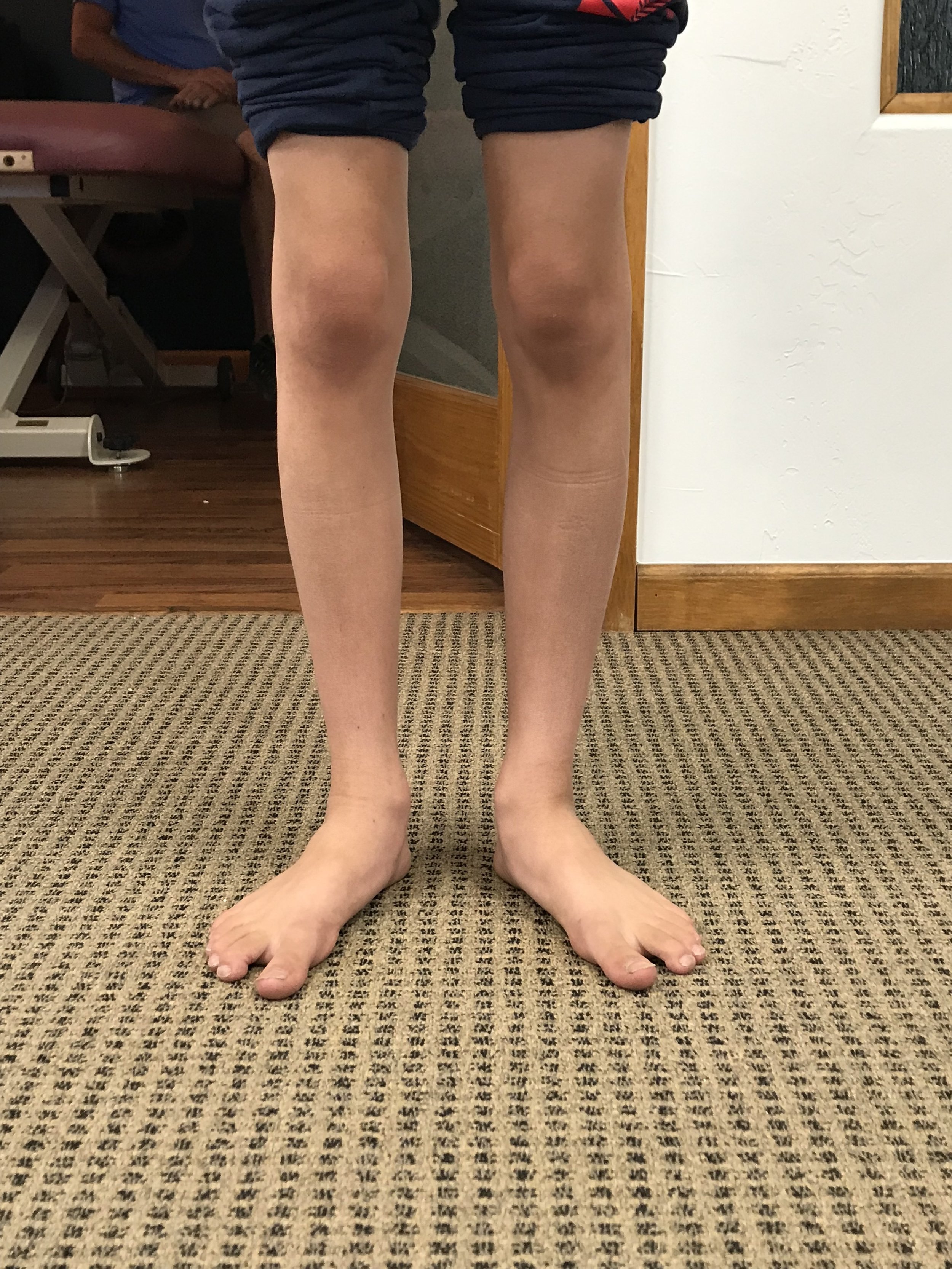
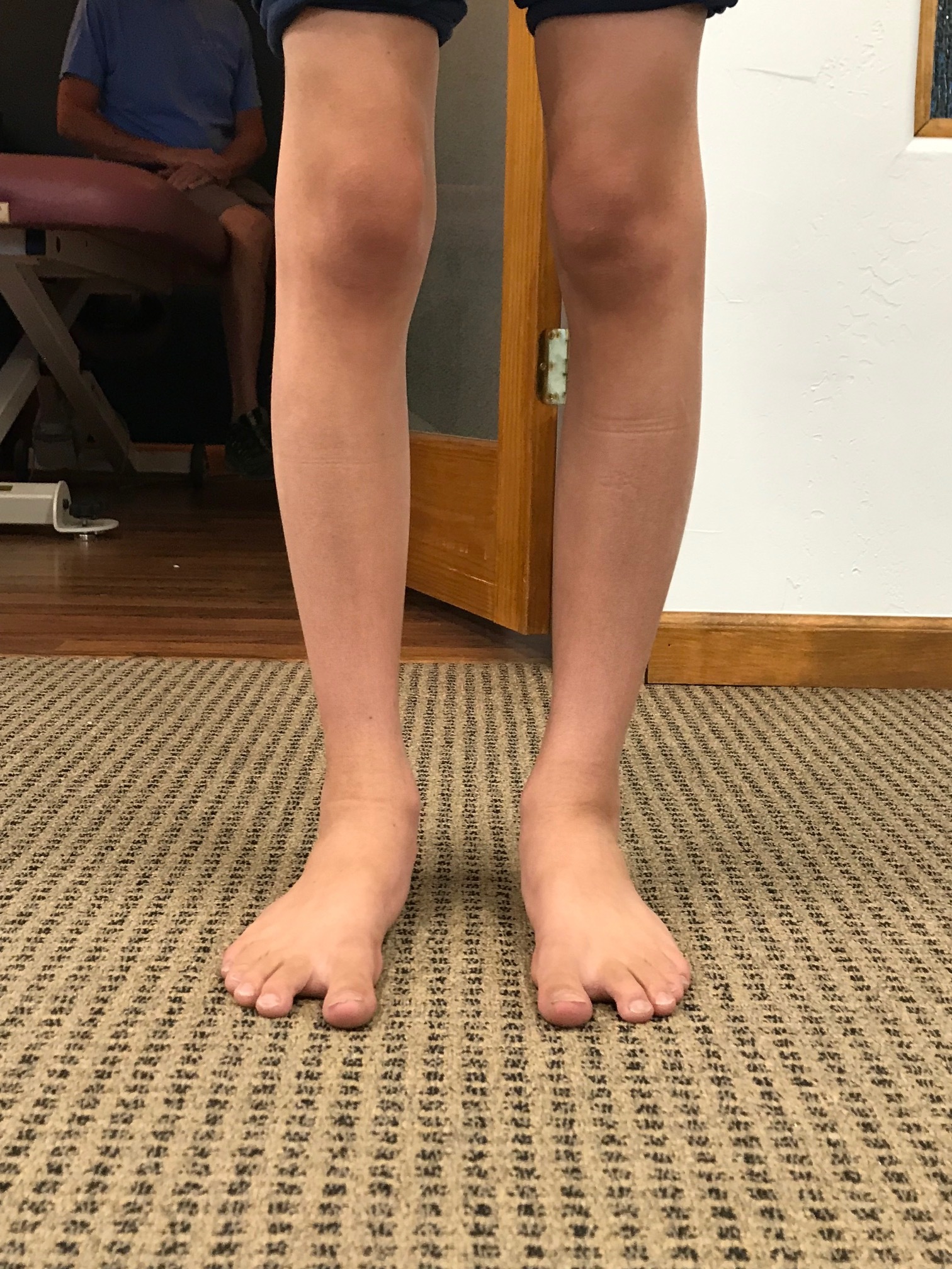
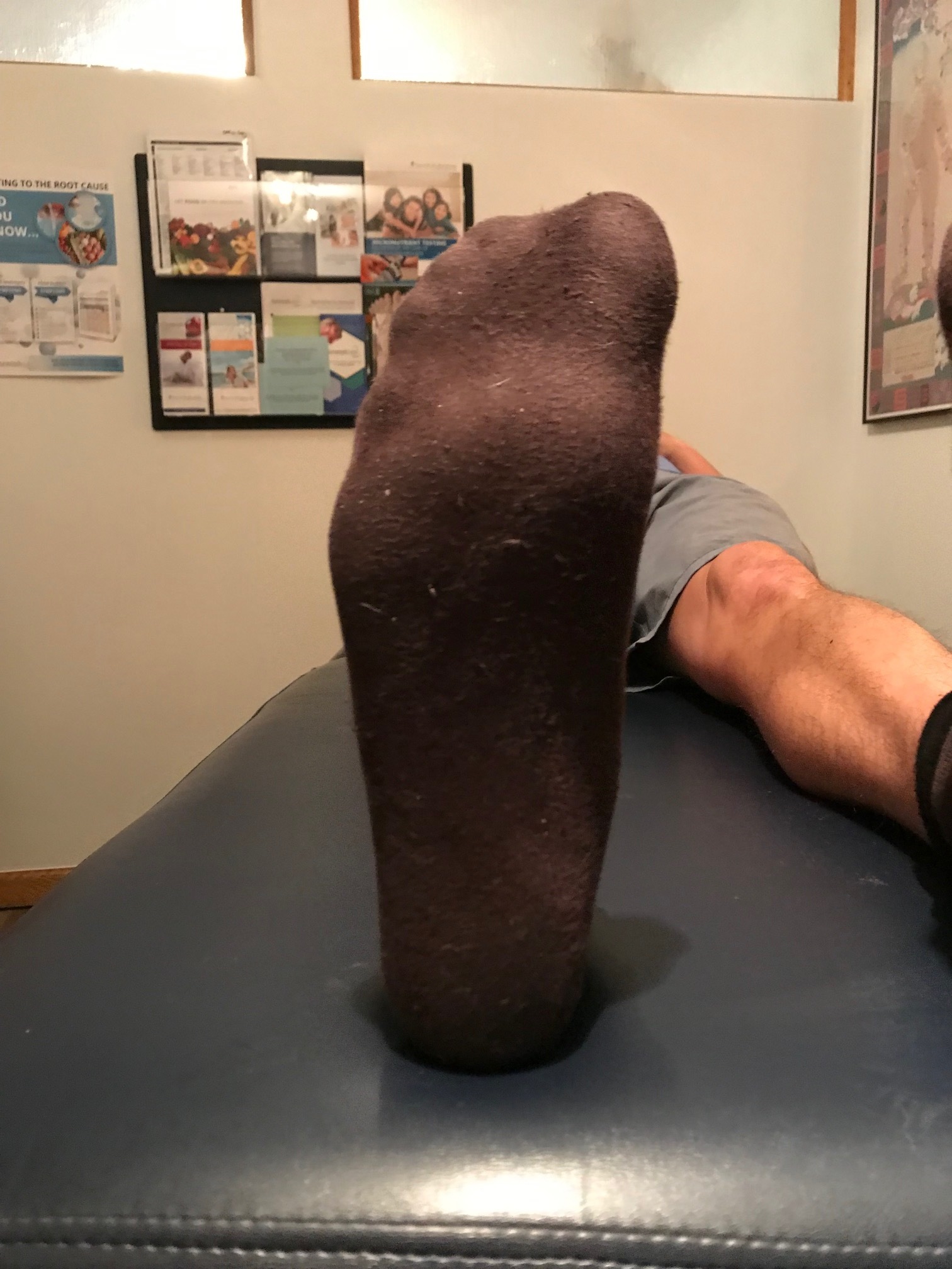
Internal tibial torsion + femoral retrotorsion + twisting = low back pain

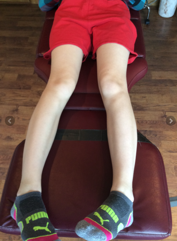
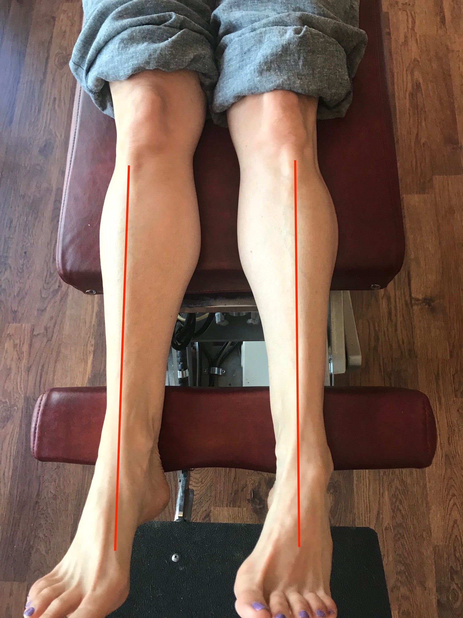
note the internal tibial torsion and lack of internal rotation of the right hip secondary to femoral retroversion

note the internal tibial torsion and lack of internal rotation of the right hip secondary to femoral retroversion

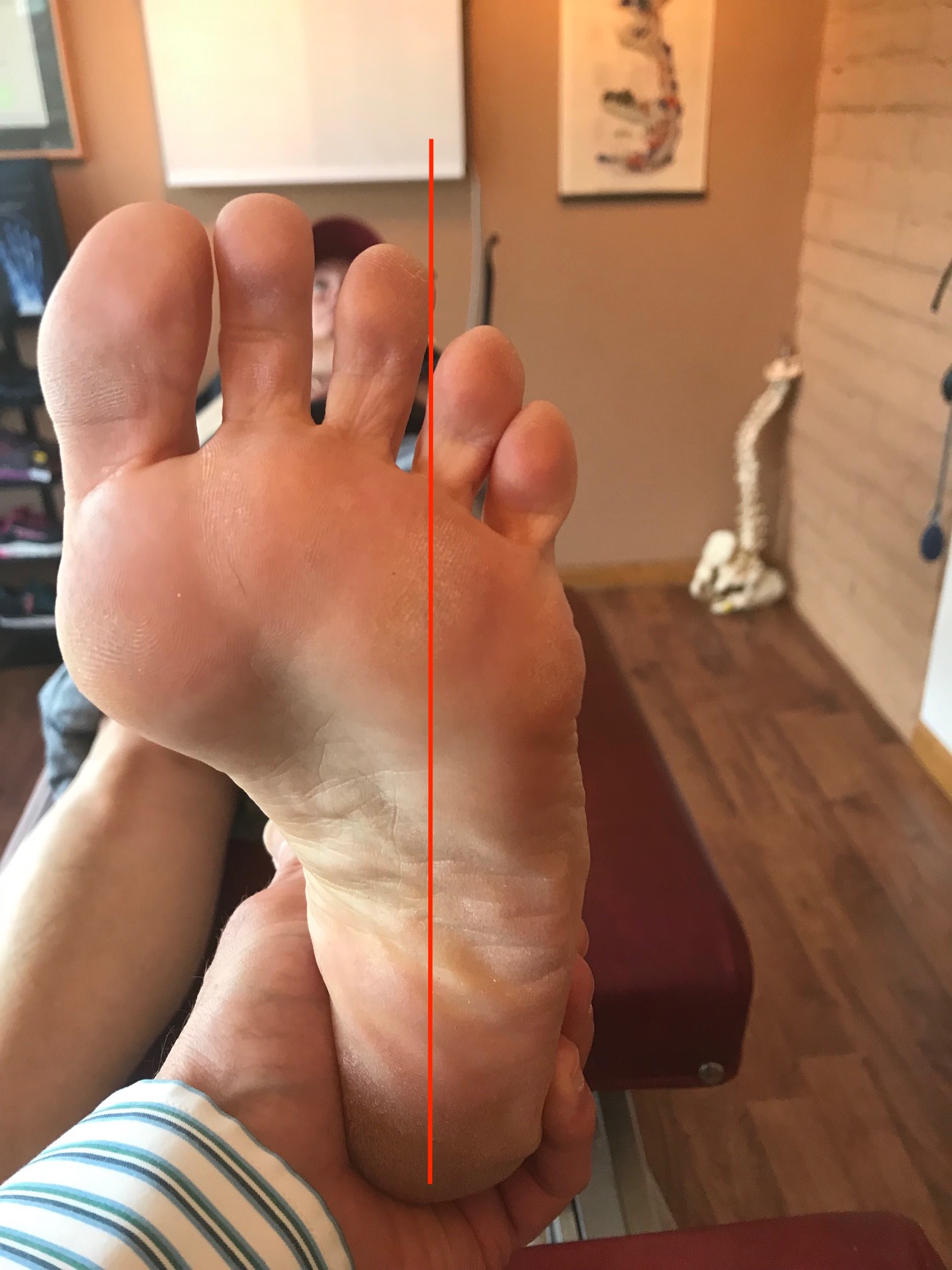
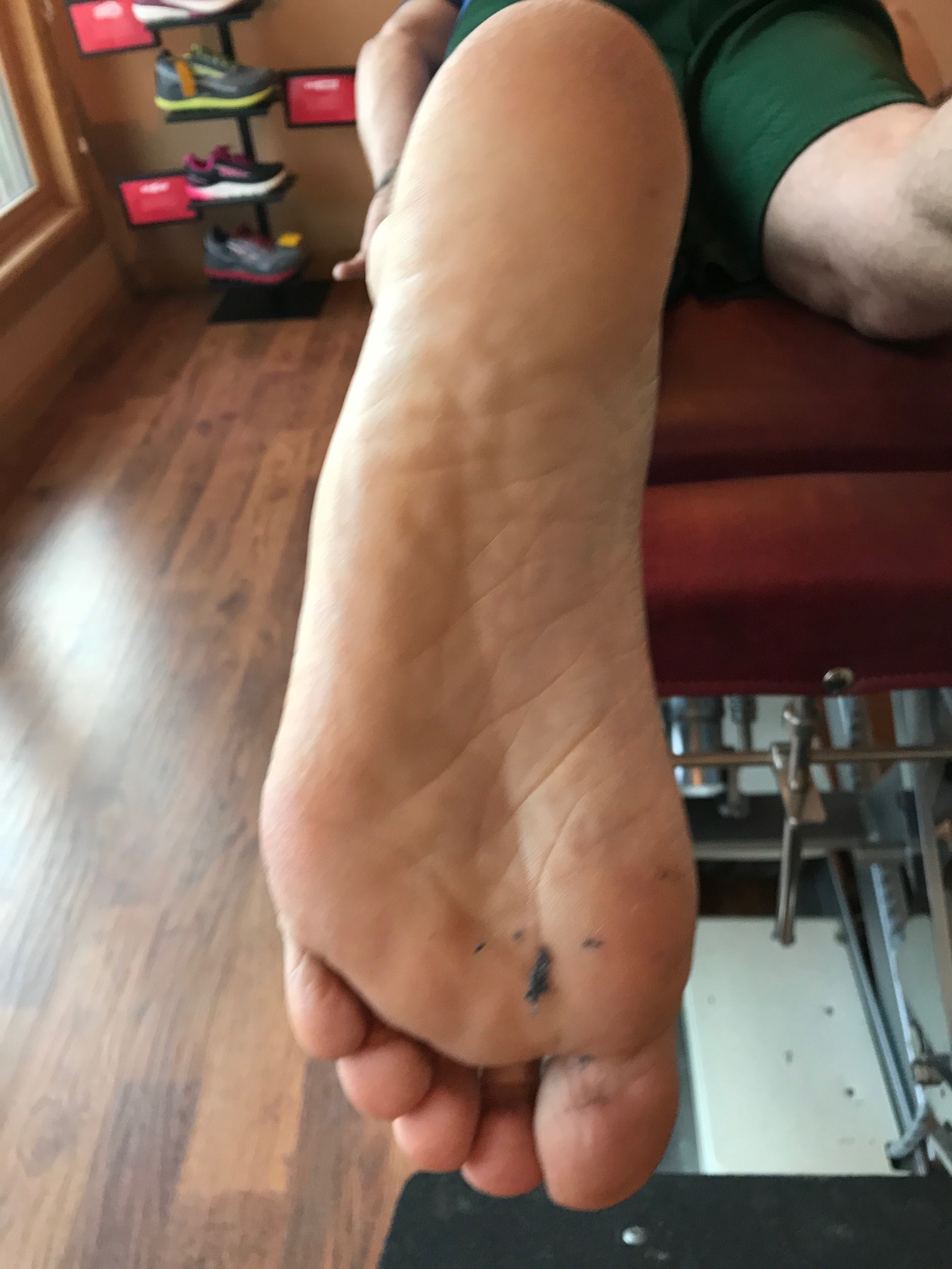
internal rotation of less than 0 on the left hip

internal rotation of less than 0 on the left hip

Here’s one that we’ve talked about before. Think about internal tibial torsion for a moment. You remember when the tibial tuberosity points straightahead and the foot points inward? 

Now combine that with femoral retro torsion. Remember that that’s when the angle of the femoral neck with the shaft is less than 8° which results in a loss of internal rotation of the hip. 

Put those two together and you have a foot that points inward and limited internal rotation of the hip on that same side. 

Stand on your right leg with your foot turned in. Twist your body to the right. Can you see how this is internal rotation of the right hip?

If people have to create internal rotation of the hip, then the motion has to occur somewhere. If it is not available at the hip and the lower extremity is fully internally rotated it has to occur north of the hip which is usually in the lumbar spine. Now think about when people are lifting things. Often times they do not do a hip hinge and reverse or flatten their lumbar lordosis, opening up the facet joints which allows more rotation. Do you remember that rotation of the lumbar spine is limited to about 5° unilaterally? Where do you think that hip rotation is going to occur? Hmmmm... Probably north of the hip. 

Better teach them to spin that weight bearing leg and foot out into some external rotation to create that needed range of motion, increase the amount of internal rotation of that hip or have them keep their shoulders and hips in the same plane when rotating.

Dr Ivo Waerlop, one of The Gait Guys 

#backpain #hipinternalrotation #internaltibialtorsion #retrotorsion #femoralretrotorsion 

The case of the lateral thigh, leg and knee pain

note how the bottom of the heel cup is rounded

note how the bottom of the heel cup is rounded

IMG_7238.jpg

This gal  came to see us with pain in the lateral thigh, knee, and lateral lower leg on the left. It has happened recently with skiing and alleviated only temporarily with acupuncture of the vastus lateralis and peroneal groups. She has been skiing with a foot bed (please see picture).

Evaluation reveals mild bilateral external tibial torsion, a right anatomically short  leg tibial, and bilateral medial knee fall (knee is medial of the sagittal plane) L>R during weight-bearing when the feet are pointing straight ahead. There is moderate loss of the medial longitudinal arches bilaterally, greater on the left.

We remember that when a patient has external tibial torsion, when the knees points straight the feet point to the outside. 

Translated to skiing, the feet need to be straightahead which brings the knee(s) inside of the sagittal plane. 

We remember that often times with leg length discrepancy is, the longer leg side will “pronate ” in attempt to shorten extremity and the shorter leg side “supinate”, in an attempt to lengthen the extremity.

Putting this all together:  the patient is pronating bilaterally, left greater than right with medial knee fall. The ski shop put the footbed you see in the picture in both of the patients boots. You can see that it is extremely rounded at the heel and, because orthotics are shank dependent devices, a round heel like this will just roll into pronation as there is more weight on the medial longitudinal arch. This makes the entire foot bed assembly relatively ineffective and increases the valgus moment at the knee, stressing the vastus lateralis as it is trying to pull the knee to midline as well as the peroneal group as it is trying to do the same from a closed chain position as well as supinate the foot. 

We placed a 3 mm sole lift under the right foot and added a post to the bottom the left orthotic to allow it to sit flat. This did not correct the problem completely and we needed to add a Morton’s toe extension (post under the first metatarsal) to invert the foot and bring the knee out into the sagittal plane.

Dr Ivo Waerlop, one of The Gait Guys 

#footbeds #kneepain #orthotics #skibootfit #thighpain #legpain #quadriceps

Two things we hope you see right away when viewing this video.

.

We talked about this woman in yesterday‘s post and, when viewing from the front, we wanted to bring out a few salient points

Notice how her foot progression angle is diminished bilaterally. Normally the foot will “toe out“ somewhere between 12 and 20° when the foot hits the ground and hers are nearly straightahead. This can be due to numerous things such as femoral retro torsion, internal tibial torsion, or subtalar version. All of these things will often have the foot point medially when examining a patient on the table and placing the knee in the sagittal plane. In this particular case, she has internal tibial torsion.

The next thing we would like you to look at is noting how her knee falls “outside“ the sagittal plane. In other words, instead of the knees pointing straight ahead, the point slightly out laterally. This is a cardinal sign of internal tibial torsion, especially in a gait analysis.

So what’s a clinician to do?

In this particular case, there’s nothing really to “fix“ as these are hard deformities that are often congenital. Our job is to help the patient to compensate and the best way possible so that they can remain pain and as injury free as possible. We discussed remediation in the post yesterday, So please refer back to that for what we did


Dr Ivo Waerlop, one of The Gait Guys

#invertedforefoot #forefootsupinatus #forefootvarus #pronation #forefoot #gaitanalysis #decreasedprogressionangle #toeingin

Internal tibial torsion puts pressure on the outside of the foot

IMG_7174.jpg
IMG_7175.jpg
IMG_7176.jpg

Take a look at these pictures. This is also a good reason to always look at the insoles. Take a good look. Can you see the increase printing on the lateral aspect of the right foot?

You’ll note that he has internal tibial torsion on the right side. This often presents with a forefoot supinatus and results in pressuring of the lateral column of the foot and an inability to descend the first ray. Note that the footbed on the right shows increased pressure of the lateral column and a lack of pressure under the head of the first.

Stand up and put the weight on the outside of your right foot. Can you feel how the toes on right side pressing more in an attempt to shift the center of gravity medially while it offloading the toes on the left foot? This is also represented in the foot beds.

Yet another great reason to not only look at the wear on the outside, but also on the inside of your clients/patients shoes.

Dr Ivo Waerlop, one of The Gait Guys

#footbeds #internaltibialtorsion #lateralfootpressure #insoles

A novel way to look at functional internal rotation of the hips

As clinicians (and coaches) we are often trying to figure out different ways to functionally assess internal rotation of the hips. How many times does the patient/client “appear“ to have appropriate internal rotation on the table only to find out that they don’t functionally and vice versa.

Take a look at it this gentleman who is a ski instructor. We are trying to simulate the standard side to side ski motion in a way that would be functionally appropriate. Keep in mind that he has bilateral internal tibial torsion and bilateral femoral retro torsion. When he began care at our office he had 5° external rotation on the right and about 8° external rotation on the left as his FULL AMOUNT OF INTERNAL ROTATION AVAILABLE to him bilaterally.

Treatment consisted largely of hip mobilization, Therapeutic exercises to emphasize internal rotation of the hips such as hip helicopters, airplanes and supine “chairs“ with internal rotation and adduction utilizing a ball between the knees; we also did acupuncture/needling of the hip capsules as well as anterior fibers of the gluteus medius and minimus. He now has about 5° internal rotation on the right now and a little less than 5 on the left. Note how the motion is clearly visualized in this video below.

Do you have other novel ways to test internal rotation of the hips functionally? Leave a comment or drop us a line and let us know

Feel like you want more? Join us this Wednesday evening on onlinece.com for Biomechanics 326: 6 MST

Dr Ivo Waerlop, one of The Gait Guys.

#functionaltesting #functionalmovement #hip #internalrotation #femoralretrotorsion #femoralretroversion #thegaitguys

Unilateral calcaneal valgus: what can it mean?

right calcaneal valgus

right calcaneal valgus

Take a good look at this picture and what do you see? Do you see the calcaneal valgus on the right side. What runs through your mind?

Possibilities for causing this condition, as well as the clinical implications are numerous.

The short list should include:

  • A shorter leg on the contralateral side: often times we will pronate more on the longer leg side to compensate for a short leg on the opposite

  • Increased rear foot and/or fore foot pronation on the valgus side. Laxity of the spring ligament or incompetency of the musculature which helps to maintain your arch (tibialis posterior, foot intrinsics, tibialis anterior to name a few) often causes more collapse on the effected side

  • A lack of available rearfoot eversion on the contralateral side. It may be that the increase calcaneovalgus is normal and the opposite side is more rigid.

  • If you were seeing this in the middle of the gait cycle it could be that that is their strategy to get around a loss of hip extension or ankle rocker

  • External tibial torsion on that side. Go ahead, stand up and spin your right foot into external rotation and keep your left foot with a normal progression angle. Can you see how your arch collapses to a greater degree on the side with the external torsion? Remember that pronation is dorsiflexion, eversion and abduction.

  • Internal tibial torsion on the contralateral side. Internal tibial torsion puts the foot into supination which makes it into more of a rigid lever rather than mobile adapter.

    And the list goes on…

    Next time you see a unilateral deformity like this, hopefully some of these things run through your mind and will help you to pinpoint where the problem actually is.

    Dr Ivo Waerlop, one of The Gait Guys

    #calcanealeversion #rearfootvalgus
    #lowerextremitydeformities

Obligate Pathomechanics

Much of what we see in gait analysis is secondary to the anatomical and physiological constraints exhibited by a patient. Take a look at this gentleman running. At first glance, you may be saying “yup, crossover gait, strengthen the gluteus medius complex“.

Now let’s talk about his physical exam. He has “windswept biomechanics“, With external tibial torsion on the right and internal table torsion on the left. There is no significant difference or increase in his Q angles bilaterally. He has a forefoot supinatus on the right side (I.e his forefoot is inverted with respect to his rear foot). He has limited plantar flexion of the first Ray complex on the right.

Now watch the video again with this in mind. Can you understand that if he’s unable to get his first ray to the ground he’s going to have any sort of hike your push off, in order to get it to the Ground he’ll need to mediately rotate his lower extremity and increase the valgus angle on that side. External tibial torsion (when you drop a plum line from the tibial tuberosity, it passes medial to a line passing to the long axis of the second metatarsal) compounds this. Stand up, rotate your right foot to the outside, keep it there and walk forward. Do you see how your knee has to go to the inside to push off your big toe?

Yes, he has a crossover gait but it is obligate and a direct function of his inability to descend the first ray, at least partly due to his forefoot supinatus and his external tibial torsion on the right.

Obligate pathomechanics. Coming to a patient in your office or one of the folks you are coaching soon.

We will be talking about foot types and pathomechanics tonite, October 16th, 2019, on our 3rd Wednesday’s teleseminar on onlinece.com: Biomechanics 314

5 pacific, 6 mountain, 7 central, 8 eastern

Dr Ivo Waerlop, one of The Gait Guys

3 clues that someone has internal tibial torsion

Watch this video a few times through and see what you notice. There are three clues that this patient has internal tibial torsion, can you find them?

He presented with right sided knee pain, medial aspect of the patella and medial joint line as well as tenderness over the medial joint line and pes anserine. Lower extremity musculature test strong and 5/5 save for his semi tendinosis on the right which tested 4/5.He has diminished endurance bilaterally in the external obliques

1. Note how his knees, right greater than left, fall outside the sagittal plane

2. Note the decreased progression angle of both feet during forward motion

3. Note how he toes off in supination, right greater than left.

This patient’s knee pain is coming from irritation of the pes anserine, particularly semitendinosus and his inability to recruit his abdominals sufficiently so, instead of the usual pattern of recruiting iliopsoas or rectus femoris, he chooses his sartorius, gracious and semi tendinosis.

Pay attention to how the new tracks, the progression angle as well as if they tow off in pronation, neutral, or supination in that can offer subtle clues to internal tibial torsion.

Dr Ivo Waerlop, one of The Gait Guys

#internaltibialtorsion #gaitanalysis #thegaitguys

https://vimeo.com/365342814

External tibial torsion and lower back pain

How can external tibial torsion and lower back pain possibly be related? Let’s take a quick look at the anatomy and see how.

knees neutral, note external rotation of the right foot and decreased progression angle

knees neutral, note external rotation of the right foot and decreased progression angle

Remember the external tibial torsion is present if we drop a plumbline from the tibial tuberosity and it passes between the first and second metatarsals or more medially. This increases the progression angle of the foot. This occurs due to “over rotation" of the lower extremity during development, often exceeding the 1.5 degrees per year of external rotation per year up to age 15 or occurring for a longer period of time, up to skeletal maturity. It can be uni or bilateral.

note when the foot is neutral, the knee points inward

note when the foot is neutral, the knee points inward

Often, due to the increased progression angle, people will try to "straighten their feet" (ie, decrees their progression angle) to move forward in the sagittal plane. This places the knees to the inside of the sagittal plane which causes medial knee fall and sometimes increased mid and forefoot pronation. This results in increased medial spin of the thigh bilaterally which increases the lumbar lordosis. Combine this with a sway back or anterior pelvic tilt and you have increased pressure on the lumbar facet joints. The facets are designed to carry approximately 20% of the load put in these circumstances are often called upon to carry the much more. This often results in facet imbrication and lower back pain. You can strengthen the abdomen all you like but if you do not change the attitude of the foot, a will often develop lower back pain, especially when the abs fatigue. Now think about if the deformity is unilateral; this will often cause asymmetrical rotation of the pelvis in a clockwise or counter clockwise direction.

So, what can you do you?

Since external tibial torsion is a "hard deformity", we can influence how the bone grows before skeletal maturity but after that will not change significantly with stretching or exercise.

  • You can teach them to walk with an increase in progression angle (ie “duck footed”). This will often keep the knee in the sagittal plane and can be surprisingly well tolerated

  • You can use a foot leveling orthotic or arch support to bolster the arch and change the mechanics of the foot, causing external rotation of the tibia which will often result in a decrease in progression angle in compensation while still keeping the knee in the sagittal plane

  • You could place a full length varus wedge in the shoe which, by inverting the foot, externally rotates the tibia which the person will often compensates for by decreasing there progression angle to keep the knee and the sagittal plane


Dr Ivo Waerlop, one of The Gait Guys


#tibialtorsion #lowbackpain #LBP #progressionangle


Sometimes it’s OK for “toes in“ squats

We hear from folks and also read on a lot of blogs and articles about whether your toes should be in or out for squats or other types of activities. The real answer is “it depends”.

What it depends on is the patient’s specific anatomy. That means we need to pay attention to knees and hips and things like femoral and tibial torsion‘s. It’s paramount to keep the knees in the sagittal plane, no matter what the lower extremity orientation is.

When somebody has external tibial torsion (i.e. when you drop a plumbline from there to view tuberosity it passes medial to the line between the second and third or second metatarsal) then having your feet and externally rotated position places the knees in sagittal plane. Having the patient go “toes in” with this type of anatomy will cause both knees to for medially and create patellofemoral tracking issues.

Likewise, like the patient in the video, (Yes, I know I say “external tibial torsion“ at the beginning of the video but the patient has internal tibial torsion as you will see from the remainder of the video) when somebody has internal tibial torsion (I.e. when you drop a plumbline from the tibial tuberosity it passes lateral to the second metatarsal or a line between the second and third metatarsal) you would need to point the toes inward to keep the knees in the sagittal plane as demonstrated in the video. You can also see in the video when her feet are placed “toes out“ they fall outside sagittal plane laterally which creates patellofemoral tracking issues like it was in this particular patient.

So, knees in or knees out? It depends…

Dr. Ivo Waerlop, one of The Gait Guys

#internaltibialtorsion #externaltibialtorsion #kneepain #kneesin #kneesout #squats #thegaitguys

External tibial torsion or femoral retrotorsion?

IMG_6727.JPG

This young lad presents to your office complaining of bilateral knee discomfort at the medial aspect, just below the patella, particularly when ascending and descending stairs and hills. You narrow it down to abnormal patellar tracking and 2 possibilities of who is driving the bus, but which is it?

Torsions of an extremity are said to exist when they measure two or more standard deviation‘s outside of normal. In external tibial torsion, the shaft of the tibia over rotates more than it’s 1.5° per year from zero at birth to greater than 19°. You are left with a foot that is has an increased progression angle and a center of gravity falls medial to the foot causing abnormal patellar tracking.

Femoral retro torsion is said to exist when the head of the femur over reduces from its 35° angle at birth to less than 8° resulting in severely limited internal rotation of the hips bilaterally. The lower extremity is often externally rotated to compensate.

An easy differential for the 2 is to drop a plumbline from the tibial tuberosity through the foot. This line normally passes through the second or between the second and third metatarsal‘s. If it falls medial to that it is eternal tibial torsion and lateral to that most likely internal tibial torsion or potentially a metatarsus varus or forefoot adductus.

IMG_6759.jpg


Another differential would be to perform “Craigs test” and measure how much internal and external rotation of the femur there is at the femoral acetabular articulation.

An easier way to put it is; those with femoral retrotorsion have less hip internal rotation and often increased amounts of external rotation; often they can’t even get past zero, never mind the requisite 4-6 degrees for normal gait. Those with increased internal rotation and diminished external rotation most likely have femoral antetorsion.

IMG_6726.jpg


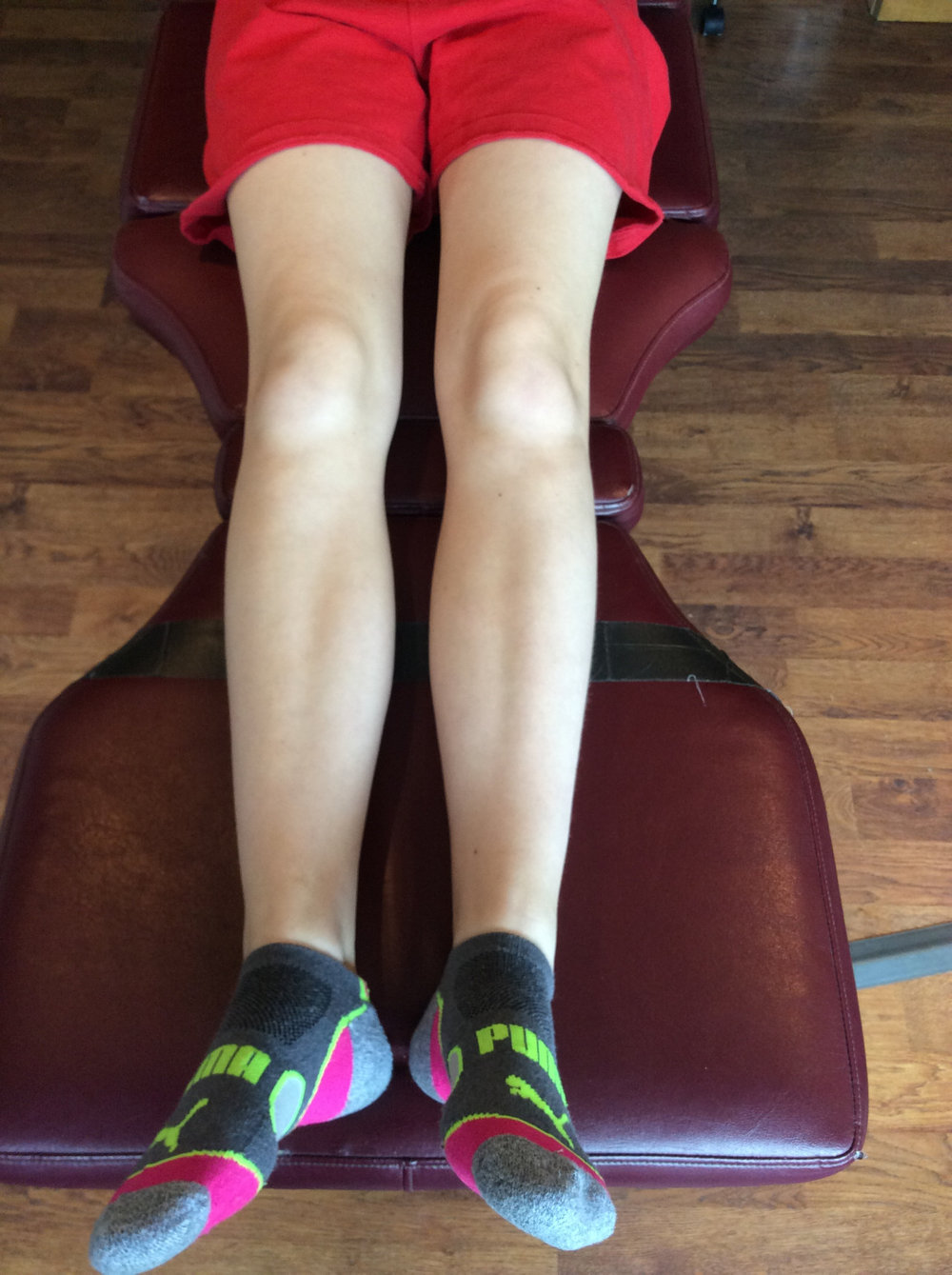
So, Which is it? When his knees are Straightahead, his feet point out; when his feet are straightahead, his knees point inward. A plumbline from the tibial tuberosity passes medial to the second metatarsal. Looking at the pictures, you can see that he is external tibial torsion along with a sandal thong deformity that we talked about last week.

Dr Ivo Waerlop, one of The Gait Guys.

#externaltibialtorsion #outturnedfoot #increasedprogressionangle #kneepain #thegaitguys

Sixth toe disease...That growth on the outside of your foot… Or on somebody’s foot is coming to see you…

IMG_6704.JPG

You know what we’re talking about. That extra growth on the lateral aspect of the foot that happens way too often and many of your clients. A Taylor’s bunion or sometimes referred to as a “bunionette”. 

What is the usual fix?

Usually in a ski boot or hiking boot, they blow out the lateral side of the shoe. This is usually not a good fix because most of these folks have internal tibial torsion and somewhat of a forefoot supinatus/varus.

IMG_6706.JPG
IMG_6707.JPG

The internal tibial torsion places the knee outside the saggital plane and an arch support without a forefoot valgus post will just push it further out, creating a conflict at the knee. The forefoot supinatus and/or varus places them on the outside of the foot as well. Remember, most of these folks are ALREADY on the outside of the foot and the foot wants to migrate laterally...so creating more space just means it migrates farther. Good thought, doesn’t work that way.

IMG_6710.JPG

So what did we do?

  • We created a valgus post for the forefoot (see picture above) tapering from lateral to medial and to help “push“ the distal aspect of the first ray down (because there was motion available that was not being used)

  • We gave him exercises to help descend the first ray like the extensor hallucis brevis exercise, toe waving as well as peroneus longus exercises

  • We gave him plenty of balance and coordination work

    Dr Ivo Waerlop, one of The Gait Guys


#6thtoe #internaltibialtorsion #forefootvarus # forefootsupinatus #gaitanalysis #thegaitguys


We’ve told you once and we will tell you again…

Folks with femoral retro torsion often experience lower back pain with twisting movements

This left handed hydrology engineer Presented to the office with an acute onset of lower back pain following “swinging a softball bat”. He comments that he always “hit it out of the park“ and hit “five home runs“ in the last game prior to his backs demise.

note the internal tibial torsion. drop a plumbline from the tibial tuberosity. it should pass through the 2nd met or between the 2nd and 3rd met shafts

note the internal tibial torsion. drop a plumbline from the tibial tuberosity. it should pass through the 2nd met or between the 2nd and 3rd met shafts

note the internal tibial torsion. drop a plumbline from the tibial tuberosity. it should pass through the 2nd met or between the 2nd and 3rd met shafts

note the internal tibial torsion. drop a plumbline from the tibial tuberosity. it should pass through the 2nd met or between the 2nd and 3rd met shafts

He presented antalgic with a pelvic shift to the left side, flexion of the lumbar spine with 0° extension and a complete loss of the lumbar lordosis. He could not extend his lumbar spine past 0° and was able to flex approximately 70. Lateral bending was approximately 20° on each side. Neurological exam negative. Physical exam revealed bilateral femoral retro torsion as seen above. Note above the loss of internal rotation at the hips of both legs, thus he has very limited internal rotation of the hips. Femoral retroversion means that the angle of the neck of the femur (also known as the femoral neck angle) is less than 8°, severely limiting internal rotation of the hip and often leading to CAM lesions.

Stand like you’re in a batters box and swing like you’re left handed. What do you notice? As you come through your swing your left hip externally rotates and your right hip must internally rotate. He has no internal rotation of the right hip and on a good day, the lumbar spine has about 5° of rotation with half of that occurring at the lumbosacral junction. Guess what? The facet joints are going to become compressed!

bisect the calcaneus. the line should fall though the 2nd metatarsal or between the 2nd and 3rd met shafts

bisect the calcaneus. the line should fall though the 2nd metatarsal or between the 2nd and 3rd met shafts

bisect the calcaneus. the line should fall though the 2nd metatarsal or between the 2nd and 3rd met shafts

bisect the calcaneus. the line should fall though the 2nd metatarsal or between the 2nd and 3rd met shafts

Now combine that with bilateral 4 foot adductus (see photos above). His foot is already in supination so it is a poor shock observer.

Go back to your “batters box“. Come through your swing left handed. What do you notice? The left foot goes into a greater amount of pronation in the right foot goes into a greater amount of supination. Do you think this is going to help the amount of internal rotation available to the hip?

When folks present with lower back pain due to twisting injuries, make sure to check for femoral torsions. They’re often present with internal tibial torsion, which is also present in this individual.

Remember a while ago we said “things occur in threes”. That goes for congenital abnormalities as well: in this patient: femoral retro torsion, internal tibial torsion and forefoot adductus.

What do we do? Treat locally to reduce inflammation and take steps to try to improve internal rotation of the hips bilaterally as well as having him externally rotate his right foot when he is in the batteries box to allow him to "create" more internal rotation of the right hip.

Dr Ivo Waerlop, one of The Gait Guys

#internalrotation #hipproblem #femoraltorsion #femoralversion #retroversion #retrotorsion #thegaitguys

Holy twisted tibias Batman! What is going here in this R sided knee pain patient?

Screen Shot 2019-06-10 at 12.28.38 PM.png

In the 1st picture note this patient is in a neutral posture. Note how far externally rotated her right foot is compared to the left. Note that when you drop a plumbline down from the tibial tuberosity it does not pass-through or between the second and third metatarsals. Also note the incident left short leg

Screen Shot 2019-06-10 at 12.28.56 PM.png

In the next picture both of the patients legs are fully externally rotated. Note the large disparity from right to left. Because of the limited extra rotation of the right hip this patient most likely has femoral retro torsion. This means that the angle of her femoral head is at a greater than 12° angle. We would normally expect approximately 40° of external Rotation. 4 to 6° is requisite for normal gait and supination.

Screen Shot 2019-06-10 at 12.28.47 PM.png

In the next picture the patients knees are fully internally rotated you can see that she has an excessive amount of internal rotation on the right compare to left, confirming her femoral antetorsion.

Screen Shot 2019-06-10 at 12.28.38 PM.png

When this patient puts her feet straight (last picture), her knees point to the inside causing the patello femoral dysfunction right greater than left. No wonder she has right-sided knee pain!

Because of the degree of external tibial torsion (14 to 21° considered normal), activity modification is imperative. A foot leveling orthotic with a modified UCB, also inverting the orthotic is helpful to bring her foot somewhat more to the midline (the orthotic pushes the knee further outside the sagittal plane and the patient internally rotate the need to compensate, thus giving a better alignment).


a note on tibial torsion. As the fetus matures, The tibia then rotates externally, and most newborns have an average of 0- 4° of internal tibial torsion. At birth, there should be little to no torsion of the tibia; the proximal and distal portions of the bone have little angular difference (see above: top). Postnatally, the tibia should twist outward (externally) a total of 15 degrees until adult values are reached between ages 8 and 10 years of 23° of external tibial torsion (range, 0° to 40°).

Wow, cool stuff, eh? Dr Ivo Waerlop, one of The Gait Guys

#tibialtorsion #tibialversion #kneepain #thegaitguys #gaitanalysis

Ode to the Popliteus

Remember the popliteus? To recap, it contracts at the initial contact phase of the walking gait cycle, to act as an accessory PCL (look HERE

https://www.thegaitguys.com/search?q=popliteus&f_collectionId=57d4982c91b18610c6ee3e0f

to read about that), then contracts eccentrically to slow the rate of internal rotation of the femur on the tibia until midstance, so as not to macerate the meniscus; It then contracts concentrically to accelerate the external rotation of the femur on the tibial plateau so it rotates faster then the tibia, to protect the meniscus as well. So, internal rotation of the femorotibial complex from initial contact to midstance and external rotation of the complex from midstance to preswing. Got it?

Now look at the video of this gal with L sided medial knee pain and past history of a left tibial plateau fracture in her youth. Do you see it? Hmmmm; doesn’t look like internal rotation does it? Don’t see it? Remember that the whole complex SHOULD be internally rotating until the swing phase leg passes the stance pase leg. See it now? Considering that the popliteus tested weak on the clinical exam, does this surprise you?

Agreed that there are many factors initiating internal rotation (and thus pronation) of the stance phase leg from initial contact to midstance, like plantar flexion, adduction and eversion of the talus, contraction of the lower leg anterior compartment muscles, eccentric contraction of the quads and hamstrings, just to name a few, can you see how (a least theoretically) one bad player can ruin the team?

Yes, popliteus rehab, along with abdominal core and foot core endurance exercises are in her future.

Dr Ivo Waerlop, one of The Gait Guys

#popliteus #kneepain #kneeproblem #thegaitguys #gaitanalysis

Neuroma! Triple Threat....

Can you guess why this patient is developing a neuroma on the left foot, between the 3rd and 4th metatarsals?

IMG_6220.jpg
IMG_6218.jpg
IMG_6219.jpg

This gal presented to the office with pain in the left foot, in the area she points to as being between the 3rd and 4th metatarsals. It has been coming on over time and has become much worse this spring with hiking long distances, especially in narrower shoes. It is relieved by rest and made worse with activity.

Note the following:

  • She has an anatomical short leg on the left (tibial)

  • internal tibial torsion on the left

  • left forefoot adductus (see the post link below if you need a refresher)

Lets think about this.

The anatomical short leg on the left is causing this foot to remain in relative supination compared the right and causes her to bear weight laterally on the foot.

The internal tibial torsion has a similar effect, decreasing the progression angle and again causing her to bear weight laterally on the foot, compressing the metatarsals together.

We have discussed forefoot adductus before here on the blog. Again, because of the metararsal varus angle, it alters the forces traveling through the foot, pushing the metatarsals together and irritating the nerve root sheath, causing hypertrophy of the epineurium and the beginnings of a neuroma.

In this patients case, these things are additive, causing what I like to a call the “triple threat”.

So, what do we do?

  • give her shoes/sandals with a wider toe box

  • work on foot mobility, especially in descending the 1st ray on the left

  • work on foot intrinsic strength, particularly the long extensors

  • treat the area of inflammation with acupuncture

Dr Ivo Waerlop, one of The Gait Guys

#forefootadductus #metatarsusadductus #neuroma #gaitanalysis #thegaitguys #internaltibialtorsion

3 things

Its subtle, but hopefully you see these 3 things in this video.

I just LOVE the slow motion feature on my iPhone. It save me from having to drag the video into Quicktime, slow it down and rerecord it.

This gal has a healing left plantar plate lesion under the 2nd and 3rd mets. She has an anatomical leg length deficiency, short on the left, and bilateral internal tibial torsion, with no significant femoral version. Yes, there are plenty of other salient details, but this sketch will help.

  1. 1st if all, do you see how the pelvis on her left dips WAY more when she lands on the right? There is a small amount of coronal plane shift to the right as well. This often happens in gluteus medius insufficiency on the stance phase leg (right in this case), or quadratus lumborum (QL) deficiency on the swing phase leg (left in this case) or both. Yes, there are other things that can cause this and the list is numerous, but lets stick to these 2 for now. In this case it was her left QL driving the bus.

  2. Watch the left and right forefeet. can you see how she strikes more inverted on the left? this is a common finding, as the body often (but not always) tries to supinate the shorter extremity (dorsiflexion, eversion and adduction, remember?) in an attempt to “lengthen” it. Yes, there is usually anterior pelvic tilt accompanying it on the side, because I knew you were going to ask : )

  3. Look how her knees are OUTSIDE the saggital plane and remain there in her running stride. This is commonly seen in folks with internal tibial torsion and is one of the reasons that in our opinion, these folks should not be put medially posted, torsionally rigid, motion control shoes as this usually drive the knees FURTHER outside the saggital plane and can macerate the meniscus.

Yep, lots more we could talk about on this video, but in my opinion, 3 is a good number.

Dr Ivo Waerlop, one of The Gait Guys

#thegaitguys #gaitanalysis #footpain #gaitproblem #internaltibialtorsion #quadratuslumborum #footstrike

https://vimeo.com/329212767

Things seem to come in 3's...

Things tend to occur in threes. This includes congenital abnormalities. Take a look this gentleman who came in to see us with lower back pain.

Highlights with pictures below:

  • bilateral femoral retrotorsion

  • bilateral internal tibial torsion

  • forefoot (metatarsus) adductus

So why LBP? Our theory is the lack of internal rotation of the lower extremities forces that motion to occur somewhere; the next mobile area just north is the lumbar spine, where there is limited rotation available, usually about 5 degrees.

Dr Ivo Waerlop, one of The Gait Guys.

#tibialtorsion #femoraltorsion #femoralretrotorsion #lowbackpain #thegaitguys #gaitproblem
this is his left hip in full internal rotation. note that he does go past zero.

this is his left hip in full internal rotation. note that he does go past zero.

full internal rotation of the right hip; note he does not go past zero

full internal rotation of the right hip; note he does not go past zero

note the internal tibial torsion. a line dropped from the tibial tuberosity should go through the 2nd metatarsal or between the 2nd and 3rd.

note the internal tibial torsion. a line dropped from the tibial tuberosity should go through the 2nd metatarsal or between the 2nd and 3rd.

ditto for the keft

ditto for the keft

a line bisecting the calcaneus should pass between the 2nd and 3rd metatarsal shafts. If talar tosion was present, the rearfoot would appear more adducted

a line bisecting the calcaneus should pass between the 2nd and 3rd metatarsal shafts. If talar tosion was present, the rearfoot would appear more adducted

less adductus but still present

less adductus but still present

look at that long flexor response in compensation. What can you say about the quadratus plantae? NO bueno…

look at that long flexor response in compensation. What can you say about the quadratus plantae? NO bueno…

Ditto!

Ditto!

Motion control Shoes + Internal Tibial Torsion = Knee Pain

Thinking about putting a motion control shoe under that foot to control pronation? You had better make sure you make friends with the knee, as it will often (depending on the compensation) be placed OUTSIDE the SAGGITAL PLANE. Like Dr Allen has said many times before , the knee is basically a hinge joint placed between 2 ball and socket joints, and it is usually the one to start grumbling...

Learn more as Dr Ivo Waerlop of The Gait Guys explains in this brief video

#gait #Gaitanalysis #gaitguys #thegaitguys #kneepain #motioncontrolshoes #internaltibialtorsion

https://vimeo.com/154496722