The 4 Factors of Heel Rise.

Screen Shot 2019-01-14 at 12.48.19 PM.png
Screen Shot 2019-01-14 at 12.48.08 PM.png

These SHOULD all happen to have appropriate heel rise and forward progression

1. active contraction of the posterior compartment of the calf

2. passive tension in the posterior compartment of the calf

3. knee flexion and anterior translation of the tibia ankle rocker

4. the windlass mechanism

a problem with any one of these (or more collectively) can effect heel rise, usually causing premature heel rise.

ask yourself:

  • Do you think the posterior compartment is actively contracting? not enough or too much? Remember the medial gastrocnemius adducts the heel at the end of terminal stance to assist in supination. Don't forget about the tibialis posterior as well as the flexor digitorum longs and flexor hallucinate longus.

  • Does there appear to be increased passive tension in the posterior compartment? How visible and prominent are their calf muscles?

  • Do they have forward progression of the body mass?

  • How is his windlass mechanism? Good but not good enough.

Dr Ivo Waerlop. One of The Gait Guys…

#gait, #gaitanalysis, #continuingeducation, #limp, #casestudy, #gaitparameters, #heelrise, #prematureheelrise, #windlassmechanism

Two out of Three ain't Bad...But sometimes it is

image credit: https://commons.wikimedia.org/wiki/File:Meatloaf_(1).jpg

image credit: https://commons.wikimedia.org/wiki/File:Meatloaf_(1).jpg

“What do you mean my plantar fasciitis is due to my hip?”

I recently saw a 60 YO male patient with right-sided plantar fasciitis of approximately 1-1/2 months duration. It began insidiously with pain located at the medial calcaneal facet on the right hand side. He had localized tenderness in this area with some spread distally towards the metatarsal heads. He has ankle dorsiflexion was relatively symmetrical with mild impairment on the right compared to left but only approximately 2 degrees. He had hip extension is 0 degrees on the affected side and 10 degrees on the affected side. Sacroiliac pathomechanics were present as well with the loss of flexion and extension. He had a slight leg length discrepancy, short on the symptomatic side.

So what is going on?

Moving forward in the sagittal plane requires a few things:

Adequate hip extension

Adequate ankle dorsiflexion

Adequate hallux dorsiflexion with an intact Windlass mechanism

He has a diminished step length going from right to left. Because of the lack of hip extension, the motion needs to occur somewhere. His ankle dorsiflexion is almost sufficient but less sufficient on the right (symptomatic) side than it is on the left. He has adequate hallux dorsiflexion but lacks adequate hip extension. Like the song goes, begin "Two of of three ain’t bad". However in this case, it is bad. He has an intact windlass mechanism. In fact, a little too intact. This is causing a tug at the medial calcaneal facet, creating an insertional tendinitis that we know as "plantar fasciitis".

So we did we do?

  • Manipulated the right sacroiliac joint

  • Gave him lift she/spread/reach exercises

  • Gave him shuffle walk exercises

  • Worked on hip flexor lengthening

  • Treated the plantar fascial insertion locally with acupuncture and laser therapy

Dr Ivo Waerlop, one of The Gait Guys

#gait, #gaitanalysis,#thegaitguys, #anklerocker#halluxdorsiflexion, #plantarfascitis

Supination, anyone?

Pronation gets all the press; but what about its counterpart, supination? There could not be one without the other. If anything, supination is at least as, if not more important to create propulsion.

Pronation is dorsiflexion, eversion and abduction of the foot. It provides shock absorption. Supination is plantar flexion, inversion and adduction. It helps the foot become a rigid lever so we can GO (Like in Theo Selig’s “Go Dig Go” ).

external leg rotation supination.gif

Locking of the lateral column of the foot (4th and 5th metatarsal, cuboid and calcaneus) is a necessary prerequisite for normal force transmission through the foot and ultimately placing weight on the head of the 1st metatarsal for proper (high gear) toe off . Locking of the lateral column minimizes muscular strain as the musculature (soleus, peroneus longus and brevis, EHL, EDL, FDL and FHL) is usually not strong enough to perform the job on its own.

external rotary moment.gif

This process is initiated by the opposite leg going into swing phase, which initiates dorsiflexion, inversion and abduction of the talus

The peroneus longus tendon aids this process by wrapping around the cuboid (the brevis attaches to the base of the 1st metatarsal) on its way to insert onto the base of the 1st metatarsal. When the peoneus longus contracts, it dorsiflexes and everts the cuboid, which, along with the soleus (which plantar flexes and inverts the subtalar joint) allows dorsiflexion of 4th and 5th metatarsals and “locks” the lateral column. Without this mechanism, there is no locking. Without locking, there is no supination. Without supination, there is little rigidity and inefficient propulsion.

The calcaneo cuboid locking mechanism. Another cool thing you learned about gait today from The Gait Guys.

tumblr_n92mssK8su1qhko2so2_r1_1280.gif
tumblr_n92mssK8su1qhko2so3_r1_1280.gif

So you want to do a Gait Analysis: Part 4

This is the 4th in a multi part series. If you missed part 1, click here. For part 2, click here, part 3, click here

These are the basics, folks. We hope this is a review for many.

A quick review of the walking gait cycle components:

There are two phases of gait: stance and swing

Stance consists of:

  • Initial contact
  • Loading response
  • Midstance
  • Terminal stance
  • Pre-swing

Swing consists of:

  • initial (early) swing
  • mid swing
  • terminal (late) swing

today, lets explore Terminal stance

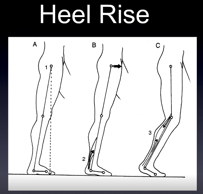
Terminal stance is one of the last stages of stance phase. Following midstance, where maximal pronation should be occurring, the stance phase foot should now begin supinating, initiated by the the opposite foot in swing phase moving forward of the center of gravity. 

Lets look at what is happening here at the major anatomical areas:

Foot

  • Supination begins from the opposite, swing phase leg (see above)
  •  the calcaneus inverts to neutral
  •  the center of gravity of the foot raises from its lowest point at midstance
  • The lower leg should begin externally rotating (as it follows the talus)
  • The thigh should follow the lower leg and should also be externally rotating; sometimes to a greater extent due to the shape and size of the medial condyle of the femur (which is larger than the lateral)
  • these actions are perpetuated by the gluteus maximus and posterior fibers of the gluteus medius, as well as posterior compartment of the lower leg including the flexor digitorum longus, flexor hallucis longus, peroneus longus and tibialis posterior
Ankle
  • The ankle should be 5 degrees dorsiflexed and in ankle rocker
  • the calcaneocuboid locking mechanism should be engaging to assist the peroneus longus in getting the head of the 1st metatarsal to the ground

Knee

  • near or at full extension. This is perpetuated by the quadriceps and biceps femoris, contracting concentrically and attenuated by the semi membranosis and tendonosis. The popliteus contracts eccentrically as soon as the knee passes midstance to keep the rates of external rotation of the tibia and femur in congruence.

Hip

  • The hip should be extending to 10 degrees.

Can you picture what is happening? Try and visualize these motions in your mind. Can you understand why you need to know what is going on at each phase to be able to identify problems? If you don’t know what normal looks like, you will have a tougher time figuring out what is abnormal.

Ivo and Shawn. Gait and foot geeks extraordinaire. Helping you to build a better foundation to put all this stuff you are learning on.

pictured used with permission from Foot Orthoses and Other Conservative Forms of Foot Care

The Windlass Mechanism of the Plantar fascia. What is a Windlass anyway?
After yesterdays post, we thought we might provide more insight to the Windlass mechanism and low and behold; we found AN ENTIRE PAPER on it! Wow, were we thrilled since there …

The Windlass Mechanism of the Plantar fascia. What is a Windlass anyway?

After yesterdays post, we thought we might provide more insight to the Windlass mechanism and low and behold; we found AN ENTIRE PAPER on it! Wow, were we thrilled since there is not a ton of decent stuff out on this topic (yes, we are a little geeky, but then again so are you if you are reading this !).

A Windlass is the tightening of a rope or cable around a pivot point. The plantar fascia acts like a cable between the calcaneus and its distal insertion into the proximal phalanyx at the metatarsal phalangeal joints. When the toes are dorsiflexed (as in forefoot rocker from yesterdays post, see bottom diagram above), the heel and toes SHOULD become approximated, as the plantar fascia shortens from its winding around the metatarsal head, contributing to supination of the foot.

To be accurate, this concept of the Windlass mechanism is quite complex because the dorsiflexion of the great toe also shortens the length of the flexor hallucis brevis.  And we know that the sesamoid bones under the big toe are embedded in its tendon. Their repositioning as the Windlass engages will drive the sesamoids under the metatarsal, elevating it, and shift this joints eccentric axis.  But this is a complex story and post better left for another day.  Simply put, this is a complex joint, do not let anyone fool you otherwise.  Don’t beleive us ? Ask any bunion (or God forbid a fusion) surgery patient how they are doing.  You will get the point then. 

Here’s the link to the article (we know you want to read it ! )

http://www.ncbi.nlm.nih.gov/pmc/articles/PMC385265/

The Windlass Mechanism. Just another fascinating foot fact from The Gait Guys.