Podcast 84: Toe Walkers, Hip Impingment & Olympic Lifting Shoes

Plus: pulmonary edema syndrome in Triathlete swimmers, truths about olympic lifting shoes and more !

Show sponsors:

www.newbalancechicago.com

A. Link to our server: 

http://traffic.libsyn.com/thegaitguys/pod_84f.mp3

Direct Download: 

http://thegaitguys.libsyn.com/podcast-84

Other Gait Guys stuff

B. iTunes link:

https://itunes.apple.com/us/podcast/the-gait-guys-podcast/id559864138

C. Gait Guys online /download store (National Shoe Fit Certification and more !) :

http://store.payloadz.com/results/results.aspx?m=80204

D. other web based Gait Guys lectures:

Monthly lectures at : www.onlinece.com   type in Dr. Waerlop or Dr. Allen,  ”Biomechanics”

Show notes:

Are Triathletes Really Dying of Heart Attacks?

 
We had some stuff on FB last week about head positioning during running.  Alot of people tried to simplify it.  There is more to it. Here is another perspective.
 
Toe Walking children
 
Olympic lifting shoes ? or Converse Chuck Tailors ?
 
Journal of Foot and Ankle Research | Abstract | The associations of leg lean mass with foot pain, posture and function in the Framingham foot study
http://www.jfootankleres.com/content/7/1/46/abstract
 
Hip Impingements
 
Achilles oddity: Heeled shoes may boost load during gait | Lower Extremity Review Magazine
http://lermagazine.com/news/in-the-moment-rehabilitation/achilles-oddity-heeled-shoes-may-boost-load-during-gait
 
Toe Walking in Children. Do you know what you are dealing with ? Part 2
So you have now ruled out possible Autism-spectrum, possible CMT (Charcot-Marie Tooth Disease), CP (Cerebral Palsy), MD (Muscular Dystrophy) in your young toe walking individual…

Toe Walking in Children. Do you know what you are dealing with ? Part 2

So you have now ruled out possible Autism-spectrum, possible CMT (Charcot-Marie Tooth Disease), CP (Cerebral Palsy), MD (Muscular Dystrophy) in your young toe walking individual.  Now you have been left with the aftermath foggy diagnosis of “Idiopathy Toe Walking”, that doesn’t leave you as a parent or clinician with much to work with or likely to be confident about. Let us try to help make things clearer and give you some other cognitive options to entertain. New research in recent years has brought new light onto the issue and we wanted to use today’s blog post as a platform to share it with you. 

In a previous week’s “Part 1” blog post & video (link) you can see in the gait on the video that nothing appears to be terribly abnormal in the foot structure (from what we can tell), the client is merely remaining in the plantarflexed posture and forefoot weight bearing.  This is highly ineffective gait and can be very fatiguing let alone to mention the sustained loading into the posterior compartment and plantarflexor mechanism (gastrosoleus-achilles) not to mention the sustained forefoot loading response on the foot bones and joints. Remember, the tibialis posterior and long toe flexors are close neighbors with capabilities of plantarflexion moments, so there are possible clinical manifestations there as well not to mention the obvious (especially to long-time Gait Guys readers) deficits that will be found in functional ankle dorsiflexion, ankle rocker and S.E.S. (skill, endurance, strength) of the anterior compartment mechanism (tibialis anterior, long toe extensors, peroneus tertius).  Even if this client were to go into normal heel strike and stance phases right now, they would have lots of work to do to restore the anterior-posterior compartment balance, the 3 foot rockers (heel, ankle and forefoot) abd posterior compartment length to avoid functional pathology not to mention the timely coordination of all these events. 

Idiopathic toe walking is suggested to be as prevalent as 12%. Toe walking is categorized when there is an absence, or at least a limitation, of heel strike during initial walking gait contact phase. We are not referring to, at all, forefoot running principles. Neuromotor maturation comes about via the suppression of the primitive reflexes/windows and appearance of the postural reflexes and responses. Delays or subtractions of these windows/reflexes may cause challenges in the normal development and maturation of the central and/or peripheral nervous systems.  With toe walking, the clinical window most studies suggest is to begin investigation after 3 years of age when the primitive motor patterns should have solidified and the gait and postural patterns have begun to layer on top of those primitive reflexes.  Remember though, the primitive patterns are not sequentially fixed, meaning that infants move in and out of these reflexes until they become skilled and permanent.  It is not until they are fixed that the postural patterns, which are volitional, can be gradually built. This should bring some deeper thoughts to your mind right now.  Is toe walking behavior a missed primitive window or a non-volitional postural window? These kids are not doing this by choice, anyone who has worked with these types of cases knows this very well, and we have seen our share. 

In the literature and clinics a plethora of things have been tried and discussed (ie. serial casting, botulinum toxin, surgical tendon lengthening, gait retraining, orthoses/orthotics, night splints, day splints and the like). Keep in mind that only one of the above is addressing a functional change via cognitive and higher brain center demand, “gait retraining”. The others are passive forced attempts.  But is gait training enough ? And how far back into primitive and postural gait pattern training do you have to go? Gait training certainly does something as eluded to by two research papers we posted on our Facebook page in previous weeks. See those references below.

“For both feet, contact time of the heel was increased after the training period, whereas contact time of the forefeet decrease. Also positive changes in the active range of joint motion of the ankle (dorsal extension) were observed in both feet. These positive effects were visible also in the follow–up assessment.” -Pelykh study

Daily intensive gait training may influence the elastic properties of ankle joint muscles and facilitate toe lift and heel strike in children with CP. Intensive gait training may be beneficial in preventing contractures and maintain gait ability in children with CP.” - Willerslev-Olsen study

So what else could be going on here ? Is this neurodevelopmental ? Yes, for sure.  But where did things go awry ?  And how do we fix it ? Remember, the development of primitive and postural reflexes is supposed to occur proximal to distal (ie. from core to hand/foot).

In a recent study in the Journal of Child Neurology,  

“for the first time, motor and sensory challenges presenting in healthy children with an idopathic toe walking gait have been identified.These challenges imply an immaturity or mild impairment at the cerebellum or motor cortex level.”

As the article suggested, the research did not render direct cause(s) for the gait pattern, rather some very viable theories on the topic. They found that only the areas of balance, upper body coordination and bilateral coordination were areas found to be problematic in the toe walkers. These 3 components require the integration of the tactile, vestibular and proprioceptive systems as a team. Diving deeper into how these 3 outputs are linked, there is a required “mix of occulomotor control and cues together with subtle and gross postural adjustments” (3). As Williams et al (3) suggested, “they are skills requiring the coordination of movements in which each side of the body moves simultaneously or in sequence”.  Kind of sounds like some topics on Arm Swing/Leg swing and also on the topic of phasic/antiphasic gait we have discussed over and over again here on TGG and in recent podcasts (82) doesn’t it ?  It was proposed that perhaps idiopathic toe walkers negotiate their sensory challenges by unconsciously engaging toe walking behavior to change or challenge these inputs.  Here were some of the proposed thoughts from the Williams study.

“The tactile receptors of the skin may be stimulated through pressure at the ball of the foot or lessened by a reduction of surface contact by raising the heel off the ground. Proprioceptive input may be changed at the knee, ankle and even toe joints by unconsciously repositioning of the foot posture.  The vestibular input may be increased by the vertical stimulation of the bouncy type gait that results from toe walking.”(3) Williams

It seems clear from the Williams study that these children demonstrate a number of sensory needs that motivate toe walking to alter (increase or decrease) or improve sensory input.  The study also suggests that the toe walking gait is an attempt to modify input on postural stimuli during gait to serve diminished postural and position awareness.

The findings of this study are important.  Our most recent blog posts and podcasts (Nov 2014) have discussed some of the components to build, control and coordinate gait on a higher neurologic level. The Williams article seems to support these discussions, that some pathologic gaits are initiated on a neurologic level as opposed to biomechanical at the foot and ankle level.  This sounds like the work offered by “the functional neurologist”, graduates of the Carrick Institute for Graduate Studies ! (carrickinstitute.com)

Have a great day gait brethren !

Shawn and Ivo, The Gait Guys

References:

1. Eur J Phys Rehabil Med. 2014 Oct 9. [Epub ahead of print]

Treatment outcome of visual feedback training in an adult patient with habitual toe walking.

NeuroRehabilitation. 2014 Oct 15. [Epub ahead of print]

2. Gait training reduces ankle joint stiffness and facilitates heel strike in children with Cerebral Palsy.

3. Is idiopathic toe walking really idiopathic ? The motor skills and sensory processing abilities associated with idiopathic toe walking gait.  J Child Neurol 2014, 29:71 Williams, C. , Curtin, Wakefield and Nielsen
More Tricks for stretching, part 3

We have been talking about ways to enhance stretching, talking about taking avvantage of reciprocal inhibition (please see part 1 here) and autogenic  (or post isometric) inhibition (please see part 2 here). 
Befo…

More Tricks for stretching, part 3

We have been talking about ways to enhance stretching, talking about taking avvantage of reciprocal inhibition (please see part 1 here) and autogenic  (or post isometric) inhibition (please see part 2 here). 

Before we talk about this next one, we need to give you a little background (neurologically speaking). 

Take a look at the picture above and note the posturing of the baby in the 2 positions. These neurological reflexes (or postures) are called symmetrical tonic neck reflexes or responses (STNR’s for short) and were described in animals and men by Magnus and de Kleyn in 1912 (1). This work was later studied and reported by by Arthur Simons in 1916  (2) and later by Francis Walshe in 1923 (3). These were later made popular by Berta and Karl Bobath in the 70’s (who studied Walshes work), whom they are often attributed to (4). 

You next question is “Do these persist into healthy adulthood”? and the answer is a resounding YES (5).

Take a look at the picture above again and note the following: 

  • When the neck is flexed, the fore limbs flex (and the muscles facilitating that, bicep, brachialis, anterior deltoid are contracting) and the hind limbs are extending (relatively), with the glutes maximus, quadriceps, foot dorsiflexors contracting.

  • Note that when the head is extended, the forelimbs are extended and the hind limbs flexed. Think about the muscles involved. Upper extremity tricep, anconeus, posterior deltoid, lower back extensors, hamstrings and foot plantar flexors facilitated.

The reflex is based on the mechanoreceptors in the neck articulations and muscles and are frequently used by us and many others in the rehabilitation field. Generally speaking, looking up facilitates things which make you extend above T12, and flex below T12. Looking down facilitates flexion above T12 and extension below. 

We would encourage you at this point to “assume” these positions and feel the muscles which are active and at rest.

So, how can we take advantage of these while stretching? 

Think about your head position:

  •  If you are standing up and hinging at the hips to stretch your hamstrings (notice we did not say “bent at the waist”; there is a BIG difference in shear forces applied to your lumbar spine) you would probably want your neck bent forward, as this would fire your quads which would in turn ALSO inhibit your hamstrings, in addition to the STNR inhibiting the hamstring. 

  • If you were in a hip flexor stretch position, you would want you head up, looking at the ceiling to take advantage of the reflex. 

We are confident you can think of many more applications of this reflex and trust that you will, as it can apply to both upper and lower extremity stretches. Just remember that this reflex is symmetrical and will affect BOTH sides. Of course, there are reflexes that only effect things unilaterally, but that is the subject of another post. 

The Gait Guys. Helping make you better at what you do for yourself and others and assisting you on using the neurology that God gave you. 

  1. http://www.worldneurologyonline.com/article/arthur-simons-tonic-neck-reflexes-hemiplegic-persons/#sthash.6QS3Eat3.dpuf 
  2. Simons A (1923) Kopfhaltung and Muskeltonus. Ges.Z. Neurol.Psychiatr. 80: 499-549.
  3. Walshe FMR (1923) On certain or postural reflexes in hemiplegia, with special reference to the so-called “associated movements.” Brain 46: 1-37. 
  4. Janet M. Howle . Symmetrical Tonic Neck Reflex in Neuro-developmental Treatment Approach: Theoretical Foundations and Principles of Clinical Practice.   NeuroDevelopmental Treatment, 2002  p 341 ISBN 0972461507, 9780972461504
  5. Bruijn SM1, Massaad F, Maclellan MJ, Van Gestel L, Ivanenko YP, Duysens J. Are effects of the symmetric and asymmetric tonic neck reflexes still visible in healthy adults?Neurosci Lett. 2013 Nov 27;556:89-92. doi: 10.1016/j.neulet.2013.10.028. Epub 2013 Oct 21.

Podcast 83: Gait & Brain Injury, and Compression Wraps Theories

Plus: Rocker Shoes, Knee Replacements, and Strong Ankles

Show sponsors:

www.newbalancechicago.com

www.lemsshoes.com

A. Link to our server: 

http://traffic.libsyn.com/thegaitguys/pod_83ff.mp3

Direct Download: 

http://thegaitguys.libsyn.com/podcast-83

B. iTunes link:

https://itunes.apple.com/us/podcast/the-gait-guys-podcast/id559864138

C. Gait Guys online /download store (National Shoe Fit Certification and more !) :

http://store.payloadz.com/results/results.aspx?m=80204

D. other web based Gait Guys lectures:

www.onlinece.com   type in Dr. Waerlop or Dr. Allen,  ”Biomechanics”

Show notes:

Texting on the Loo

Hepatic encephalopathy: effect of liver failure on brain function.

http://www.nature.com/nrn/journal/v14/n12/fig_tab/nrn3587_F1.html

Invalid video embed.

The Bouncy Gait: Premature heel rise gait. Taking another look.

This is a great video example of a premature heel rise during gait. You should be able to clearly see it on the left foot (and this was toned down after we brought it to his awareness!).  The heel rise occurs early in the stance phase of gait, instead of the late stance phase.

We have talked about this bouncy type vertically oriented gait many times in blog posts and in our podcasts.  This is a pretty prevalent problem in the world, mostly because so many people have impaired ankle rocker/dorsiflexion from weak anterior compartments and short/tight posterior compartments.  None the less, for the majority, this is a pathologic gait pattern and it will impart undue stress into the posterior mechanism (calf-achilles complex). Just think about it, this person is going vertical at or prior to the tibia achieving 90degrees (perpendicular to the ground) instead of continuing to progress the tibia to 110+ degrees to enable normal timely pronation and foot biomechanical events.  This is not a normal gait. Period. This will change the function of the entire posterior chain upward. 

If you want to see another great example  from the frontal plane, check out this cute video representation of a vertial/premature heel rise bouncy gait. 

This gait style is caused by a premature heel rise from joint range limitation and/or from premature engagement of the gastrosoleus (and sometimes even the long toe flexors, you will see them hammering and curled in many folks). It can be a learned habitual pattern and nothing more, we have even seen it even in child-parental gait modeling in our offices. These people will never get to NORMAL full late-midstance of gait (without biomechanical compromise) and thus never achieve full hip extension nor adequate ankle dorsiflexion / ankle rocker. The gait cycle is an orchestrated symphony of timely events and when one or several timely events are omitted or impaired the mechanics are passed into other areas for compensation. This vertical gait style is very inefficient in that the gluteals cannot adequately power into hip extension into a forward progression drive, because the calf is prematurely generating vertical movement through ankle plantarflexion.  This strategy is sometimes deployed because the person actually is significantly ankle dorsiflexion (ankle rocker) deficient.  Meaning, they hit the limitations of dorisflexion and in order to progress forward they first have to go vertical.  This vertical motion, because they are moving into ankle plantarflexion, re-buys more ankle dorsiflexion range which then can be used if they so choose. Obviously, the remedy is to find the functional deficit, remove it and retrain the pattern.  There are a whole host of other problems that go with this compensation pattern but we wanted our mission to stay focused today.  Remember, this is usually a subconscious motor pattern compensation. Is it like the toe walking issue we talked about last week (post link here) ? It is similar in some ways and can have primitive and postural motor pattern implications. We will follow up the “Idiopathy Toe Walking Gait: Part 2” shortly but we wanted to strategically put this blog post ahead of it, because there are similar characteristics and implications. Trust us, there is a method to our madness :)

Shawn and Ivo

The Gait Guys

More effective stretching, Part 2
Last week we looked at one (of many) methods to make stretching more effective, utilizing a neurological reflex called “reciprocal inhibition” If you missed that one, or need a review, click here. 
Another way to ge…

More effective stretching, Part 2

Last week we looked at one (of many) methods to make stretching more effective, utilizing a neurological reflex called “reciprocal inhibition” If you missed that one, or need a review, click here

Another way to get muscles to the end range of motion is to utilize a technique called “post isometric relaxation”. Notice I did not say to lengthen the muscle; to actually add sarcomeres to a muscle you would need to use a different technique. Click here to read that post.

Contracting a muscle before stretching is believed to take advantage of a post isomteric inhibition (sometimes called autogenic inhibition), where the muscle is temporarily inhibited from contracting for a period immediately following a isometric contraction. This has been popularized by the PNF stretching techniques, such as “contract hold” or “contract relax” . EMG studies do  jot seem to support this and actually show muscle activation remains the same (1, 2) or increased after contraction (3-6). Perhaps it is due to an increased stretch tolerance (7,8). 

The technique was 1st described by Mitchell, Morgan and Pruzzo in 1979 (9). These gents felt it was important to utilize a maximal contraction (using 75-100% of contractile force) to get to have the effect. It was later shown by Feland and Marin (10) that a more minimal, submaximal contraction of 20-60% accomplished the same thing.  Lewit felt that a less forceful contraction offers the same results, and combined respiratory assists (inspiration facilitates contraction, expiration facilitates relaxation) with this technique (11). Interestingly, there are bilateral increases in range of motion with this type of stretching, indicating a cross over effect (12). Regardless of the mechanism, the phenomenon happens and we can take advantage of it. 

This is how you do it: 

  • Bring the muscle to its end range (maximum length) without stretching, taking up the slack. This should be painless, as this will elicit a different neurological reflex that may actually increase muscle tone. 
  • resist with a minimal isometric contraction (20-60%) and hold for 10 seconds.  You can inspire to enhance the effect.
  • relax and exhale slowly. It is important to wait and feel the relaxation. Stretch through the entire period of the relaxation. You should feel a lengthening of the  muscle.
  • repeat this 3-5 times

This technique can also be used with the force of gravity offering isometric resistance. In a hamstring stretch, you could lean forward while maintaining the lumbar lordosis and allowing the weight of the upper body to provide the stretch. 

Wasn’t that easy? Now you have another tool in your toolbox for yourself or your clients.

The Gait Guys. Giving you useful information and explanations in each and every post.

  1. Magnusson SP, Simonsen EB, Aagaard P, Sorensen H, Kjaer M. A mechanism for altered flexibility in human skeletal muscle. J Physiol. Nov 15 1996;497 (Pt 1):291–298
  2. Cornelius WL. Stretch evoked EMG activity by isometric coontraction and submaximal concentric contraction. Athletic Training. 1983;18:106–109
  3. Condon SM, Hutton RS. Soleus muscle electromyographic activity and ankle dorsiflexion range of motion during four stretching procedures. Phys Ther. Jan 1987;67(1):24–30 
  4. Mitchell UH, Myrer JW, Hopkins JT, Hunter I, Feland JB, Hilton SC. Neurophysiological reflex mechanisms’ lack of contribution to the success of PNF stretches. J Sport Rehabil. 2009;18:343–357 
  5. Youdas JW, Haeflinger KM, Kreun MK, Holloway AM, Kramer CM, Hollman JH. The efficacy of two modified proprioceptive neuromuscular facilitation stretching techniques in subjects with reduced hamstring muscle length. Physiother Theory Pract. May 2010;26(4):240–250 
  6. Osternig LR, Robertson R, Troxel R, Hansen P. Muscle activation during proprioceptive neuromuscular facilitation (PNF) stretching techniques. American journal of physical medicine. Oct 1987;66(5):298–307
  7. Mahieu NN, Cools A, De Wilde B, Boon M, Witvrouw E. Effect of proprioceptive neuromuscular facilitation stretching on the plantar flexor muscle-tendon tissue properties. Scandinavian journal of medicine & science in sports. Aug 2009;19(4):553–560 
  8. Mitchell UH, Myrer JW, Hopkins JT, Hunter I, Feland JB, Hilton SC. Acute stretch perception alteration contributes to the success of the PNF “contract-relax” stretch. J Sport Rehabil. May 2007;16(2):85–92
  9. Mitchell F Jr., Moran PS, Pruzzo NA: An Evaluation of Osteopathic Muscle Energy Procedures. Pruzzo, Valley Park, 1979.  
  10. Feland JB, Marin HN. Effect of submaximal contraction intensity in contract-relax proprioceptive neuromuscular facilitation stretching. Br J Sports Med. Aug 2004;38(4):E18.
  11. Lewit K: Postisometric relaxation in combination with other methods of muscular facilitation and inhibition. Man Med, 1986, 2:101-104.
  12. Markos PD. Ipsilateral and contralateral effects of proprioceptive neuromuscular facilitation techniques on hip motion and electromyographic activity. Phys Ther. Nov 1979;59(11):1366–1373

Podcast 82: Phasic vs Antiphasic Gait, Cross Over Gait & more.

Show sponsors:

www.newbalancechicago.com

www.lemsshoes.com

A. Link to our server: 

http://traffic.libsyn.com/thegaitguys/pod_82final.mp3

Direct Download: 

http://thegaitguys.libsyn.com/podcast-82-phasic-vs-antiphasic-gait-cross-over-gait-more

B. iTunes link:

https://itunes.apple.com/us/podcast/the-gait-guys-podcast/id559864138

C. Gait Guys online /download store (National Shoe Fit Certification and more !) :

http://store.payloadz.com/results/results.aspx?m=80204

D. other web based Gait Guys lectures:

www.onlinece.com   type in Dr. Waerlop or Dr. Allen,  ”Biomechanics”


Show notes:

 
Blog posts we reviewed:
 

Muscle Activity Differences in Forefoot and Rearfoot Strikers
http://www.runresearchjunkie.com/muscle-activity-differences-in-forefoot-and-rearfoot-strikers/

www.runnersworld.com/injury-treatment/forward-lean-while-running-might-reduce-knee-pain?cid=social33696857

Weight-Bearing Ankle Dorsiflexion Range of Motion—Can Side-to-Side Symmetry Be Assumed?
http://www.natajournals.com/doi/abs/10.4085/1062-6050-49.3.40

extras for this piece:

and you can use this to substantiate it: http://www.ncbi.nlm.nih.gov/pubmed/23997389

Effect of step width manipulation on tibial stress during running. J Biomech. 2014 Aug 22;47(11):2738-44. doi: 10.1016/j.jbiomech.2014.04.047. Epub 2014 May 21.

Invalid video embed.

This Client went Phasic in their Gait. Do you know what that means ? We do, and so does McGill, Liebenson, Cook and many others.

Long ago on this blog we showed and discussed a video (link) that discussed Stu McGill's research of the human movements of Georges St-Pierre and David Loiseau. The basic tenets of that video were that the hips and shoulders are used for power production and that the spine-core are used for creating stiffness and stability for the ultimate power transmission through the limb.  He made it clear that if power is generated from the spine, it will suffer. 

Here on TGG we have long talked about phasic and antiphasic motions of the arms and shoulder-pelvic blocks during gait and locomotion/sport activity.  Many of our 1000+ blog writings and 80 podcasts have talked about spine pain and how spine pain clients reduce the antiphasic rotational (axial) nature of the shoulder girdle and pelvic girdle. In the video above, we see anything but antiphasic gait, to be clear, this is a classic representation of a phasic gait. This is pathologic gait, the frontal plane sway is exaggerated and necessary because there is no axial antiphasic motion.  There is essentially frozen arm and torso movements. This client has a long standing history of severe spine trauma and pain, their central pattern generators (CPG) had to make this motor pattern choice in an attempt to avoid pain and negotiate force streams across trauma zones. If you are curious and wish to go deeper down this rabbit hole, read the 30+ articles we have produced more specifically on arm swing and locomotor phasics, just click here.

In these types of cases, the client subconsciously makes the subcortial pattern choice (overrides the normal CPG) to rotate them as a solid unit to reduce spine rotation, axial loading and compression.  We could say that quite often spine pain disables the normal arm-leg pendulums via altering the shoulder-torso and hip-pelvis phasics and the CPG that dictates them. Normally, the spine and core must present sufficient amounts of recruited stiffness, yet mobility where necessary, to enable the locomotive power and velocity generated by movements of the shoulders and hips. These are the two main portals of limb movement off of the spine/core.  These principles holds true in gait and sport. For and interesting example, in human gait the psoas is not entirely a hip flexor initiator when it comes to leg swing, it is a huge hip flexion perpetuator. The initial hip flexion in human gait comes from derotating the obliqued pelvis, via abdominal contraction, on a stiff and stable spine.  Once the pelvis rotation is initiated, the femur can further pendulum forward (via contraction of the psoas and other muscles) on the forward accelerated pelvis in the hip joint proper creating an energy efficient movement (the towel flick/whip effect). This premise holds true in gait, running, kicking etc.  This is a solid principle of effective and efficient human locomotion. This principle also holds true for a punch or throwing an object, the stable torso/spine provides a stable anchor upon which to accelerate the arm in order to create a high velocity limb movement with power.  But here is where we get annoyed much of the time.  (Soap box Tangent coming up) How often do you read articles about tight ITBand, tight psoas, tight piriformis and the like ?  As a “diagnosis” these are weak and they are the “go to diagnosis or cause” of the unseasoned clinician, trainer, coach, therapist. If we all are to be really good at our job, we must go beyond what we see in someone’s gait (since it is the compensation) and go beyond the CNS neuroprotective strategy of tightness/shortness when there is weakness or motor pattern failure.  This does not mean that you cannot, or should not, incorporate restoration methods and principles to restore length-tension relationships in your client, it means you have to resolve ALL of the problems, including the aberrant CPG they have set up as a protective default to avoid injury or further injury. 

In the case above, returning the discussion to arm and leg swing, one must understand clearly that faulty arm swing patterns and lack of antiphasic torso and pelvis oscillation is a product of surgery,  trauma and more so, pain. The client is avoiding the antiphasic presentation (hence, he is phasic) for a reason and coaching more arm swing would be just about the dumbest intervention, so don’t be “that guy”. We know this is an altered motor pattern choice, not a new fixed set point. We know this because on clinical examination the range is available, we know because we examined for it, it is just not being used.  In an example of this same principle, in this case talking hip ranges of motion, McGill discusses the same in his paper*:

“Despite the large increases in passive hip ROM, there was no evidence of increased hip ROM used during functional movement testing. Similarly, the only significant change in lumbar motion was a reduction in lumbar rotation during the active hip extension maneuver (p < 0.05). These results indicate that changes in passive ROM or core endurance do not automatically transfer to changes in functional movement patterns. This implies that training and rehabilitation programs may benefit from an additional focus on grooving new motor patterns if newfound movement range is to be used.”

Think about that next time you stretch, or are stretched by someone. As we have said before, just because you increase someone’s range of motion, does not mean they will be able to incorporate that range of motion into a movement pattern, or compensation pattern for that matter. It is only ¼ of the equation: Range of Motion,  Skill (or proprioception),  Endurance (or the proportion of slow twitch muscle) and Strength (the proportion of fast twitch muscle). There is our S.E.S. mnemonic again.

In this video case, lack of NORMAL antiphasic spinal motion (torso and pelvis moving opposite one another) is noted. Without the obliqued pelvis the swing and stance phases will be impaired. The psoas may have to become more of a hip flexor initiator, AS WELL AS the perpetuator of limb swing, because there is no pelvic obliquity from the antiphasic principles to drive it from. And so, when you see this fella in your office with bilateral tight psoas/hip flexor complex and tight quadriceps mechanisms with resultant impaired glutes and hip extension, please do not begin lengthening them as your point of initiation.  They are that way because he has gone phasic in his gait.  Change the motor patterns that drive this as best as possible, restore any weaknesses that are contributory to, or initiate, these motor patterns and then, if needed, encourage some progressive new length-tension in these muscle groups as improved motor patterning evolve to allow for it.  You are likely going to have to go back and reteach and restore primitive and postural sensory motor windows in these cases, so be patient, be kind, be wise. Oh, and do not forget that with impaired hip function, there will most likely be impaired ankle rocker,  you are going to need a wide angled lens to see, capture and remedy this lads problems.

On another note, can you imagine what this client’s video gait analysis would show and interpret ? Let alone the diagnostics and recommendations that could come from it?  What about the appearance of their foot pressures across a dynamic foot pressure plate (or God forbid a static one !), surely what is seen at the foot is this client’s problem (not !) And forgive those poor fools who recommend a shoe for this client based off of just those mediums alone.  Without a complete hands-on clinical examination to correlate gait cycle observances, any recommendations for this case will be traumatic on many levels. 

Today’s bottom line……. read, learn, think, stay hungry, be wise.

Shawn and Ivo, The Gait Guys

* Improvements in hip flexibility do not transfer to mobility in functional movement patterns.  Moreside, Janice: McGill, Stuart

link: http://journals.lww.com/nsca-jscr/Fulltext/2013/10000/Improvements_in_Hip_Flexibility_Do_Not_Transfer_to.1.aspx

Making your stretching more effective. 
While I was making linguine and clam sauce for my family, one of my favorite foods that I haven’t had in quite some time( and listening to Dream Theater of course) I was thinking about this post.  Then I remem…

Making your stretching more effective. 

While I was making linguine and clam sauce for my family, one of my favorite foods that I haven’t had in quite some time( and listening to Dream Theater of course) I was thinking about this post.  Then I remembered about voice recognition on my iMac.  Talk about multitasking!

What do you agree that stretching is good or not, you or your client still may decide to do so possibly because of the “feel good” component. Make sure to see this post here on “feel good”  part from a few weeks ago. 

If you do decide to stretch, make sure you take advantage of you or your clients neurology.  There are many ways to do this. One way we will discuss today is taking advantage of what we call myotatic reflex.

The myotatic reflex is a simple reflex arc. The reflex begins at the receptor in the muscle (blue neuron above) : the muscle spindles (nuclear bag or nuclear chain fibers). This sensory (afferent) information then travels up the peripheral nerve to the dorsal horn of the spinal cord where it enters and synapses in the ventral horn on an alpha motor neuron.  The motor neuron (efferent) leaves the ventral horn and travels back down the peripheral nerve to the contractile portion of the myfibrils (muscle fiber) from which the the sensory (afferent) signal came (red neuron above).  This causes the muscle to contract. Think of a simple reflex when somebody taps a reflex hammer on your tendon. This causes the muscle to contract and your limb moves.

Nuclear bag and nuclear chain fibers detect length or stretch in a the muscle whereas Golgi Tendon organs tension. We have discussed this in other posts here.   With this in mind, slow stretch of a muscle causes it to contract more, through the muscle spindle mechanism.

Another reflex that we should be familiar with is called reciprocal inhibition. It states simply that when one muscle (the agonist) contracts it’s antagonist is inhibited (green neuron above).  You can find more on reciprocal inhibition here.

Take advantage of both of these reflexes?   Try this:

  • do a calf stretch like this: put your foot in dorsiflexion, foot resting on the side of the doorframe.
  • Keep your leg straight.
  • Grab the the door frame with your arms and slowly draw your stomach toward the door frame. 
  • Feel the stretch in your calf; this is a slow stretch. Can you feel the increased tension in your calf? You could fatigue this reflex if you stretched long enough. If you did, then the muscle would be difficult to activate. This is one of the reasons stretching seems to inhibit performance. 
  • Now for an added stretch, dorsiflex your toes and try to bring your foot upward.  Did you notice how you can get more stretch your calf and increased length? This is reciprocal inhibition at work!

There you have it, one neurological tool of many to give you increased length.The next time you are statically stretching, take  advantage of these reflexes to make it more effective.

 The Gait Guys. Teaching you more  about anatomy, physiology, and neurology with each and every post. 

image from :www.positivehealth.com

pronation

Here is an abstract you should look at.
Br J Sports Med. 2014 Mar;48(6):440-7. doi: 10.1136/bjsports-2013-092202. Epub 2013 Jun 13.

Foot pronation is not associated with increased injury risk in novice runners wearing a neutral shoe: a 1-year prospective cohort study.

http://www.ncbi.nlm.nih.gov/pubmed/23766439
And then there is this article we came cross at Runner’s World online. Here is the article “Five things i learned about buying running shoes”.
In all fairness we do not think the article was meant to teach or say much, but we do feel like it robbed 2 minutes of our productive life, at least it was entertaining.
So it is our turn now, let us serve you some real meat.  Here are some loose thoughts on why shoe fit and research has limitations in our opinion, mostly commentary on the first article and why you need to takes its commentary with a grain of salt.
The problem lies in the knowledge base. Most researchers just do not seem to know enough about the foot types , osseous torsions, the kinetic chain, and the like, to do an ALL ENCOMPASSING study. Plus, such a study would be an infinite nightmare. This is where a clinician is needed, to draw upon all of the issues at hand, not just some of the issues.  
For example, in this study, they just looked at arch heights and their determination as to whether the foot was pronating to a degree  (foot-posture index and categorized into highly supinated (n=53), supinated (n=369), neutral (n=1292), pronated (n=122) or highly pronated (n=18).)
No where did they talk about foot types such as the very common forefoot variants of varus and valgus let along their compensated and uncompensated forms. No where were there discussions of tibial or femoral torsion or the possibly necessary foot pronation needs to bring the knee joint back to the sagittal plane. Plus, just because a foot is flat, doesn’t truly mean it is over pronated. It may be flat because of genetics, we have talked about genetic trends here in previous blog posts.  We see plenty of flat competent feet in our clinics. The may appear flat or over pronated , but that is not the case for many people. The FUNCTION must be examined, and this does not come from visual inspection or from gait analysis video. We always say “what you see in someones gait or foot function is often their compensation around other issues, it is not their problem”.
Shawn and Ivo, the gait guys

Podcast 81: Gait, critical, pure and essential principles

This week’s show sponsors: 

www.newbalancechicago.com

www.lemsshoes.com

A. Link to our server: 

http://traffic.libsyn.com/thegaitguys/pod_81f.mp3

Direct Download: 

http://thegaitguys.libsyn.com/podcast-81-gait-critical-pure-and-essential-principles

B. iTunes link:

https://itunes.apple.com/us/podcast/the-gait-guys-podcast/id559864138

C. Gait Guys online /download store (National Shoe Fit Certification and more !) :

http://store.payloadz.com/results/results.aspx?m=80204

D. other web based Gait Guys lectures:

www.onlinece.com   type in Dr. Waerlop or Dr. Allen,  ”Biomechanics”

______________

Today’s Show notes:

Show Sponsors:
 

* Gait Guys online /download store (National Shoe Fit Certification and more !) :

http://store.payloadz.com/results/results.aspx?m=80204

* Other web based Gait Guys lectures:

www.onlinece.com   type in Dr. Waerlop or Dr. Allen,  ”Biomechanics”

 
Show Notes and links:
 
Forget Cheetah Blades. This Prosthetic Socket Is a Real Breakthrough
http://www.wired.com/2014/10/forget-cheetah-blades-prosthetic-socket-real-breakthrough
 
Rebuilding and Regenerating Damaged Knees: The Future Has Arrived!
http://www.huffingtonpost.com/nicholas-dinubile-md/rebuilding-and-regenerati_b_6043374.html
 
the foot gym:
 
From a reader:
Thanks for sharing all the great information over the years. I would like to pose to you some simple questions. How do you decide what area/s are relevant to the issue a patient presents? How do you decide what is “normal” given anatomical variations, history of injuries, torsion’s, etc., and if pain is present, why would you address biomechanics, since pain is a neurological phenomenon not a biomechanical phenomenon?
This may not be that simple but would like to hear what you have to say on these topics.
Thank you,
Joe 
 
COMPARISON OF ISOMETRIC ANKLE STRENGTH BETWEEN FEMALES WITH AND WITHOUT PATELLOFEMORAL PAIN SYNDROME
http://www.ncbi.nlm.nih.gov/pmc/articles/PMC4196327/
 
the drawbacks of technology
One way compensations develop
We have all had injuries; some acute some chronic. Often times injuries result in damage to the joint or articulation;  when the ligament surrounding a joint becomes injured we call this a “sprain”. 
Joints are blessed …

One way compensations develop

We have all had injuries; some acute some chronic. Often times injuries result in damage to the joint or articulation;  when the ligament surrounding a joint becomes injured we call this a “sprain”. 

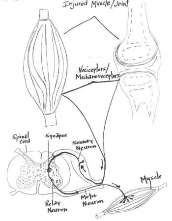
Joints are blessed with four types of mechanoreceptors.  We have covered this in many other posts (see here and here).  These mechanoreceptors apprise the central nervous system of the position (proprioception or kinesthesis) of that body part or joint via the dorsal column system or spinocerebellar tracts. Damage to these receptors can result in a mismatch or inaccuracy of information to the central nervous system (CNS). This can often result in further injury or a new compensation pattern. 

Joints have another protective mechanism called arthrogenic inhibition (see diagram above). This protective reflex turns off the muscles which cross the joint. This was described in a few great paper by Iles and Stokes in the late 80’s an early 90’s (vide infra). Not only are the muscles inhibited, but it can also lead to muscle wasting; there does not need to be pain and a small joint effusion can cause the reflex to occur. 

If the muscles are inhibited and cannot provide appropriate afferent (sensory) and efferent (motor) information to the CNS, your brain makes other arrangements to have the movement occur, often recruiting muscles that may not be the best choice for the job. We call this a “compensation” or “compensation pattern”. An example would be that if the glute max is inhibited (a 2 joint muscle, with a larger attachment to the IT band and a smaller to the gluteal tuberosity; it is a hip extender, external rotator and adductor of the thigh), you may use your lumbar erectors (multi joint muscles; extensors and lateral rotators of the lumbar spine) or hamstrings (2 joint muscles; hip extenders, knee flexors, internal and external rotators of the thigh)  to extend the hip on that side, resulting in aberrant mechanics often observable in gait, which may manifest itself as a shortened step length, increased vertical displacement of the pelvis, lateral shift of the pelvis or increase in step height, just to name a few. Keep this up for a while and the new “pattern” becomes ingrained in the CNS and that becomes your new default for that motion.

Now to fix the problem, you not only need to reactivate the muscle, but you need to retrain the activity. Alas, the importance of doing a thorough exam and thorough rehab to fix the problem.

Often times, the fix is much more involved than figuring out what the problem is (or was). Take your time and do a good job. Your clients and patients will appreciate it!

Ivo and Shawn, the gait guys

Young A, Stokes M, Iles JF : Effects of joint pathology on muscle. Clin Orthop Relat Res. 1987 Jun;(219):21-7

Iles JF, Stokes M, Young A.: Reflex actions of knee joint afferents during contraction of the human quadriceps. Clin Physiol. 1990 Sep;10(5):489-500.

image from: http://chiroeco.com/chiro-blog/results-to-referrals/2013/04/03/neurology-based-simplified-musculoskeletal-assessment/