The “ banana foot”

IMG_7018.jpg

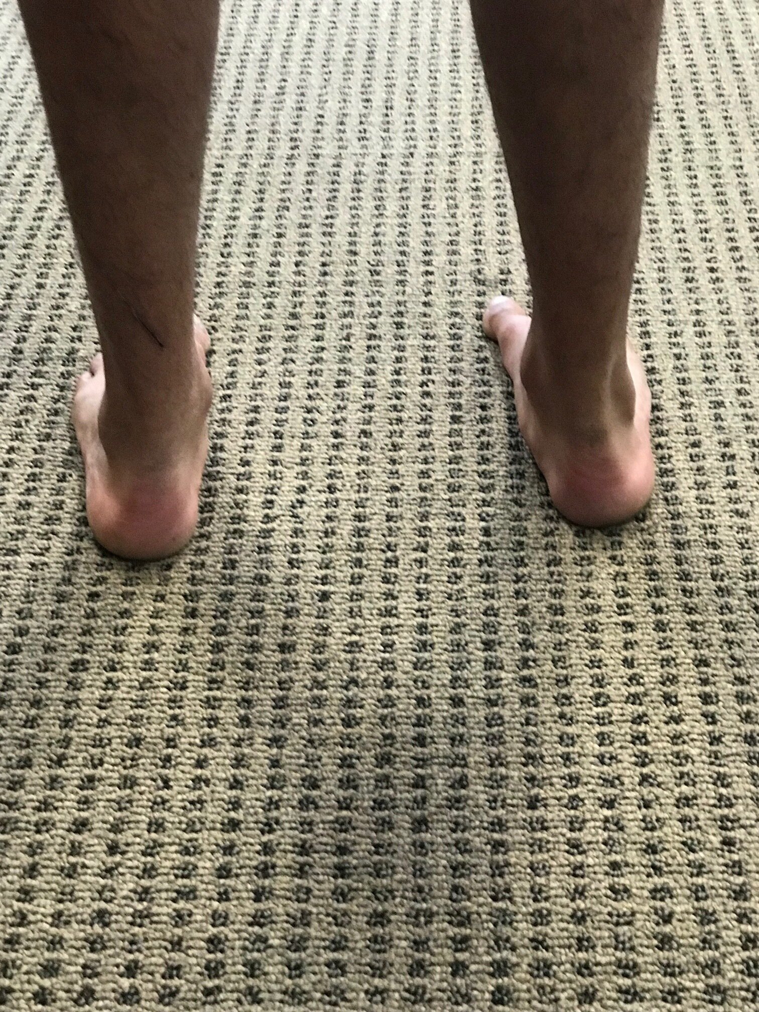
So, you see at foot that looks like this and what do you think? What are some of the biomechanical characteristics of people with the foot that when, you bisect the calcaneus, the line passing forward passes lateral to the second metatarsal or a line between the second and third?

This condition can be congenital, in conditions like forefoot adductus or compensatory.

The first thing that springs to mind when we see deformities like this is “things usually occur in threes“. So we would expect to see other anatomical and/or genetic abnormalities. An adducted forefoot, like you see here, often occurs as the result of lack of internal rotation of the hip on that side so therefore will often be present with conditions like internal tibial torsion and femoral retrotorsion, which we often, but not always, see together. Because of the increased gait and foot progression angle in these individuals, the forefoot compensates and adducts to bring the center of gravity more to midline.

Feet like this are often, but not always, cavus and rigid. If it remains in relative supination (plantarflexion, abduction and inversion) it is an excellent level but poor shock absorber.

Forefoot adduction can also be a compensation pattern if an individual is unable to get the head of their first ray completely down to the ground. It could be a true forefoot varus or more commonly, a forefoot supinatus; either results in an inability to get the first ray down. This often causes the foot to adduct in compensation, and, due to the tarsal articulations, often raises the base of the first metatarsal increasing the inclination angle of the first ray. This frequently leads to limited dorsiflexion of the first metatarsophalangeal articulation.

So what is a clinician to do?

Ensure that the mechanics of the foot are clean through manipulation and mobilization

Make sure there are appropriate flexors/extensor ratios of skill, endurance, and strength of the foot musculature both intrinsically and extrinsically. This means making sure that the long flexors and extensors are in some degree of balance.

Work on balance and coordination of the lower extremity. This can be impeded if they’re unable to get ahead of the first right down to the ground. Exercises for the peroneus longus, extensor hallucis brevis and short flexors of the foot will often help with this.

“Banana foot”. Coming to your clinic, or a clinic near you. Maybe today…

Dr. Ivo Waerlop, one of The Gait Guys.

#forefootadductus #bananafoot #supination #thegaitguys

Unilateral calcaneal valgus: what can it mean?

right calcaneal valgus

right calcaneal valgus

Take a good look at this picture and what do you see? Do you see the calcaneal valgus on the right side. What runs through your mind?

Possibilities for causing this condition, as well as the clinical implications are numerous.

The short list should include:

  • A shorter leg on the contralateral side: often times we will pronate more on the longer leg side to compensate for a short leg on the opposite

  • Increased rear foot and/or fore foot pronation on the valgus side. Laxity of the spring ligament or incompetency of the musculature which helps to maintain your arch (tibialis posterior, foot intrinsics, tibialis anterior to name a few) often causes more collapse on the effected side

  • A lack of available rearfoot eversion on the contralateral side. It may be that the increase calcaneovalgus is normal and the opposite side is more rigid.

  • If you were seeing this in the middle of the gait cycle it could be that that is their strategy to get around a loss of hip extension or ankle rocker

  • External tibial torsion on that side. Go ahead, stand up and spin your right foot into external rotation and keep your left foot with a normal progression angle. Can you see how your arch collapses to a greater degree on the side with the external torsion? Remember that pronation is dorsiflexion, eversion and abduction.

  • Internal tibial torsion on the contralateral side. Internal tibial torsion puts the foot into supination which makes it into more of a rigid lever rather than mobile adapter.

    And the list goes on…

    Next time you see a unilateral deformity like this, hopefully some of these things run through your mind and will help you to pinpoint where the problem actually is.

    Dr Ivo Waerlop, one of The Gait Guys

    #calcanealeversion #rearfootvalgus
    #lowerextremitydeformities

3 clues that someone has internal tibial torsion

Watch this video a few times through and see what you notice. There are three clues that this patient has internal tibial torsion, can you find them?

He presented with right sided knee pain, medial aspect of the patella and medial joint line as well as tenderness over the medial joint line and pes anserine. Lower extremity musculature test strong and 5/5 save for his semi tendinosis on the right which tested 4/5.He has diminished endurance bilaterally in the external obliques

1. Note how his knees, right greater than left, fall outside the sagittal plane

2. Note the decreased progression angle of both feet during forward motion

3. Note how he toes off in supination, right greater than left.

This patient’s knee pain is coming from irritation of the pes anserine, particularly semitendinosus and his inability to recruit his abdominals sufficiently so, instead of the usual pattern of recruiting iliopsoas or rectus femoris, he chooses his sartorius, gracious and semi tendinosis.

Pay attention to how the new tracks, the progression angle as well as if they tow off in pronation, neutral, or supination in that can offer subtle clues to internal tibial torsion.

Dr Ivo Waerlop, one of The Gait Guys

#internaltibialtorsion #gaitanalysis #thegaitguys

https://vimeo.com/365342814

Whaddaya Think of these Shoes?

Would you put YOUR patient/client/own feet in them?

Dr Ivo Waerlop, one of The Gait Guys, discusses a common manufacturers defect to look out for, especially in people with rear foot problems. You have to watch out for manufacturers defects in shoes : )

LEARNING OPPORTUNITY THIS WEDNESDAY NIGHT, MAY 15TH

Biomechanics 308
online.com 5 PST, 6 MST, & CST, 8 EST

#gait #thegaitguys #shoeproblem #manufacturersdefect#footproblem

https://vimeo.com/335772235

Is your (or your athletes) cleat neutral or in varus?

Cleats are often the athletes primary interface with the ground and are responsible for transmitting the forces from the core and appendicular muscles down to the ground. The construction of the cleat as well as its characteristics (such as a forefoot varus cant in the forefoot, like this one here) can make all the difference in the world in athletic performance.

Dr Ivo Waerlop, one of The Gait Guys

#gait #thegaitguys #forefoot #varus #valgus #gaitanalysis #cleatproblems #cleatconstruction

Knee braces and long legs?

Knee brace fixed at a zero to 5 degree flexion angle, creating a long leg? 

We know that the knee is supposed to flex during stance phase, usually around 20-25 degrees (depending on speed and weight, increases in bot increases the flexion requirement) to create dampening from vertical oscillation of the pelvis. What happens if they cannot flex? This creates a virtual "long leg" on that side this will usually result in:

  • Increased vertical translation of the pelvis upward on the braced side and

  • A compensation to make up for this "long leg; circumduction in this case,  but it could be any of the other compensations that we have talked about in posts here on the blog. 


work arounds? They are tough as each can create their own set of problems

  • allow more flexion in the knee on the braced side (not always possible)

  • place a full length sole lift on the opposite side to make up for the difference

  • use crutches

  • use a skateboard : )

we are sure you have some as well that you would LOVE to share with us

Dr Ivo Waerlop, one of The Gait Guys

#shortleg #LLD #compensations #legbrace #gaitproblem #thegaitguys


K ShamaeiGS SawickiAM Dollar Estimation of quasi-stiffness and propulsive work of the human ankle in the stance phase of walking - PloS one, 2013 - journals.plos.org

MORAIS FILHO, Mauro César; REIS, Renata Albertin dos  and  KAWAMURA, Cátia Myuki.Evaluation of ankle and knee movement pattern during maturation of normal gait. Acta ortop. bras. [online]. 2010, vol.18, n.1 [cited  2019-04-25], pp.23-25.

Whoa! Dangerous shoes ahead....

IMG_5734.jpg
IMG_5736.jpg
IMG_5735.jpg

Holy smokes ! Can you believe this?

Take a look at these BRAND NEW, just out of the box pair of Brooks Cadence shoes. We do not usually see many manufacturer defects from this brand. Looks like someone might have been asleep at the “upper goes on the midsole” machine

Check out the varus cant to the rearfoot of the right shoe. Now look at the forefoot valgus cant to the left shoe. This would not be a great shoe for someone who has too much rear foot eversion and midfoot pronation on the right and and uncompensated forefoot valgus on the left, but we do not think it was designed for that specific, small niche market.

Think of the biomechanical implications on a "neutral" foot. Placing the right rear foot in varus would effectively halt or slow pronation in the rear foot and midfoot of that foot. This could be a good thing for an over pronator but, in a neutral foot, this would cause them to toe off in supination on that side resulting in low gear push off and biomechanical insufficiency, not to mention the increased external rotation of the lower extremity and lack of shock absorption from 1 of the 4 mechanisms of shock absorption left (mid foot pronation, ankle dorsiflexion, knee flexion, thumb flexion, contralateral drop of the pelvis). Now, imagine if that same person had internal tibial torsion. Talk about placing the knee outside of the sagittal plane ! Can you say macerated meniscus?

And now the left shoe. Look at the valgus cant! If you had and uncompensated forefoot valgus, where the forefoot is everted with respect to the rear foot or a forefoot varus, where they had adequate range of motion to allow the first ray to descend, then this could be a good thing, otherwise they are toeing off in too much pronation. This could be a real problem for a midfoot pronator or someone with large amounts of external tibial torsion, because they commonly toe off in too much pronation and low gear to begin with, as this shoe would accelerate pronation from midfoot to the forefoot

The bottom line? Look at your patients/clients shoes, as well as your own before purchasing them and examined for manufacturer defects. The upper should sit squarely on the midsole and the shoe should not rock or tip from side to side.

TGG

Pain on the outside of one leg, inside of the other. 

Whenever you see this pattern of discomfort, compensation is almost always at play and it is your job to sort it out. 

This patient presents with with right sided discomfort lateral aspect of the right fibula and in the left calf medially. Pain does not interfere with sleep.  He is a side sleeper 6 to 8 hours. His shoulders can become numb; left shoulder bothers him more than right.

PAST HISTORY: L shoulder surgery, rotator cuff with residual adhesive capsulitis. 

GAIT AND CLINICAL EVALUATION: see video. reveals an increased foot progression angle on the right side. Diminished arm swing from the right side. A definite body lean to the right upon weight bearing at midstance on that side.

He has external tibial torsion bi-lat., right greater than left with a right short leg which appears to be at least partially femoral. Bi-lat. femoral retrotorsion is present. Internal rotation approx. 4 to 6 degrees on each side. He has an uncompensated forefoot varus on the right hand side, partially compensated on the left. In standing, he pronates more on the left side through the midfoot. Ankle dorsiflexion is 5 degrees on each side. 

trigger points in the peroneus longus, gastroc (medial) and soles. 

Weak long toe extensors and short toe flexors; weak toe abductors. 

pathomechanics in the talk crural articulation b/l, superior tip/fib articulation on the right, SI joints b/l

WHAT WE THINK:  

1.    This patient has a leg length discrepancy right sided which is affecting his walking mechanics. He supinates this extremity as can be seen on video, especially at terminal stance/pre swing (ie toe off),  in an attempt to lengthen it; as a result, he has peroneal tendonitis on the right (peroneus is a plantar flexor supinator and dorsiflexor/supinator; see post here). The left medial gastroc is tender most likely due to trying to attenuate the midfoot pronation on the left (as it fires in an attempt to invert the calcaneus and create more supination). see here for gastroc info

2.    Left shoulder:  Frozen shoulder/injury may be playing into this as well as it is altering arm swing.

WHAT WE DID INITIALLY (key in mind, there is ALWAYS MORE we can do):    

  •  build intrinsic strength in his foot in attempt to work on getting the first ray down to the ground; EHB, the lift/spread/reach exercises to perform.
  • address the leg length discrepancy with a 3 mm sole lift
  • address pathomechanics with mobilization and manipulation. 
  • improve proprioception: one leg balancing work
  • needled the peroneus longus brevis as well as medial gastroc and soles. 
  • follow up in 1 week to 10 days.

Pretty straight forward, eh? Look for this pattern in your clients and patients

tumblr_o7p469lND91qhko2so1_1280.jpg
tumblr_o7p469lND91qhko2so2_1280.jpg
tumblr_o7p469lND91qhko2so3_1280.jpg
tumblr_o7p469lND91qhko2so4_1280.jpg
tumblr_o7p469lND91qhko2so6_1280.jpg
tumblr_o7p469lND91qhko2so8_1280.jpg
tumblr_o7p469lND91qhko2so9_1280.jpg
tumblr_o7p469lND91qhko2so5_r1_1280.png

Pain on the outside of the leg? Could it be your orthotic? What you wear on your feet amplifies the effect of the orthotic.

This woman presented with right-sided pain on the outside of her leg after hiking approximately an hour. She noticed a prominence of the arch in her right orthotic. She hikes in a rigid Asolo boot ( see below). Remember that footwear amplifies the effect of an orthotic!

In the pictures below you can see the prominent arch. The orthotic has her “over corrected” so that she toes off in varus on that side. The rigid footwear makes the problem worse. The peroneus group is working hard (Especially the peroneus longus)  to try and get the first Ray down to the ground.

The “fix” was to soften the arch of the orthotic and grind some material out. Look at the pictures where the pen is pointing to see how some of the midsole material was taken out. Notice how I ground it somewhat medial to further soften the arch.

She felt better much better after this change and is now a “happy hiker” :-)


Got Arm Swing?

We have written many times about arm swing. Click here for some of our posts here on Tumblr.

Here we are again at the beach. Look at the beautiful difference in arm swing from side to side in the guy carrying the bag. Makes you want to tell him to use a backpack, eh?

Never mind what it does to his gait

  • decreased arm swing on the carrying side
  • increased step length on the left side
  • increased thigh flexion of the left side
  • increased body lean and head tilt to right side (Take a look at this paper)

think about the increased metabolic cost. Think about what this  type of input (increased amplitude of movement unilaterally) is doing to your cortex!

keep your movements symmetrical, folks!

The Gait Guys

tumblr_mvcjetUi171qhko2so1_540.png
tumblr_mvcjetUi171qhko2so2_540.png

Hip Abduction moment?

This was a great question we received, so we thought we would make a post of it, so everyone could benefit.

“@GregLehman: @KineticRev @TheGaitGuys do you guys have a link to your thoughts on how an ER leg allows the quads to create a hip abductor moment? Thanks”

First of all, What IS a hip abduction moment?

In posts, we often refer to a “moment”, meaning almost literally, a few seconds where a certain motion occurs. When are watching someone from behind and see their heel adduct as they get to terminal stance and pre swing (just before they toe off), you are seeing an “adductory moment” of the heel, sometimes referred to as an “adductory twist”.

Now lets think about the hip. Have you ever seen a framing square used by a carpenter? It is an “L” shaped device to make sure things are square (like hanging a door). The hip is kind of like this. It is shaped like an “L” with the neck and head forming the shorter side of the “L” and the femoral shaft forming the longer side. If you imagine the short side of the square attached to the pelvis and now hinging that away from the body, you have abduction of the hip. Normally, this task is tended to (primarily) by the middle fibers of the gluteus medius and posterior fibers of the gluteus minimus, assisted by the quadratus lumborum on the opposite side.

How can the quad be involved?

We remember that the quadriceps has four parts, the vastus lateralis, vastus intermedius and vastis medialis (collectively called “the vasti’) and the rectus femoris.

The rectus femoris proximal attachments are at the anterior inferior iliac spine (this is called the straight or anterior head) and the superior lip of the acetabulum (called the reflected or posterior head) Please see the top of the 2nd picture above, you can see the 2 heads. The distal attachment, after blending with the vasti, is into the patellar tendon and ultimately the tibial tuberosity.

The rectus is an accessory hip flexor and knee extensor, though it not normally a prime mover for either of these motions. It’s amount of action depends on the position of both the knee and hip.  When the knee is flexed, the rectus has less mechanical advantage, because it is placed in a lengthened position; same goes if the hip is extended.  It will be shortened if the hip is flexed and if the knee is extended at the same time, will have a mechanical disadvantage.

Now think about the direction of travel of each of the heads.

The “straight” head actually runs more obliquely from lateral to medial from its proximal attachment (AIIS) to the distal attachment (blending with vasti and patellar tendon); the refelected head runs a similar course, but not as oblique. If you were to externally rotate the thigh (remember, some folks may have an externally rotated foot due to external tibial torsion), it would actually give these heads more mechanical advantage (when the knee is relatively extended, such as at heel strike/ initial contact and toe off/ preswing) as abductors (remember to think from the ground up, closed chain, so the distal attachments are acting more like the origin); thus, the abductor moment we have talked about.


There you have it @Greglehman. Thanks for the great question.


The Gait Guys. Uber Gait Aficionado’s Extraordinaire. Come and learn with us. Watch us on Youtube; follow us on Facebook and Twitter, see many of our downloads on our payloadz site by clicking here.


All material copyright 2013 the Gait guys/ The Homunculus Group. All rights reserved; don’t make us call Lee.

tumblr_mnhlmqYQkw1qhko2so1_1280.jpg
tumblr_mnhlmqYQkw1qhko2so2_1280.jpg

What foot type do we have here?


OK, so this gentlemen comes in with knee pain, L > R and an interesting “jog” in his gait from midstance to toe off (ie, the 2nd half of his gait cycle). 

A few questions for you:

Q: What foot type does he have?

A: Forefoot valgus, L > R. The forefoot is everted with respect to the rear foot. Need to brush up? click here and here for a refresher

Q: What is the next question you should be asking?

A: Is it a rigid deformity (ie the 1st ray is “stuck” in plantar flexion or flexible (ie, the 1st ray can move into dorsiflexion. Hint: look for a callus under the base of the big toe in a rigid deformity

Q: Which is the best type of shoe for this person? Motion control, guidance or neutral?

A: most likely, neutral. A motion control shoe will usually keep the foot in more relative inversion, and that may be a bad thing for this person. Mobility is key, so a flexible shoe would probably be best.

Q: Would a conventional or zero drop shoe be appropriate?

A: A conventional shoe, with a higher ramp delta, will most likely accentuate the deformity (especially if it is a rigid deformity). This is for at least 2 reasons: 1. plantar flexion is part of supination (due to the higher heel; remember plantar flexion, inversion and adduction) and this will make the foot more rigid. 2. The medial side of the foot will be hitting the ground 1st; if the 1st ray is in plantar flexion, this will be accentuated. 


The Gait Guys. Foot Nerds to the max. Convincing you to join forces with us in spreading the word and gait literacy. LIke this post? tell others! Don’t like this post? Tell us!

Need to know more? Take our National Shoe Fit Program and get certified! email us at thegaitguys@gmail.com for details.

tumblr_mavtgyFcvj1qhko2so1_1280.jpg
tumblr_mavtgyFcvj1qhko2so2_1280.jpg

What do you see? A pedograph analysis

You have heard us time and time again talking about pedographs. When our site finally relaunches, we will have a link for you to be able to purchase one if you like. They really are indispensable and are a window to the gait cycle. In a retail setting, they are an excellent sales tool. With practice, they are a valuable asset to your skill set. As you look at more and more of them, they become easier to interpret in light of what you are seeing when you evaluate the individual.

So what do we see here?

Let’s divide the foot into 3 sections: the rear foot, the mid foot and the fore foot.

First of all, are they symmetrical? Look carefully at the fore foot on each side. NO! the right foot looks different than the left, so we are looking at asymmetrical pathology.

Let’s start at the rear foot: Left looks relatively normal; Right shows some artifact from either the sock, pants being stepped on, or perhaps a heel smudge. More pressure at the medial calcaneal facet on the right as well (more ink = more pressure). The shapes are relatively symmetrical, so equal amounts of rear foot pronation (or motion)

Next up, the mid foot. similar shapes, more pressure and printing on the right. Why? Increased weighting, maybe a leg length deficiency.

How about the fore foot? Lots going on here.

Let’s start with the right foot.

The forces should be coming across from right to left (lateral to medial). See the gap in printing between the lesser metatarsal heads and the big toe? Can you see how the printing under the great toe is further back than you would expect? This tells you the force is behind the head of the 1st metatarsal, not on it. This is a cardinal sign of a partially compensated forefoot varus (in other words, the individual can only partially get the head of the 1st metatarsal down to the ground). this printing is due to the soft tissue around the toe being pressed into the ground.

How about those toes? See the dark printing at the most medial aspect of the great toe? this is most likely caused by a callus. See how it spreads laterally? This is the area of the flexor hallucis brevis insertion, and ink here means it is firing. Now look at the increased printing of the 2nd and 4th toes. They are gripping (via flexor digitorum longus) to attempt to stabilize the foot.

How about the left foot? Different than the right. A similiar pattern for mets 2-4 that we saw on the right BUT look at the at 1st metatarsal! WOW, is it printing alot! This means that 1st met head is being driven into the ground pretty hard. It is probably accompanied by pain. This persons 1st metatarsal is making a medial tripod, but perhaps too much so. You usually see this type of printing in someone who has an uncompensated fore foot valgus (forefoot everted with respect to rear foot) or a plantar flexed 1st ray deformity (in other words, the 1st metatarsal is “stuck:” in a downward position).

How about the gripping of the 4 lesser toes? Trying to stabilize that foot, no doubt, as it will be trying to tip to the outside (rather than the inside, like we often see).

What about that big toe? This results from the foot being turned outward and the individual rolling off of the medial aspect of the great toe. It is too far medial and toward the edge of the big to for the flexor hallucis brevis tendon.

Lots of info. Were you able to see most of what we were talking about? Perfect practice makes perfect!

Want to know more? Get a pedograph! Want to find out more about interpretation? We literally wrote the book. Get your copy by clicking here.

The Gait Guys. Spread the feet, spread the word! Increasing the understanding of gait, one post at time.

all material copyright 2012 The Gait Guys/The Homunculus Group. If you want to use our stuff, please ask and give us credit.