Tibial torsion and the effect on progression angle

more tibial torsion = a change in progression angle.

How does tibial torsion impact the development of the foot progression angle?

Stratifying the data by Foot Progression Angle (FPA) revealed there were significant differences in tibial torsion among the groups and provided evidence that tibial torsion influences the direction and magnitude of the FPA. Offsetting torsions between the tibia and femur were more common in people with higher and lower FPA and had clearer patterns where the tibia tended to follow the direction of the FPA.

So, got that? The foot progression angle follows the tibial torsion...

Why do we care?

the greater the “kickstand” angle to the foot, the more we progress through the mid foot (rather than from the lateral aspect of the heel, up the lateral column, across the transverse metatarsal arch and through the 1st ray). This causes more mid foot pronation and more medial knee fall, resulting in gait inefficiency and often times in our experiences, increased knee pain.

Gait Posture. 2016 Sep;49:426-30. doi: 10.1016/j.gaitpost.2016.08.004. Epub 2016 Aug 3.
The rotational profile: A study of lower limb axial torsion, hip rotation, and the foot progression angle in healthy adults.
Hudson D1.

Video Gait Case: internal tibial torsion

Real time VIDEO of internal tibial torsion (at least highly suspect, the guy wouldn't stop for me to examine him :) Knee appears in the sagittal plane, foot, well, not so much.

Need a refresher on tibial torsion ? Here ya go !
https://thegaitguys.tumblr.com/post/43648553025/yes-we-are-all-twisted-part-3-continued-if

Subtle clues. Helping someone around their anatomy

tumblr_n9jt5dtWlc1qhko2so1_1280.jpg
tumblr_n9jt5dtWlc1qhko2so3_1280.jpg

This patient comes in with low back pain of years duration, helped temporarily with manipulation and activity. Her exam is relatively benign, save for increased lumbar discomfort with axial compression in extension and extension combined with lateral bending. Believe it or not, her abdominal and gluteal muscles (yes, all of them) test strong (no, we couldn’t believe it either; she is extremely regular with her exercises). She has bilateral internal tibial torsion (ITT) and bilateral femoral retro torsion (FRT). She has a decreased progression angle of the feet during walking and the knees do not progress past midlilne. There is a loss of active ankle rocker with gait, but not on the exam table; same with hip extension. 

We know she has a sweater on which obscures things a bit, but this is what you have to work with. Look carefully at her posture from the side. The gravitational line should pass from the earlobe, through the shoulder, greater trochanter and through or just anterior to the lateral malleolus.

In the top picture, can you see how her pelvis is anterior to this line? Do you see how it gets worse when she lifts her hands over her head (yes, they are directly over head)? This can signify many things, but often indicates a lack of flexibility in the lumbar lordosis; in this case, she cannot extend her lumbar spine further so she translates her pelvis forward. Most folks should have enough range of motion from a neutral pelvis and enough stability to allow the movement to occur without a significant change. Go ahead, we know you are curious, go watch yourself do this in a mirror and see if YOU change.

Looking at the this picture, can you pick out that she has a genu valgus? Look at the hips and look at the tibial angle.

Did you note the progression angle (or lack of) in her feet? This is a common finding (but NOT pathognomonic) in patients with internal tibial torsion. Notice the forefoot adductus on the right foot?

So what do we think is going on?

  • ITT and FRT both limit the amount of internal rotation of the thigh and lower leg. Remember you NEED 4 degrees of each to walk normally. Most folks have significantly more
  • if you don’t have enough internal rotation of the lower extremity, you will need to “create” it. You can do this by extending the lumbar spine (bottom picture, right) or externally rotating the lower extremity
  • Since her ITT and FRT are bilateral, she flexes the pelvis and nutates the pelvis anteriorly.
  • the lumbar facet joints should only carry 20% of load
  • she is increasing the load and causing facet imbercation resulting in LBP.

What did we do?

  • taught her about neutral pelvic positioning, creating more ROM in the lumbar spine
  • had her consciously alter her progression angle of her foot on strike, to create more available ROM in internal rotation
  • encouraged her to wear neutral shoes
  • worked on helping her to create more ankle rocker and hip extension with active drills and exercise (ie gait rehabilitation); shuffle walks, Texas walk, toes up walking, etc

why didn’t we put her in an orthotic to externally rotate her lower extremity? Because with internal tibial torsion, this would move her knee outside the saggital plane and create a biomechanical conflict at the knee and possibly compromising her meniscus.

Cool case, eh? We thought so. Keep on learning so your brain keeps expanding. If you are not growing your brain, you are shrinking it!

The Gait Guys

Some Basic Tenets of Gait

Initial contact? Loading response? Mid stance? Terminal Stance? pre swing? Are these terms that are familiar to you? Hmmm. How about the wrong orthotic for someone with internal tibial torsion? Join us in this excerpt from a recent Gait and Needling seminar


https://vimeo.com/226733697

Tibial Torsion and Genu Valgum

Join us in this brief video about tibial torsion and genu valgum in a 6-year-old

Mooney JF 3rd Lower extremity rotational and angular issues in children. Pediatr Clin North Am. 2014 Dec;61(6):1175-83. doi: 10.1016/j.pcl.2014.08.006. Epub 2014 Sep 18.

Killam PE. Orthopedic assessment of young children: developmental variations. Nurse Pract. 1989 Jul;14(7):27-30, 32-4, 36.

Kling TF Jr, Hensinger RN. Angular and torsional deformities of the lower limbs in children. Clin Orthop Relat Res. 1983 Jun;(176):136-47.

When the wrong shoe, meets the right foot

Is it any wonder that this gentleman has pain on the dorsum of this his feet?

1st of all, how about his internal tibial torsion? It is bilateral, L > R. This places the majority of his weight on the outside of his feet, keeping him somewhat supinated most of the time.

2nd: he has an anatomical leg length discrepancy on the right which is tibial (see pictures 2 and 3). This will place EVEN MORE weight on the outside of the right foot, as it will often remain in supination in an attempt to "lengthen" itself.

3rd, take a look at his shoes. Is this particular model supposed to be rear foot posted in varus? Talk about adding insult to injury! This will place this guys feet into EVEN MORE supination and EVEN MORE on the outside of his feet. maybe the right shoe is worn into more supination because of his right sided LLD?

And if that wasn't enough, this particular shoe has increased torsional rigidity through the midfoot, slowing or arresting any hope of shock absorption that he may have. 

Yikes! We sure wish more folks knew more about feet and shoes! Maybe they should think about taking the National Shoe Fit Program? Email us for more info.

tumblr_o5bqzkYA0a1qhko2so1_1280.jpg
tumblr_o5bqzkYA0a1qhko2so2_1280.jpg
tumblr_o5bqzkYA0a1qhko2so3_1280.jpg
tumblr_o5bqzkYA0a1qhko2so4_1280.jpg
tumblr_o5bqzkYA0a1qhko2so5_1280.jpg
tumblr_o5bqzkYA0a1qhko2so6_1280.jpg
tumblr_o5bqzkYA0a1qhko2so7_1280.jpg
tumblr_o5bqzkYA0a1qhko2so8_1280.jpg
tumblr_o5bqzkYA0a1qhko2so9_r1_1280.jpg

 Every foot has a story. 

 This is not your typical “in this person has internal tibial torsion, yada yada yada” post.  This post poses a question and the question is “Why does this gentleman have a forefoot adductus?”

The first two pictures show me fully internally rotating the patients left leg. You will note that he does not go past zero degrees and he has femoral retroversion. He also has bilateral internal tibial torsion, which is visible in most of the pictures. The next two pictures show me fully internally rotating his right leg, with limited motion, as well and internal tibial torsion, which is worse on this ® side

 The large middle picture shows him rest. Note the bilateral external rotation of the legs. This is most likely to create some internal rotation, because thatis a position of comfort for him (ie he is creating some “relief” and internal rotation, by externally rotating the lower extremity)

 The next three pictures show his anatomically short left leg. Yes there is a large tibial and small femoral component. 

 The final picture (from above) shows his forefoot adductus. Note that how, if you were to bisect the calcaneus and draw a line coming forward, the toes fall medial to a line that would normally be between the second and third metatarsal’s. This is more evident on the right side.  Note the separation of the big toe from the others, right side greater than left. 

Metatarsus adductus deformity is a forefoot which is adducted in the transverse plane with the apex of the deformity at LisFranc’s (tarso-metatarsal) joint. The fifth metatarsal base will be prominent and the lateral border of the foot convex in shape . The medial foot border is concave with a deep vertical skin crease located at the first metatarso cuneiform joint level. The hallux (great toe) may be widely separated from the second digit and the lesser digits will usually be adducted at their bases. ln some cases the abductor hallucis tendon may be palpably taut just proximal to its insertion into the inferomedial aspect of the proximal phalanx (1)

Gait abnormalities seen with this deformity include a decreased progression angle, in toed gait, excessive supination of the feet with low gear push off from the lesser metatarsals. 

 It is interesting to note that along with forefoot adductus, hip dysplasia and internal tibial torsion are common (2) and this patient has some degree of both. 

 His forefoot adductus is developmental and due to the lack of range of motion and lack of internal rotation of the lower extremities, due to the femoral retrotorsion and internal tibial torsion.  If he didn’t adduct the foot he would have to change weight-bearing over his stance phase extremity to propel himself forward. Try internally rotating your foot and standing on one leg and then externally rotating. See what I mean? With the internal rotation it moves your center of gravity over your hip without nearly as much lateral displacement as would be necessary as with external rotation. Try it again with external rotation of the foot; do you see how you are more likely displace the hip further to that side OR lean to that side rather than shift your hip? So, his adductus is out of necessity.

Interesting case! When you have a person with internal torsion and limited hip internal rotation, with an adducted foot, think of forefoot adductus!


1.  Bleck E: Metatarsus adductus: classification and relationship to outcomes of treatment. J Pediatric Orthop 3:2-9,1983.

2. Jacobs J: Metatarsus varus and hip dysplasia. C/inO rth o p 16:203-212, 1960

Do you have enough in the anterior tank ? Dr. Allen’s quiz question and lesson of the week.One of my favorite sayings to my clients, “Do you have enough anterior strength to achieve and maintain posterior length?”  Translation, do you have enough an…

Do you have enough in the anterior tank ? Dr. Allen’s quiz question and lesson of the week.

One of my favorite sayings to my clients, “Do you have enough anterior strength to achieve and maintain posterior length?”  

Translation, do you have enough anterior lower leg compartment strength (tibialis anterior, long toe extensor muscle group, peroneus tertius) to achieve sufficient ankle dorsiflexion in order to achieve posterior compartment length (gastric, soleus, tibialis posterior, long toe flexor muscle) ?  You see, you can either regularly stretch the calf-achilles complex or you can achieve great anterior compartment strength, to drive sufficient ankle dorsiflexion, in effect EARNING the posterior compartment length. This is a grounded principle in our offices. It is the premise of the Shuffle Walk exercise (link) and many others we implement in restoring someones biomechanics.

Now on to today’s quiz question.

In this photo, both people are just mere moments before heel strike. 

1. Who is gonna need to have more eccentric strength in the anterior compartment ? And what if they don’t have it ? Repercussions ?  

2. Who is toeing off the lateral forefoot ? 

3. Who is crossing over more and thus could have more gluteus medius weakness ?

A picture is worth a thousand words. Answers and dialogue below.

.

.

.

.

.

.

1. The lady in the high heeled shoes. If she heel strikes first, the larger longer heel on her shoe will mean she will need more of a prolonged eccentric loading of the anterior compartment to lower the forefoot to the ground. I hope she shortens her strike so she can get close to mid foot strike, it will negate most of this issue.  Repercussions? Forefoot pain, clenching/hammering of her toes from use of the long flexors to dampen loading of the metatarsal heads, and even possibly anterior shin splint like pain.

2. The lady is clearly in more lateral toe off, this is from the intoe’ing we see. This is low gear toe off. She may have limb torsion, internal tibial torsion to be specific, or insufficent external hip rotation control as a possibility. There are several possibilities here.

3. Hard to say, but the man seems to be crossing over more.

There is also no arm swing, hands are in the pockets, this is a big hit to gait economy. We have discussed these numbers in previous blog posts, the numbers are significant and real.  Step width is also a real factor, reduced step width leads to joint stacking challenges and is found with weaker hip abductors and changes in the iliotibial band length.

A picture can be worth a thousand words. I am a few short of the mark today, but I wanted to keep it short.

Dr. Shawn Allen, one of the gait guys

The Pitfalls of Motion Control Features.

Welcome to Monday, folks. Today Dr Ivo discusses why not all shoes are created equal and why you need to understand and educate your peeps about shoes!

Internal tibial torsion is when the foot is rotated internally with respect to the tibia. When the foot is straight (like when you are walking, because the brain will not let you walk too internally rotated because you will trip and fall), the knee will rotated OUTSIDE the saggital plane (knee points out). Putting a medially posted shoe on that foot rotates the foot EVEN FURTHER laterally. Since the knee is a hinge joint, this can spell disaster for the meniscus.

need to know more? email us or send us a message about our National Shoe Fit Program.

tumblr_o471xcV05d1qhko2so1_1280.jpg
tumblr_o471xcV05d1qhko2so3_1280.jpg
tumblr_o471xcV05d1qhko2so2_1280.jpg
tumblr_o471xcV05d1qhko2so4_1280.jpg

Holy twisted tibias Batman! What is going here in this R sided knee pain patient?

In the 1st picture note this patient is in a neutral posture. Note how far externally rotated her right foot is compared to the left. Note that when you drop a plumbline down from the tibial tuberosity it does not pass-through or between the second and third metatarsals. Also note the incident left short leg
In the next picture both of the patients legs are fully externally rotated. Note the large disparity from right to left. Because of the limited extra rotation of the right hip this patient most likely has femoral retro torsion. This means that the angle of her femoral head is at a greater than 12° angle. We would normally expect approximately 40° of external Rotation. 4 to 6° is requisite for normal gait and supination.

In the next picture the patients knees are fully internally rotated you can see that she has an excessive amount of internal rotation on the right compare to left, confirming her femoral antetorsion.

When this patient puts her feet straight (last picture), her knees point to the inside causing the patello femoral dysfunction right greater than left. No wonder she has right-sided knee pain!

Because of the degree of external tibial torsion (14 to 21° considered normal), activity modification is imperative. A foot leveling orthotic with a modified UCB, also inverting the orthotic is helpful to bring her foot somewhat more to the midline (the orthotic pushes the knee further outside the sagittal plane and the patient internally rotate the need to compensate, thus giving a better alignment).

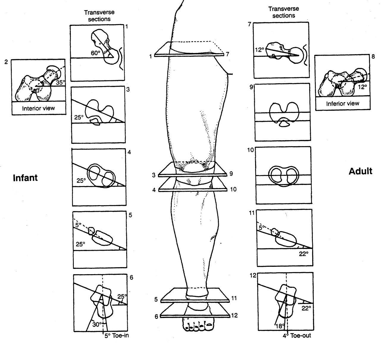
a note on tibial torsion. As the fetus matures, The tibia then rotates externally, and most newborns have an average of 0- 4° of internal tibial torsion. At birth, there should be little to no torsion of the tibia; the proximal and distal portions of the bone have little angular difference (see above: top). Postnatally, the tibia should twist outward (externally) a total of 15 degrees until adult values are reached between ages 8 and 10 years of 23° of external tibial torsion (range, 0° to 40°). more cool stuff on torsions here

Wow, cool stuff, eh?

Got Motion Control? Sometimes too much of a good thing is a bad thing!

Welcome to Monday and News You can Use, Folks.

Today we look at short video showing what someone with internal tibial torsion looks like in a medially posted (ie motion control) running shoe. Note how the amount of internal rotation of the lower leg decreases when the shoe is removed and when he runs. Be careful what shoes you recommend, as a shoe like this is likely to cause damage down the road.

You can follow along listening to Dr Ivo’s commentary. This was filmed at a recent seminar he was teaching.

tumblr_nxo5gvpjDh1qhko2so1_1280.jpg
tumblr_nxo5gvpjDh1qhko2so2_1280.jpg

Now THERE”S some internal tibial torsion!

So, this gent came in to see us with L sided knee pain after it collapsed with an audible “pop” during a baseball game. He has +1/+2 laxity in his ACL on that side. He has subpatellar and joint line pain on full flexion, which is limited slightly to 130 (compared to 145 right)

 We know he has internal torsion because a line drawn from the tibial tuberosity dropped inferiorly does not pass through or near the plane of the 2nd metatarsal (more on tibial torsions here)

What would you do? Here’s what we did:

  • acupuncture to reduce swelling
  • took him out of his motion control shoes (which pitch him further outside the saggital plane)
  • gave him propriosensory exercises (1 leg balance: eyes open/ eyes closed; 1 legged mini squats, BOSU ball standing: eyes open/eyes closed)
  • potty squats in a pain free range
  • ice prn
  • asked him to avoid full flexion

Is it any wonder he injured his knee? Imagine placing the FOOT in the saggital plane, which places the knee FAR outside it; now load the joint an twist, OUCH!

The “Dodgy Foot”, a UK runner’s dilemma.We get “help me” emails from all over the world on a regular basis. Recently we received this photo from a runner in Oxford, UK,  The runner was frustrated, explaining a “dodgy foot”.  We like the word. dodg·y…

The “Dodgy Foot”, a UK runner’s dilemma.

We get “help me” emails from all over the world on a regular basis. Recently we received this photo from a runner in Oxford, UK,  The runner was frustrated, explaining a “dodgy foot”.  We like the word. 

dodg·y    däjē/

-dishonest or unreliable; potentially dangerous; of low quality.

We can guarantee you that the solution here to this runner’s form issue is not wholly at the foot which appears “in toed” and slanted and appears ready to kick the back of the right heel, not to mention the knees that are about to brush together.  Thus, merely working on their foot strike would be so remedial and corrupt that it would a crime. 

Ivo and I do not take on cases via the internet because we cannot give all the information because we cannot examine the client, many do offer such services but people are not being given the whole story and we pledged long ago not to be part of the problem.  Anyone who recommends exercises from things they see on a video gait analysis are basically doing the same disservice in our opinion. But sometimes, as in this case, their inquiry is simple, there is a photo or video and it allows us to highlight an important component of an individuals gait which can lead them on a road to appropriate discovery. This is one of those cases.  I will not be presenting a solution, because I do not have the examination information I need, but I will propose a solid thought process that further investigation may afford progress towards resolution.

This is a non-pathologic cross over gait in my mind until proven otherwise, there may be other sources, causes and components, but when it quacks like a duck you’d be silly not to check for webbed feet. This runner even confirmed upon questioning that the left foot scuffs the inside of the right ankle/shin often, both sides scuff in fact but more left shoe on right shin. No Einsteinian epiphany there. 

  • This means a narrow swing through  (adducting) left limb. 
  • This means stance and swing phase gluteus medius communication problems. 
  • This means swing leg foot targeting problems. 
  • This often suggests right, but sometimes both right and left, frontal plane pelvis sway problems which means pelvis control is challenged which means core lumbar stability control is challenged. 
  • This means adaptive arm swing changes from the clean norm.  
  • This does NOT mean this runner has pain, or pain yet, or maybe never will have pain but there are many determinants of that which I will discuss below. 

But, make no mistake, this is flawed gait mechanics. The left swing leg is clearly targeting a more medial placement, meaning limb adduction (active or passive or both is to be determined) and this is a product of the cross over gait (unfamiliar with the cross over gait ? SEARCH our blog for the term, you will need a few hours of free time to get through it all).  Some would call the cross over gait a lazy gait, but I would rather term it an efficient gait taken too far that it has now become a liability, a liability in which they can no longer stabilize frontal plane sway/drift. A wider gait on the other hand, as in most sprinters, is less efficient but may procure more power and the wider base is more stable affording less frontal plane drift. Just go walk around your home and move from a very narrow line walking gait to a wide gait and you will feel a more powerful engagement of the glutes. Mind you, this is not a fix for cross over gaits, gosh, if it was only that simple !

This runner must investigate whether there is right frontal plane drift, and if it is in fact occurring, find the source of the drift.  It can come from many places on either limb. (This client says they are scuffing both inside ankles, which is not atypical and so we likely have drift on both right and left). We have discussed many of them here in various places on the blog over the years. Now as for “Why” the foot looks in toed, well that can also come from many places. Quite simply the adducted limb once it leaves toe off can look like this. But, perhaps it is also a product of insufficient external rotation maintenance occurred during that left stance phase, affording more internal rotation which is being unchecked and observed here during early swing.  Remember though, if this is in fact a cross over gait result, in this gait the limb approaches the ground unstacked (foot is too far inside a left hip joint plumb line) the foot will greet the ground at a far lateral strike and in supination.  Pronation will thus be magnified and accelerated, if there is enough time before toe off. However, and you can try this on your own by walking around your home, put yourself in terminal stance at toe off. Make sure you have the foot inverted so you are toeing off the lateral toes (low gear toe off). Does this foot not look like the one in the photo ? Yes it does, now just lift the foot off the ground and you have reproduced this photo. And when combined with a right pelvis drift, the foot will sneak further medially appearing postured behind the right foot. 

Keep this in mind as well, final pronation and efficient hallux (big toe) toe off does often not occur in someone who strikes the ground on a far lateral foot. I am sure this runner will now be aware of how poorly they toe off of the big toe, the hallux.  They will tend to progress towards low gear toe off, off the lesser toes. This leaves the foot inverted and this is what you are seeing in her the photo above. That is a foot that is inverted and supinated and it carried through all the way through toe off and into early swing. It is a frequently component of the cross over gait, look for it, you will find it, often. 

Final thoughts, certainly this can be an isolated left swing phase gluteus medius weakness enabling an adducted swing limb thus procuring a faulty medial foot placement, but it is still part of the cross over phenomenon.  Most things when it comes to a linked human frame do not work in isolation.  But i will leave you with a complicating factor and hopefully you will realize that gait analysis truly does require a physical exam, and without it you could be missing the big picture problem.  What if she has a notable fixed anatomic internal tibia torsion on that left side. Yup, it could all be that simple, and that is not something you can fix, you learn to manage that one as a runner.  

* Side bar rant: Look at any google search of runners photos and you will see this type of swing limb foot posturing often, far too often.  And yes, you can take the stance that “I do it as well and i have no injuries or problems so what is the big deal?”.  Our response is often “you do have an issue, it may be anatomic or functional, but you do have an asymmetrical gait and you think it is not a problem, YET”. And maybe you will run till you are 6 feet under and not have a problem because you have accomodated over many years and you are a great compensator, yes, some people get lucky. Some people also do not run enough miles that these issues express themselves clinically so lets be fair. But some of these people are reality deniers and spend their life buying the newest brace or gadget, trying a different shoe insert, orthotic or new shoe of the month and shop over and over again for another video gait analysis expert who can actually fix their pain or problem. And then there are those who have a 45 minute home exercise program that they need to do to keep their problems at bay, managing, not fixing anything.  Or, they spend an hour a week on the web reading article after article on what are the top 4 exercises for iliotibial band syndrome for example. They shop for the newest Graston practitioner, the newest kinesio taping pattern, Voodoo bands, breathing patterns, compression socks etc.  And sometimes they are the ones that say they still dont have a problem.You get the drift.  Gosh darn it, find someone who knows what the hell they are doing and can help you fix the issues that are causing the problem.  And yes, some of the above accoutrements may be assistive in that journey. 

I have dealt with this unique toe off issue way too many times not to roll my eyes at it any longer. It is to the point that it is an automated evaluation and solution program that begins to run in my head. Once you see something enough times, you learn all of the variations and subtle nuiances that a problem can take on. But, trying to fit everyone into a similar solution model is where the novice coach, trainer or clinician will get into trouble. Trust us, it all starts with an examination, a true clinical physical examination.  If one leaves the investigatory process to a series of screens or functional movement patterns, “activation” attempts, digital gait analysis or strength tests one is juggling chainsaws and the outcome you want is often not likely to occur. There is nothing wrong with making these components part of the investigation process, but on their own, they are not enough to get the honest answer many times.  Of course, Ivo and i were not able to jump the pond and examine this runner with our own eyes and hands so today’s dialogue was merely to offer this runner some food for thought to open their mind to our thought process, in the hopes that they can find someone to help them solve the underlying problem and not merely make the gait look cleaner. Making someone’s walking or running gait look cleaner is not hard, but making it subconsciously competent and clean (without thought or effort) requires a fix to the underlying problem. We can ALMOST guarantee you that the solution here to this runner’s form issue is not wholly at the foot that looks in toed and slanted. Merely working on their foot strike would be so remedial and corrupt that it would a crime.

Dr. Shawn Allen, one of the gait guys

tumblr_nx0asr7Q1c1qhko2so1_1280.jpg
tumblr_nx0asr7Q1c1qhko2so2_1280.jpg
tumblr_nx0asr7Q1c1qhko2so3_1280.jpg

Sometimes you need to run that valgus post clear back to the heel!

A valgus post assists in pronation. Some fols have modereate to severe internal tibial torsion and need to be able to pronate more to get the knee into the saggital plane for patello femoral conflicts. They usually run from the tail of the 5th metatarsal forward, but sometimes need to run it clear back to the heel to get enough pronation to occur.

Do you know your stuff? Would you correct this child’s gait ? Give them orthotics, exercises, force correction, leave them alone ? 

Is he Internal Tibial torsioned ? Is he “pigeon toed” ,if that is the only lingo one knows, :(  Does he have femoral torsion ?  A pronation problem locally at the foot or an internal spin problem through the entire limb ? Or a combination of the above ? 

What’s your solution?

It MUST be based on the knowledge necessary to fix it, not the limits of YOUR knowledge. You can never know what to do for this lad from his gait evaluation, no matter how expensive your digital, multi-sensor, 3D multi-angle, heat sensor, joint angle measuring, beer can opening, gait analysis set up is. You can never know what to do for this lad if you do not know normal gait, normal neuro-developmental windows, normal biomechanics, know about torsions (femoral, tibial, talar etc), foot types etc.  It is a long list.  You cannot know what to do for this kid if you do not know how to accurately and logically examine them. 


Rule number 1. First do no harm.

If your knowledge base is not broad enough, then rule number one can be easily broken ! Hell, if you do not know all of the parameters to check off and evaluate, you might not even know you are breaking rule number one !  If everything looks like a weak muscle, every solution will be to “activate” and strengthen and not look to find the source of that weakness.  Muscles do not “shut down” or become inhibited because it is 10 minutes before practice or because it is the 3rd Monday of the month. You are doing your client a huge disservice if you think  you are smarter than their brain and activate muscles that their brain has inhibited for a reason. What if it were to prevent joint loading because of a deeper problem ?  If every foot looks flat and hyper pronated, and all you know is orthotics or surgery or shoe fit, guess what that client is prescribed ? If all you see is torsions, that is all you will look to treat. If all you see is sloppy “running form” and all you know is “proper running form” forcing your client into that “round peg-square hole” can also lead to injury and stacking of compensation patterns.  

One’s lack of awareness and knowledge, are one’s greatest enemies. If you don’t know something exists, because you’ve never studied or learned it, how can you be aware of it ? If you’re not spending enough time examining a client, you might not be aware of an issue even though you may be knowledgeable about the issue.
One must have both awareness and knowledge. One must also be aware that compensations are the way of the body. What you see is not your client’s problem. It is their strategy to cope.

Are you helping your client ? Hurting them ?  Adding risk to their activity ? Are you stepping beyond your skill set ?  

Rule Number 1: First do no harm. 

Shawn and Ivo

PS: we will get to this case another time, we just wanted to make a point today about the bigger problems out in the world.

the gait guys

This is part 2 of a 2 part post; with the video from the case previously discussed

please note the following in the video:

  • body lean to left during left stance phase (to clear right longer leg)
  • circumduction of right lower extremity  (to clear right longer leg)
  • lack of arm swing bilaterally (cortical involvement)
  • patient looking down while walking (decomposition of gait)
  • shortened step length (decomposition of gait)
  • increased tibial varum bilaterally

ASSESSMENT:  This patient’s short leg and internal tibial torsion impediments to her full recovery. She has increased tibial varum noted which is complicating the picture. This is causing pathomechanics and an abductory moment not only at the knee but also in the lumbar, thoracic and cervical spines.

WHAT DID WE DO?:                    

  • We attempted to do the one leg standing exercise. She needed to hold on and did not feel stable on the left hip while performing this.  This is probably more of confidence rather than ability issue. 
  • We gave her the stand/sit exercise to try to improve gluteal recruitment.
  • We also gave her the lift/spread/reach exercise to attempt to strengthen her feet.
  • A full-length 5 mm lift was cut for the left shoe  She felt more stable when walking on this.
  • She was treated with IC, PIR and manipulative therapy and neuromuscular stim of the knee as well as left hip area above, below and at the joint line of the knee as well as gluteus medius and minimus.   
  • We may need to consider building a more aggressive orthotic with a forefoot varus post depending upon her progress and response to care  

 The Gait Guys. Making it real, each and every post here on the blog.

special thanks to SZ for allowing us to publish her case, so others can learn

tumblr_nc9grebaIQ1qhko2so1_1280.jpg
tumblr_nc9grebaIQ1qhko2so2_1280.jpg
tumblr_nc9grebaIQ1qhko2so3_1280.jpg

Wow! What would you do?

This is part 1 of a 2 part post. Look for the other one a few minutes after this one with a video up top for the conclusion

PRESENTING PROBLEM: This 54 YO female patient presents with with left sided knee pain.  She had a total knee replacement (TKR) done in 2011.  She’s had a significant amount of discomfort on the medial aspect of the knee since then. She had an MRI of the hip done thinking the problem was there, and found nothing.   She is walking with a bad limp, left leg is half inch shorter than the right.  Pain is worse at night, changes with weather. 

She has knee pain on the lateral aspect (points to tibial plateau and joint line) with swelling that goes down to the ankle left side.  She has been wearing a “Good Feet” OTC orthotic on the left side which she states helps quite a bit.

Generally speaking, stretching and analgesics make the discomfort better.    Ibuprofen 400 mg. b.i.d. can take the edge off  Soft sided brace (neoprene sleeve) makes a difference as well. The hard sided brace gives her difficulty.

WORK HISTORY: She works for a preschool.  Her job involves standing and getting up and down a lot.  

FAMILY HISTORY:  She has left sided lid ptosis, this evidently is familial.  

PHYSICAL EXAM:  She stood 5’ 1” and weighed approx. 150 pounds.

Viewing the knees bi-lat., the left knee is markedly externally rotated.

She does have a left short leg; tibial and femoral.  She has bilateral tibial torsion (look at the tibial tuberosities and drop a line straight down; it should pass through the 2nd metatarsal head) and marked internal tibial torsion on the left side (>60 degrees) with femoral retrotorsion (less than 8 degree angle of femoral head with the shaft) on this side.  There is no rotation of the thigh or leg past zero degrees midline. .  She had 10 degrees of tibial varum on the left hand side.  Her Q-angle is 10 degrees on that side.  There is plantar flexion inversion of the foot.  Left lower extremity has less sensation secondary to the her TKR  surgery.

Gait evaluation reveals a fair amount of midfoot pronation noted on the left hand side in addition to an intoed gait.  She has to lean her body over to the left to get the right leg to clear.

Some mild weakness noted of hip abduction musculature left hand side gluteus medius, middle and anterior fibers. Knee stability tests were negative.

Neurologically, otherwise, she had full integrity with respect to sensation, motor strength and deep tendon reflexes in the upper and lower extremities.

Please see part 2 of this post for additional info including our assessment and what WE did.

 The Gait Guys. Making it real, each and every post here on the blog.

special thanks to SZ for allowing us to publish her case, so others can learn

tumblr_n9y3v5nRcH1qhko2so1_1280.jpg
tumblr_n9y3v5nRcH1qhko2so2_1280.jpg
tumblr_n9y3v5nRcH1qhko2so3_1280.jpg
tumblr_n9y3v5nRcH1qhko2so4_1280.jpg
tumblr_n9y3v5nRcH1qhko2so5_1280.jpg

More on the “little guy”

We have been following this little guy for some time now. If you have not been keeping up, perhaps you should read herehere and here 1st. 

So, what do we see in these latest pictures?

Top left: neutral view.

  • He enjoys flip flops; probably not the best thing for a developing kiddo, in light of the excessive engagement of the posterior compartment (and reciprocal inhibition of the anterior compartment)
  • he has some tibial varum (ie bowleggedness) L > R
  • he has some developmental genu valgum whnich appears to be improving (need a Q angle review? click here)
  • no tibial torsion present on L: for a review on torsions, click here
  • still some external tibial torsion present on R (see section below on middle shots)

Top right and bottom: full internal rotation of R thigh: compare with bottom: full internal rotation of L thigh

  • he has adequate internal rotation (4 degrees needed) but not as great as left side (see bottom shot); this represents some improvement since we started
  • he has generous internal rotation of the left thigh

Middle Left: full external rotation of right thigh

  • note the position of the knee and the position of the foot; external tibial torsion is present. for a review of torsions, click here.
  • he has limited external rotation of the right thigh (compared with the left. The knee should fall more outside the saggital plane

Middle right: full external rotation of the left thigh

  • note the position of the knee and the position of the foot; internal tibial torsion is present. 
  • he has generous external rotation of the left thigh (compared with the left)


of other significant note: most of his calcaneal valgus has resolved; longitudinal arches are improved.

What now?

  • He continues to develop normally and continues to improve since his original presentation to the office
  • Having the child continue to walk barefoot
  • Continue to wear shoes with little torsional rigidity, to encourage additional additional intrinsic strength to the feet
  • He should continue to limit “W” sitting, as this will tend to increase the genu valgus present
  • We reviewed 1 leg balancing “games” and encouraged continuing agility activities, like balance beam, hopping, skipping and jumping on each leg individually
  • added in using a push and pedal bike
  • added in heel walking exercises

Ivo and Shawn. Bald. Good looking. Extraordinary Gait Geeks. Taking the world of gait literacy by storm with each and every post.

tumblr_n9y3ea9yyg1qhko2so4_r1_1280.jpg
tumblr_n9y3ea9yyg1qhko2so3_1280.png
tumblr_n9y3ea9yyg1qhko2so2_1280.jpg
tumblr_n9y3ea9yyg1qhko2so1_1280.jpg

How do you measure tibial torsion anyway?

With all the talk on the Crossfit blog about the knees out debate, we though we would shed some light on measuring torsions, beginning with tibial torsion, since this does not seem to have been taken account of in the discussion and we feel it is germane. 

Yo may have seen some of our other posts in tibial torsion here or here; this post will serve to help you measure it. 

Looking at the top left picture: we can see that the axis of the tibial plateau and the transmalleolar axis (an imaginary line drawn through the medial and lateral malleolus) are parallel at birth (net angle zero) and progress to 22 degrees at skeletal maturity, resulting from the outward rotation of the tibia of about 1-1.5 degrees per year. This results in a normal external tibial version of about 17-18 degrees (you subtract 5 degrees for the talar neck angle, talked about in the link above). Note that this is the normal or ideal angle we would expect (hope?) to see. Go 2 standard deviations in either direction and we have external and internal tibial torsions.

You can go about taking this measurement in may ways; we will outline 2 of them. 

  1. In the upper left picture, we see an individual who has their knee flexed to 90 degrees over the side of a table while seated. This represents the tibial plateau angle. You the use a protractor to measure the angle between the tibial plateau and an imaginary line drawn through the medial and lateral malleoli. This is the transmalleolar angle. You then subtract 5 degrees from this number (remember the talar neck angle?) to get the angle of tibial version (or torsion).
  2. In the lower left and right pictures, we have the patient supine with the knees pointed upward and tibial plateau flat on the table. Then, working from inferiorly, use a goniometer to measure the angle of the transmalleolar axis. Again, we subtract 5 degrees for the talar neck.

We would encourage you to read up on torsions. This post, which we wrote over a year ago, is probably one of the most important ones on tibial torsions. 

Torsions. Important stuff, especially when you are talking about the axis of the knees in activities like a squat. Remember, the knee is a hinge between 2 multiaxial joints (hip and ankle) and will often take the brunt of the (patho)mechanics, as it has fewer degrees of freedom of movement. If you have external tibial torsion and you push your knees (angle your feet) out further, you are moving the knees outside the saggital plane. You have better have a very competent medial tripod! If you have internal tibial torsion, angling the feet out may be a good idea. Know your (or your patients/clients/athletes) anatomy!

The Gait Guys. Bald, Good Looking and Twisted. Here to help you navigate your way through better biomechanics.