Subtle clues to flexor dominance

IMG_5515.jpg

Take a close look at these photographs. Compare the prominence of the extensor tendon‘s left to right. What do you see? Do you notice the deeper furrowing of the extensor tendons on the left? Do you see the subtle increased extension of the metatarsophalangeal and requisite increased flexion of the inter-phalangeal articulations, left versus right? What about the height of the arches?

Keep a keen eye out for subtle signs. They can make a real difference in your clinical diagnosis and results…

Podcast 138 (for real). Are you fighting your own gait/running neurology?

Topics:
1. Running with the extensors. Convergence and divergence of neurons.
2. Fighting your gait neurology. The lies about the Bird dog rehab exercise.
3. ACL and ACL rehab. Surgery or no sugery. Wise? Risks ? How social media discussions might just be getting it wrong.
4. Cross over gait and lateral heel strike and ensuing problems at great toe off. A failure to medial foot tripod high gear toe off ?
5. Are the hip flexors actually hip flexors in gait ? what are your high knee drills doing? Anything good?

Key words: acl, analysis, cross, extensor, flexors, gait, heel, hip, instability, knee, over, plri, pools, problems, running, strike, surgery

Links to find the podcast:

iTunes page: https://itunes.apple.com/us/podcast/the-gait-guys-podcast/id559864138?mt=2

Direct Download:http://traffic.libsyn.com/thegaitguys/pod_138_real_-_82818_2.12_PM.mp3

Permalink URL:http://thegaitguys.libsyn.com/podcast-138-for-real

Libsyn URL: http://directory.libsyn.com/episode/index/id/6978817

Our Websites:
www.thegaitguys.com

summitchiroandrehab.com

doctorallen.co

shawnallen.net

Our website is all you need to remember. Everything you want, need and wish for is right there on the site.
Interested in our stuff ? Want to buy some of our lectures or our National Shoe Fit program? Click here (thegaitguys.com or thegaitguys.tumblr.com) and you will come to our websites. In the tabs, you will find tabs for STORE, SEMINARS, BOOK etc. We also lecture every 3rd Wednesday of the month on onlineCE.com. We have an extensive catalogued library of our courses there, you can take them any time for a nominal fee (~$20).

Our podcast is on iTunes and just about every other podcast harbor site, just google "the gait guys podcast", you will find us.

Get your foot in High Gear!

image source: Foot Orthoses and Other Conservative Forms of Foot Care. Michaud 1997; Williams an Wilkins: with permission

image source: Foot Orthoses and Other Conservative Forms of Foot Care. Michaud 1997; Williams an Wilkins: with permission

When it comes to gait, getting the 1st ray to the ground is the name of the game. When weight travels through the medial forefoot and we are able to push off the 1st ray complex, that is called "high gear push off". This was 1st discussed F Bojsen-Møller in this excellent paper (1), that just happens to be a free full text! Craig Payne, The Running Research Junkie has offered and excellent commentary on the topic here as well.

High Gear Push Off can happen when 3 conditions are met: 

  1. we have a intact visual, vestibular and kinesthetic systems that ensure we can remain upright in the gravitational plane.
  2. we have an intact calcanocuboid locking mechanism
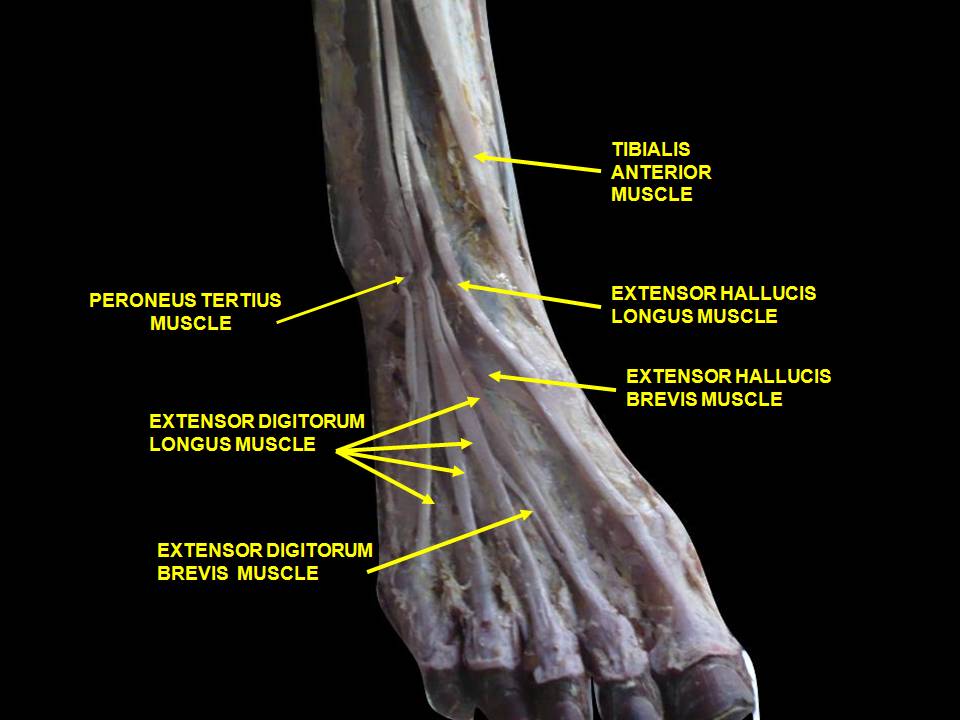
  3. we have adequate skill, endurance and strength of our extensor hallucis brevis

The 1st condition is more global and ensures that our cerebellum and vestibular apparatus are playing nice together to create balance, of the literal sort, We seek to keep our visual axes parallel and our center of gravity over our foot. Remember that the BODY will move itself AROUND the visual axes. If the axes are off, the brain will tilt the head and the body will move AROUND the head to accommodate. We have talked about that in these posts here on the blog. 

The 2nd condition, the calcaneo-cuboid locking mechanism, works in the coronal plane and relies on a functioning peroneal group, where the peroneus longus and brevis wrap around the lateral malleolus, cuboid and tail of the 5th metatarsal, crossing the foot diagonally to insert on the base of the 1st metatarsal. When working properly, its actions will be to plantar flex and everting the forefoot, lowering the 1st ray complex down and assisting the shift of the center of gravity more medial for the weight to pass through the medial foot and out through the hallux (ideally). 

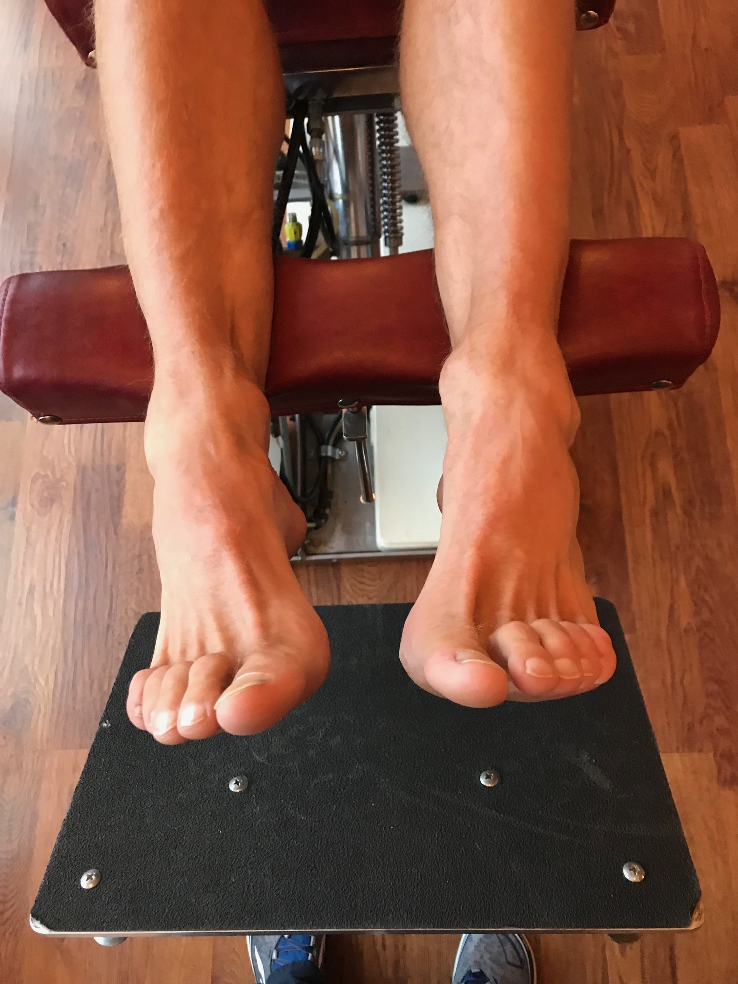
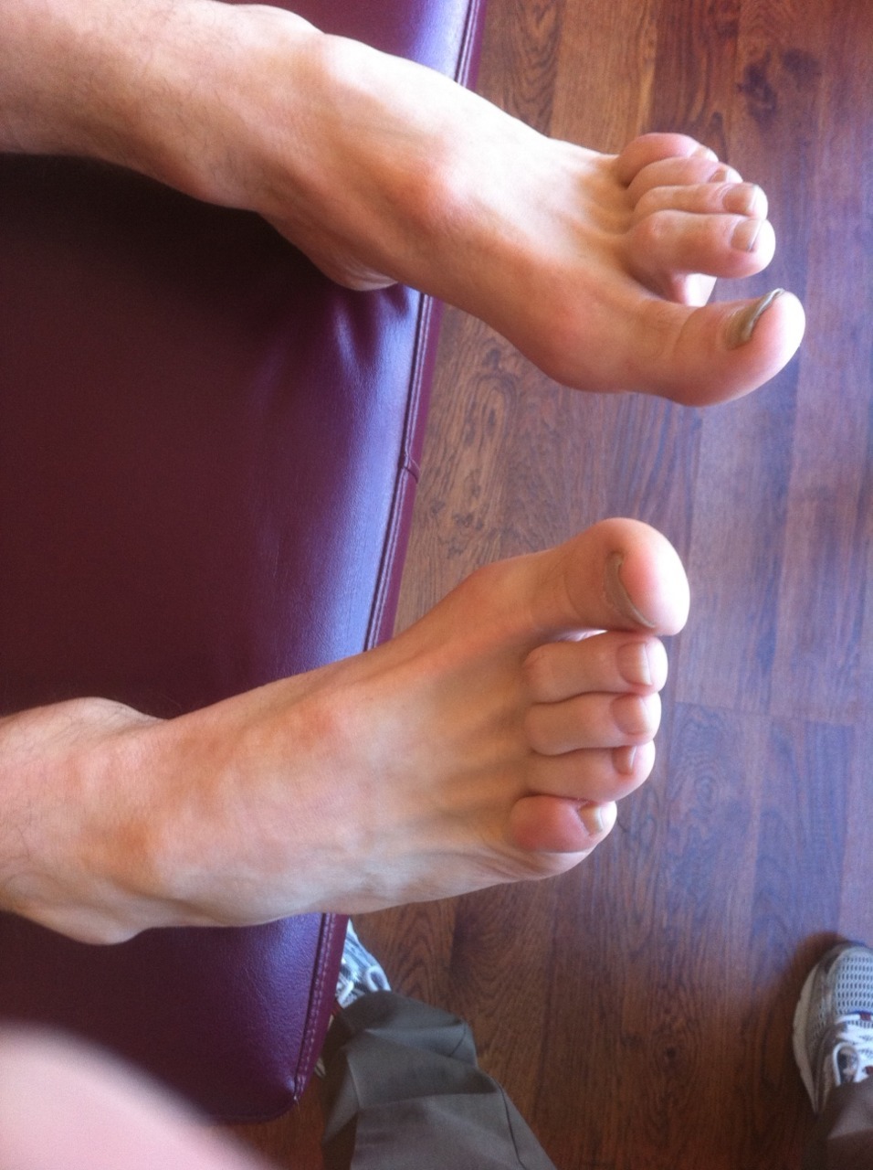
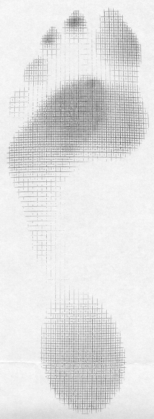
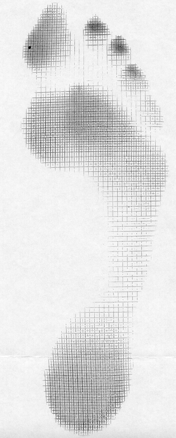
The 3rd condition, the ability to descend the 1st ray, relies on the actions of the peroneus, appropriate supination of the forefoot and ability of the extensor hallucis brevis to do its job.  Because the tendon travels behind the axis of rotation of the 1st metatarsal phalangeal joint, in addition to providing extension of the proximal phalynx of the hallux, it can also provide a downward moment on the distal 1st metatarsal (when properly coupled to and temporally sequenced with the flexor hallicus brevis and longus). If the axis of motion for the 1st metatarsal phalangeal joint moves posteriorly, to behind (rather than under) the joint, the plantar pressures increase at MTP’s 4-5 and decrease at the medial mid foot, moving you into low gear push off.  If moved even further posteriorly, the plantar pressures, and contact time in the mid foot and hind foot (2). For more on the extensor hallucis brevis, see our post here.

As you can see, high gear is desirable over low gear push off, but sometimes circumstances or biomechanics do not permit. High gear push off ensures the forefoot is dorsiflexed and everted with respect to the rearfoot and the calcaneocuboid and talonavicular joint axes are perpendicular to one another, giving us a rigid lever to push off of as the center of gravity moves medially across the foot. In low gear push off, the foot is inverted and plantarflexed and the stress falls on the lesser metatarsals and lateral stabilizing complex of the ankle, moving the center of gravity laterally, in addition to the calcaneocuboid and subtalar joint axes being more parallel,  creating a less rigid lever for push off and decreased mechanical efficiency.


1. Calcaneocuboid joint and stability of the longitudinal arch of the foot at high and low gear push off. J Anat. 1979 Aug; 129(Pt 1): 165–176.  link to free full text: https://www.ncbi.nlm.nih.gov/pmc/articles/PMC1233091/

2. van der Zwaard BC1, Vanwanseele B, Holtkamp F, van der Horst HE, Elders PJ, Menz HB Variation in the location of the shoe sole flexion point influences plantar loading patterns during gait. J Foot Ankle Res. 2014 Mar 19;7(1):20.


What did you notice? The Devil is in the details...

 Cavus foot? Loss of the transverse arch? Prominence of extensor tendons?

The question is: Why?

It’s about reciprocal inhibition. The concept, though observed in the 19th century, was not fully understood and accepted until it earned a Nobel prize for its creditor, Sir Charles Sherrington, in 1932. Simply put, when a muscle contracts, its antagonist is neurologically inhibited, So when your bicep contracts, your tricep is inhibited. This holds true whether you actively contract the muscle or if the muscle is irritated (causing contraction).

So how does this apply to this foot?

We see prominence of the extensor tendons (particularly the extensor digitorum brevis EDB; the longus would have caused extension at the distal interphalangeal joint). The belly of the muscle is visible, telling us that it is active. It is neurologically linked to the flexor digitorum brevis (FDB). This muscle, in turn, has slips which attach it to the abductor hallucis brevis (AHB) medially and the abductor digiti minimi (ADM) laterally. These muscles together form 2 triangles (to be discussed in another post) on the bottom of the foot, which lend to the stability of the foot and the arches, especially the transverse.

When the EDB fires, it inhibits the FDB, (which, in addition to flexing the MTP’s, assists in maintaining the arch). The EDB has an effect which drops the distal heads of the metatarsals as well (Hmm, think about all the people with met head pain) Now, look at the course of the tendons of the EDB. In a cavus foot, there is also a mild abductory moment, which flattens the arch. Conversely, the FDB in a cavus foot would serve to actually increase the arch, and would have a ,mild adductory moment. Net result? A flattened transverse arch.

Now look at the Flexor digitorum longus, overactive in tbis foot (as evidenced by the flexion of the distal interphalangeal joints, mild adduction of the toes (due to the change of direction of pull in a cavus foot) and lowering of the met heads due to hyperextesnion at the MTP joints ). This mm is reciprocally linked with the extensor digitorum longus. The prominence of the extensor tendons is do to increased activity of the EDB (go ahead, extend all your fingers and look at the tendons in your hand. Now flex the  DIP and IP joints and extend the MTP; see how they become more prominent?).

Reciprocal inhibition. It’s not just for dinner anymore…

We are and remain; The Gait Guys

The Beef on the EDL.....

We have long been promoting appropriate function of the long extensors of the toes  here, in our practices, our lectures, on Youtube, in our book......You get the idea. Lets take a closer look at this often weakened and overlooked muscle.

We remember that the EDL lies mostly in the superior and somewhat lateral part of the anterior compartment of the lower leg, comprising approximately the upper 2/3 from under the lateral tibial plateau and fibula, and from the interosseus membrane. It lies under the tibialis anterior, and the extensor hallucis longus lies below it. Its tendons pass inferiorly and travel under the extensor retinaculum and attaches to the base of the distal phalanges of toes 2-4. These muscles act from initial contact to loading response to help eccentrically lower the foot to the ground and ensure smooth heel rocker and most likely attenuate the speed of initial pronation as the talus glides anteriorly on the calcaneal facets and again from terminal stance through initial swing to provide compression of the metatarsal phalangeal and interphalangeal joints, to offset the long flexors (which are often overactive) and create clearance for the toes during swing.  

jhowardL.gif
jhowardR.gif

What does it look like when the long extensors don’t work so well? Have a look at the pedograph on the right (pair J howard r). what do we see? First we notice the lack of printing under the head of the 1st metatarsal and increased printing of the second metatrsal head. Looks like this individual has a forefoot supinatus, or possibly a forefoot varus (cannot get the head of the 1st metatarsal to the ground, and thus a weak medial tripod, possibly insufficient extensor hallucis brevis, peroneus longus, flexor digitorum brevis, or all of the above). Next we see increased printing of the distal phalanges of digits 2-4. Looks like the long flexors are dominant, which means the long extensors are inhibited. What about the lack of printing of the 5th toe? I thought the flexors were overactive? They are, but due to the supinatus, the foot is tipped to the inside and the 5th barely contacts the ground!

How do you fix this?

  • Help make a better foot tripod using the toe wave, tripod standing and extensor hallucis brevis exercises.
  • Make sure the articulations are mobile with joint mobilization, manipulation and massage.
  • How about dry needling and acupuncture to improve function?
  • Make sure the knee and hip are functioning appropriately.
  • Put them in footwear that will allow the foot to function better (a less rigid, less ramp delta shoe).
  • As a last resort, if they cannot make an adequate tripod because of lack of motivation, anatomical constraints or both, use a foot leveling orthotic.


The EHB....In all its glory...

The extensor hallucis brevis : An overlooked "miracle worker"

tumblr_n3vbw7hW5t1qhko2so2_400.jpg

The Extensor Hallicus Brevis, or EHB as we fondly call it is an important muscle for descending the distal aspect of the 1st ray complex (1st metatarsal and medial cunieform) as well as extending the 1st metatarsophalangeal joint. It is in part responsible for affixing the medial tripod of the foot to the ground.  Its motion is generally triplanar, with the position being 45 degrees from the saggital (midline) plane and 45 degrees from the frontal (coronal) plane, angled medially, which places it almost parallel with the transverse plane. With pronation, it is believed to favor adduction (1).

It arises from the anterior calcaneus and inserts on the dorsal aspect of the proximal phalynx. It is that quarter dollar sized fleshy protruding, mass on the lateral aspect of the dorsal foot.  The EHB is the upper part of that mass. It is innervated by the lateral portion of one of the terminal branches of the deep peronel nerve (S1, S2), which happens to be the same as the extensor digitorum brevis (EDB), which is why some sources believe it is actually the medial part of that muscle. It appears to fire from loading response to nearly toe off, just like the EDB; another reason it may phylogenetically represent an extension of the same muscle (2-4).

Because the tendon travels behind the axis of rotation of the 1st metatarsal phalangeal joint, in addition to providing extension of the proximal phalynx of the hallux (as seen in the child above), it can also provide a downward moment on the distal 1st metatarsal (when properly coupled to and temporally sequenced with the flexor hallicus brevis and longus), assisting in formation of the foot tripod we have all come to love (the head of the 1st met, the head of the 5th met and the calcaneus).

Why is this so important?

The central axis of a joint (sometimes called the instantaneous axis of motion) is the center of movement of that articulation. It is the location where the motion will occur around, much like the center of a wheel, where the axle attaches. In an articulation, it usually involves one bone moving around another. Lets look at an example with a door hinge.

A hinge is similar to a joint, in that it has parts with is joining together (the door and the jamb), with a “joint” in between, The axis of rotation of the hinge is at the pivot rod. When the door, hinge and jamb are all aligned, it functions smoothly. Now imagine that the hinge was attached to the jamb 1/4” off center. What would happen? The hinge would bind and the door would not operate smoothly.

Now let’s think about the 1st metatarsal phalangeal joint. It exists between the head of the 1st metatarsal and the proximal part of the proximal part of the proximal phalanyx. Normally, because the head of the 1st metatarsal is larger than the heads of the lesser ones, the center of the joint is higher (actually,almost 2X as high; 8mm as opposed to 15mm). We also remember that the 1st metatarsal is usually shorter then the 2nd, meaning during a gait cycle, it bears the brunt of the weight and hits the ground earlier than the head of the 2nd.

tumblr_lij2n4n1mK1qggnse.jpg

The head of the 1st metatarsal should slide (or should we say glide?) posteriorly on the sesamoids during dorsiflexion of the hallux at pre swing (toe off). It is able to do this because of the descent of the head of the 1st metatarsal, which causes a dorsal posterior shift of the axis of rotation of the joint. We remember that the head of the 1st descends through the conjoined efforts of supination and the coordinated efforts of the peroneus longus, extensor hallucis brevis, extensor hallucis longus, dorsal and plantar interossei and flexor hallucis brevis (which nicely moves the sesamoids and keeps the process going smoothly)(1, 5).

Suffice it to say, if things go awry, the axis does not shift, the sesamoids do not move, and the phalanyx crashes into the 1st metatarsal, causing pain and if it continues, a nice spur you can write home about!

Treating and needling this muscle is easy, as it is very accessible on the dorsum of the foot and due to the decreased receptor density, is not too uncomfortable. We like to needle the peroneus longus and short flexors as well, as they all have the function of lowering the head of the 1st ray. Check it out in this quick how to video.

1. Michaud T: Human Locomotion: The Conservative Management of Gait Related DisordersNewton Biomechanics; First Edition 2011

2. https://www.physio-pedia.com/Extensor_Hallucis_brevis

3. http://www.wheelessonline.com/ortho/extensor_hallucis_brevis

4. Becerro de Bengoa Vallejo R., Losa Iglesias M.E., Jules K.T.  Tendon Insertion at the Base of the Proximal Phalanx of the Hallux: Surgical Implications (2012)  Journal of Foot and Ankle Surgery,  51  (6) , pp. 729-733.

5. Zelik, K.E., La Scaleia, V., Ivanenko, Y.P. et al. Eur J Appl Physiol (2015) 115: 691. https://doi.org/10.1007/s00421-014-3056-x

One way to correct an dysfunctional Extensor Hallucis Brevis

The Extensor Hallicus Brevis, or EHB  (beautifully pictured above causing the  extension (dorsiflexion) of the proximal big to is an important muscle for descending the distal aspect of the 1st ray complex (1st metatarsal and medial cunieform) as well as extending the 1st metatarsophalangeal joint.

Since this muscle is frequently dysfunctional, and is one of THE muscles than can lower the head of the 1st metatarsal, along with the peroneus longus and most likely the tibialis posterior (through its attachment to the 1st or medial cunieform), needling can often assist in normalizing function and works especially well, when coupled with an appropriate rehab program. Here is one way to needle it effectively. 

What’s wrong with this picture? The model is obviously well sculpted and hopefully will paid for the toll that this exercise will be taking on her nervous system overtime. Take a close look at the picture above on the left. Look carefully and …

What’s wrong with this picture?

 The model is obviously well sculpted and hopefully will paid for the toll that this exercise will be taking on her nervous system overtime. Take a close look at the picture above on the left. Look carefully and what do you notice? Do you see it?

This exercise is neurologically incongruent.  Her right arm is flexed at the same time as her right hip. When does this ever happen in gait?

 Do you remember crossed extensor responses or tonic neck reflexes? If not, see here and here. When we walk the right arm and left leg or flexed while the left arm and right leg are extended. During a tonic neck response, and that is rotated to one side the upper and lower extremity (upper greater than lower) should extend on that side with flexion on the contralateral side.

During a tonic neck reflex, the head is rotated to one side the upper and lower extremity (upper greater than lower) should extend on that side with flexion on the contralateral side. In the picture above her torso is rotated to the left while looking straight ahead which is effectively right neck rotation and her extremities are flexed on that side.

 In the picture above her torso is rotated to the left while looking straight ahead which is effectively right neck rotation and her extremities are flexed on that side.

Who thinks of these things? Certainly not folks that are paying attention to appropriate neurology and physiology!  Oh yeah, and the ad was for massage cream. Jeez…

tumblr_n4b8plBI1q1qhko2so1_400.png
tumblr_n4b8plBI1q1qhko2so2_1280.jpg
tumblr_n4b8plBI1q1qhko2so3_1280.jpg
tumblr_n4b8plBI1q1qhko2so4_400.png
tumblr_n4b8plBI1q1qhko2so5_500.jpg

A new twist on an old exercise

Do you know the the “Bird Dog” exercise? It looks like the picture above. The upper and contralateral lower extremities are extended, the the opposite ones are flexed. Seems to make make sense, unless you think about gait and neurology (yes, as you can see, those things seem to always be intertwined).

Think about gait. Your right leg and left arm flex until about midstance, when they start to extend; the left leg and right arm are doing the opposite. At no point are the arm and opposite leg opposing one another. Hmmm.

If you look at it neurologically, it is a crossed extensor reflex (see above); again, flexion of the lower extremity is paired with flexion of the opposite upper extremity. It is very similar to a protective reflex called the “flexor reflex” or “flexor reflex afferent”.

Wouldn’t it make more sense to do a cross crawl pattern? Or maybe like the babies shown above? Seems like if that’s the way the system was programmed, maybe we should try and emulate that. Don’t we want to send the appropriate messages to our nervous system for neurological re patterning? If you are doing the classic “opposite” pattern, what is your reasoning? Can you provide a sound neurological or physiological reason?

Think before you act. Know what you are doing.

The Gait Guys. Bridging the gap between neurology and gait, so you can do a better job.

The Power of Facilitation: How to supercharge your run.

While running intervals this crisp, cool 19 degree morning, something dawned on me. My left knee was hurting from some patellar tracking issues, but only on initial contact and toe off. I gene…

The Power of Facilitation: How to supercharge your run.

While running intervals this crisp, cool 19 degree morning, something dawned on me. My left knee was hurting from some patellar tracking issues, but only on initial contact and toe off. I generally run with a midfoot strike. I began concentrating on my feet, lifted and spread my toes and voila! my knee pain instantly improved. Very cool, and that is why I am writing this today. 

Without getting bogged down in the mire of quad/hamstring facilitation patterns, lets look at what happened.

I contracted the long extensors of the toes: the extensor digitorum longus and the extensor hallicus longus; the short extensors of my toes: the extensor digitorum brevis, the extensor hallucis brevis: as well as the dorsal interossei.the peroneus longus, brevis and tertius were probably involved as well.

Do you note a central theme here? They are all extensors. So what, you say. Hmmm… 

Lets think about this from a neurological perspective:

In the nervous system, we have 2 principles called convergence and divergence. Convergence is when many neurons synapse on one (or a group of fewer) neuron(s). It takes information and “simplifies” it, making information processing easier or more streamlined. Divergence is the opposite, where one(or a few) neurons synapse on a larger group. It takes information and makes it more complicated, or offers it more options.

In the spinal cord, motor neurons are arranged in sections or “pools” as we like to call them in the gray matter of the cord. These pools receive afferent information  and perform segmental processing (all the info coming in at that spinal cord segment) before the information travels up to higher centers (like the cerebellum and cortex). One of these pools fires the extensor muscles and another fires the flexor muscles.. 

If someone in the movie theater keeps kicking the back of our seat, after a while, you will say (or do) something to try and get them to stop. You have reached the threshold of your patience. Neurons also have a threshold for firing.  If they don’t reach threshold, they don’t fire; to them it is black and white. Stimuli applied to the neuron either takes them closer to or farther from threshold.  When a stimulus takes them closer to firing, we say they are “facilitating” the neuron. If it affects a “pool” of neurons, then that neuronal pool is facilitated. If that pool of neurons happens to fire extensor muscles, then that “extensor pool” is facilitated.

When I consciously fired my extensor muscles, two things happened: 1. Through divergence, I sent information from my brain (fewer neurons in the cortico spinal pathway) to the motor neuron pools of my extensor muscles (larger groups of motor neurons) facilitating them and bringing them closer to threshold for firing and 2. When my extensor muscles fired, they sent that information (via muscle spindles, golgi tendon organs, joint mechnoreceptors, etc) back to my cerebellum, brain stem and cortex (convergence) to monitor and modulate the response.

When I fired my extensor muscles, I facilitated ALL the neuronal pools of ALL the extensors of the foot and lower kinetic chain. This was enough to create balance between my flexors and extensors and normalize my knee mechanics.

If you have followed us for any amount of time, you know that it is often “all about the extensors” and this post exemplifies that fact.

 Next time you are running, have a consciousness of your extensors. Think about lifting and spreading our toes, or consciously not clenching them. Attempt to dorsiflex your ankles and engage your glutes. It just may make your knees feel better!

The Gait Guys. Facilitating your neuronal pools with each and every post.

All material copyright 2013 The Gait Guys/ The Homunculus Group. All rights reserved. If you rip off our stuff, we will send Lee after you!