Toes Spacers, anyone?

Less pain through better mechanics?

cc3.jpg

We have been using toe separators for various foot problems like hallux valgus, hammer toes and flexor dominance/extensor weakness. Our reasoning is that through changing the angle of attachment of the muscle, you alter the mechanical advantage of that muscle and help it to work more efficiently. This seems implied in the literature with respect to foot orthoses (1-3) but we could not find any data regarding toe separators. Toe separators DO seem to reduce pain and increase function (4-6). Perhaps this is through better biomechanics, mechanical deformation, proprioceptive changes, or most likely a combination of all these factors and more.  We think clinical results speak volumes. It is nice to see more data coming out on these easy to implement clinical tools. 

What is you clinical reasoning or rationale for using these devices? We would love to hear and if you have an article for reference you could share, that would be great. 


1. Scherer PR, Sanders J, Eldredge DE, Duffy SJ, Lee RY. Effect of functional foot orthoses on first metatarsophalangeal joint dorsiflexion in stance and gait. J Am Podiatr Med Assoc. 2006 Nov-Dec;96(6):474-81.

2. Halstead J, Chapman GJ, Gray JC, Grainger AJ, Brown S, Wilkins RA, Roddy E, Helliwell PS, Keenan AM, Redmond ACFoot orthoses in the treatment of symptomatic midfoot osteoarthritis using clinical and biomechanical outcomes: a randomised feasibility study. Clin Rheumatol. 2016 Apr;35(4):987-96. doi: 10.1007/s10067-015-2946-6. Epub 2015 Apr 28.

3. Bishop C, Arnold JB, May T. Effects of Taping and Orthoses on Foot Biomechanics in Adults with Flat-Arched Feet. Med Sci Sports Exerc. 2016 Apr;48(4):689-96. doi: 10.1249/MSS.0000000000000807.

4. Chadchavalpanichaya N, Prakotmongkol V, Polhan N, Rayothee P, Seng-Iad S. Effectiveness of the custom-mold room temperature vulcanizing silicone toe separator on hallux valgus: A prospective, randomized single-blinded controlled trial. Prosthet Orthot Int. 2017 Mar 1:309364617698518. doi: 10.1177/0309364617698518. [Epub ahead of print]

5. Tehraninasr A, Saeedi H, Forogh B, Bahramizadeh M, Keyhani MR. Effects of insole with toe-separator and night splint on patients with painful hallux valgus: a comparative study. Prosthet Orthot Int. 2008 Mar;32(1):79-83. doi: 10.1080/03093640701669074.

6. Tang SF, Chen CP, Pan JL, Chen JL, Leong CP, Chu NK. The effects of a new foot-toe orthosis in treating painful hallux valgus. Arch Phys Med Rehabil. 2002 Dec;83(12):1792-5. 


The Beef on the EDL.....

We have long been promoting appropriate function of the long extensors of the toes  here, in our practices, our lectures, on Youtube, in our book......You get the idea. Lets take a closer look at this often weakened and overlooked muscle.

We remember that the EDL lies mostly in the superior and somewhat lateral part of the anterior compartment of the lower leg, comprising approximately the upper 2/3 from under the lateral tibial plateau and fibula, and from the interosseus membrane. It lies under the tibialis anterior, and the extensor hallucis longus lies below it. Its tendons pass inferiorly and travel under the extensor retinaculum and attaches to the base of the distal phalanges of toes 2-4. These muscles act from initial contact to loading response to help eccentrically lower the foot to the ground and ensure smooth heel rocker and most likely attenuate the speed of initial pronation as the talus glides anteriorly on the calcaneal facets and again from terminal stance through initial swing to provide compression of the metatarsal phalangeal and interphalangeal joints, to offset the long flexors (which are often overactive) and create clearance for the toes during swing.  

jhowardL.gif
jhowardR.gif

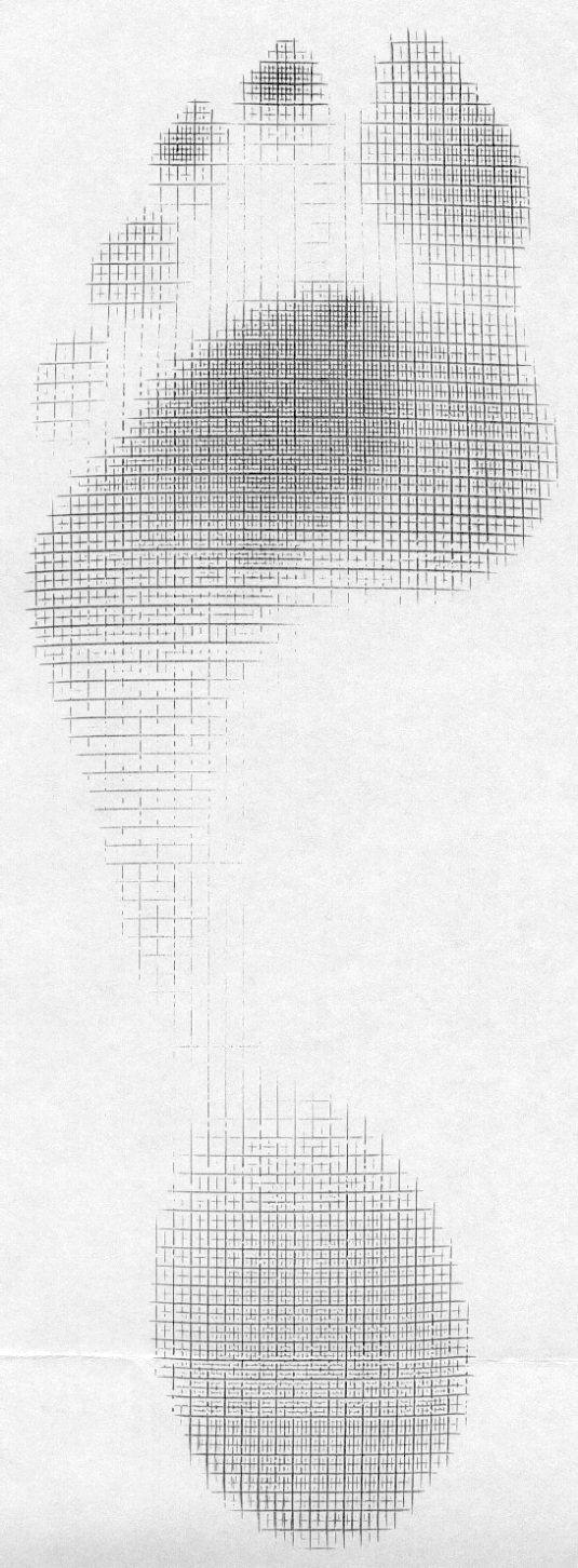
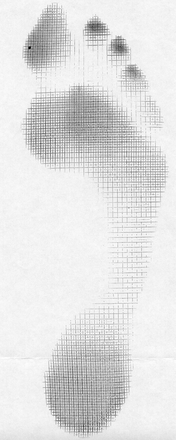
What does it look like when the long extensors don’t work so well? Have a look at the pedograph on the right (pair J howard r). what do we see? First we notice the lack of printing under the head of the 1st metatarsal and increased printing of the second metatrsal head. Looks like this individual has a forefoot supinatus, or possibly a forefoot varus (cannot get the head of the 1st metatarsal to the ground, and thus a weak medial tripod, possibly insufficient extensor hallucis brevis, peroneus longus, flexor digitorum brevis, or all of the above). Next we see increased printing of the distal phalanges of digits 2-4. Looks like the long flexors are dominant, which means the long extensors are inhibited. What about the lack of printing of the 5th toe? I thought the flexors were overactive? They are, but due to the supinatus, the foot is tipped to the inside and the 5th barely contacts the ground!

How do you fix this?

  • Help make a better foot tripod using the toe wave, tripod standing and extensor hallucis brevis exercises.
  • Make sure the articulations are mobile with joint mobilization, manipulation and massage.
  • How about dry needling and acupuncture to improve function?
  • Make sure the knee and hip are functioning appropriately.
  • Put them in footwear that will allow the foot to function better (a less rigid, less ramp delta shoe).
  • As a last resort, if they cannot make an adequate tripod because of lack of motivation, anatomical constraints or both, use a foot leveling orthotic.