Subtle clues to flexor dominance

IMG_5515.jpg

Take a close look at these photographs. Compare the prominence of the extensor tendon‘s left to right. What do you see? Do you notice the deeper furrowing of the extensor tendons on the left? Do you see the subtle increased extension of the metatarsophalangeal and requisite increased flexion of the inter-phalangeal articulations, left versus right? What about the height of the arches?

Keep a keen eye out for subtle signs. They can make a real difference in your clinical diagnosis and results…

What did you notice? The Devil is in the details...

 Cavus foot? Loss of the transverse arch? Prominence of extensor tendons?

The question is: Why?

It’s about reciprocal inhibition. The concept, though observed in the 19th century, was not fully understood and accepted until it earned a Nobel prize for its creditor, Sir Charles Sherrington, in 1932. Simply put, when a muscle contracts, its antagonist is neurologically inhibited, So when your bicep contracts, your tricep is inhibited. This holds true whether you actively contract the muscle or if the muscle is irritated (causing contraction).

So how does this apply to this foot?

We see prominence of the extensor tendons (particularly the extensor digitorum brevis EDB; the longus would have caused extension at the distal interphalangeal joint). The belly of the muscle is visible, telling us that it is active. It is neurologically linked to the flexor digitorum brevis (FDB). This muscle, in turn, has slips which attach it to the abductor hallucis brevis (AHB) medially and the abductor digiti minimi (ADM) laterally. These muscles together form 2 triangles (to be discussed in another post) on the bottom of the foot, which lend to the stability of the foot and the arches, especially the transverse.

When the EDB fires, it inhibits the FDB, (which, in addition to flexing the MTP’s, assists in maintaining the arch). The EDB has an effect which drops the distal heads of the metatarsals as well (Hmm, think about all the people with met head pain) Now, look at the course of the tendons of the EDB. In a cavus foot, there is also a mild abductory moment, which flattens the arch. Conversely, the FDB in a cavus foot would serve to actually increase the arch, and would have a ,mild adductory moment. Net result? A flattened transverse arch.

Now look at the Flexor digitorum longus, overactive in tbis foot (as evidenced by the flexion of the distal interphalangeal joints, mild adduction of the toes (due to the change of direction of pull in a cavus foot) and lowering of the met heads due to hyperextesnion at the MTP joints ). This mm is reciprocally linked with the extensor digitorum longus. The prominence of the extensor tendons is do to increased activity of the EDB (go ahead, extend all your fingers and look at the tendons in your hand. Now flex the  DIP and IP joints and extend the MTP; see how they become more prominent?).

Reciprocal inhibition. It’s not just for dinner anymore…

We are and remain; The Gait Guys

The Beef on the EDL.....

We have long been promoting appropriate function of the long extensors of the toes  here, in our practices, our lectures, on Youtube, in our book......You get the idea. Lets take a closer look at this often weakened and overlooked muscle.

We remember that the EDL lies mostly in the superior and somewhat lateral part of the anterior compartment of the lower leg, comprising approximately the upper 2/3 from under the lateral tibial plateau and fibula, and from the interosseus membrane. It lies under the tibialis anterior, and the extensor hallucis longus lies below it. Its tendons pass inferiorly and travel under the extensor retinaculum and attaches to the base of the distal phalanges of toes 2-4. These muscles act from initial contact to loading response to help eccentrically lower the foot to the ground and ensure smooth heel rocker and most likely attenuate the speed of initial pronation as the talus glides anteriorly on the calcaneal facets and again from terminal stance through initial swing to provide compression of the metatarsal phalangeal and interphalangeal joints, to offset the long flexors (which are often overactive) and create clearance for the toes during swing.  

jhowardL.gif
jhowardR.gif

What does it look like when the long extensors don’t work so well? Have a look at the pedograph on the right (pair J howard r). what do we see? First we notice the lack of printing under the head of the 1st metatarsal and increased printing of the second metatrsal head. Looks like this individual has a forefoot supinatus, or possibly a forefoot varus (cannot get the head of the 1st metatarsal to the ground, and thus a weak medial tripod, possibly insufficient extensor hallucis brevis, peroneus longus, flexor digitorum brevis, or all of the above). Next we see increased printing of the distal phalanges of digits 2-4. Looks like the long flexors are dominant, which means the long extensors are inhibited. What about the lack of printing of the 5th toe? I thought the flexors were overactive? They are, but due to the supinatus, the foot is tipped to the inside and the 5th barely contacts the ground!

How do you fix this?

  • Help make a better foot tripod using the toe wave, tripod standing and extensor hallucis brevis exercises.
  • Make sure the articulations are mobile with joint mobilization, manipulation and massage.
  • How about dry needling and acupuncture to improve function?
  • Make sure the knee and hip are functioning appropriately.
  • Put them in footwear that will allow the foot to function better (a less rigid, less ramp delta shoe).
  • As a last resort, if they cannot make an adequate tripod because of lack of motivation, anatomical constraints or both, use a foot leveling orthotic.


tumblr_nfzfwqcZBr1qhko2so1_1280.jpg
tumblr_nfzfwqcZBr1qhko2so2_1280.jpg
tumblr_nfzfwqcZBr1qhko2so3_r1_400.jpg
tumblr_nfzfwqcZBr1qhko2so4_r1_400.jpg
tumblr_nfzfwqcZBr1qhko2so6_r1_250.jpg

The case of the dropped (plantarflexed) metatarsal head. Or, “How metatarsalgia can happen”.

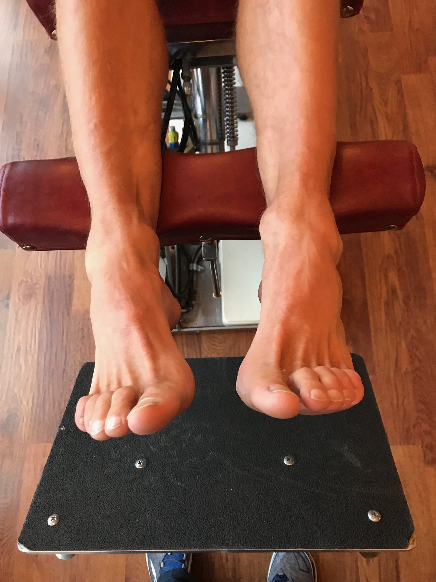
This gentleman came in with fore foot pain (3rd metatarsal head specifically), worse in the AM upon awakening, with first weight bearing that would improve somewhat during the day, but would again get worse at the end of the day and with increased activity. The began insidiously a few months ago (like so many problems do) and is getting progressively worse.

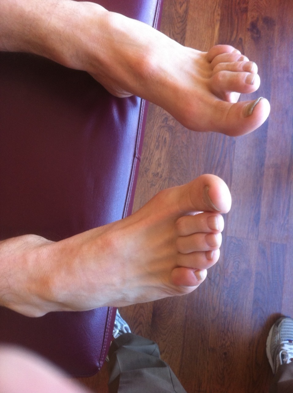
Rest and ice offer mild respite, as does ibuprofen. You can see his foot above. please note the “dropped” 3rd metatarsal head (or as we prefer to more accurately say, “plantarflexed 3rd metatarsal head”) and puffiness and prominence in that area on the plantar surface of the foot. 

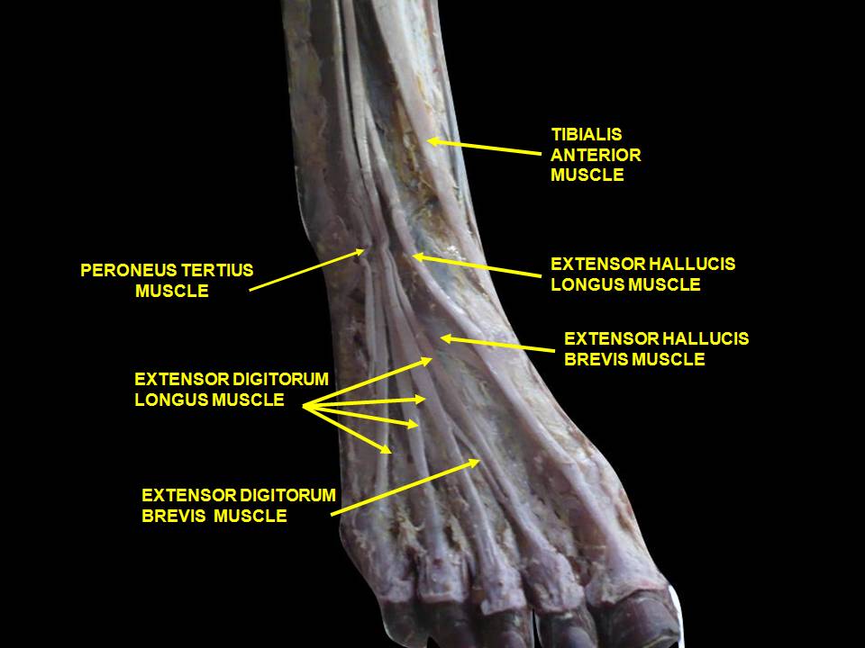
To fully appreciate what is going on, we need to look at the anatomy of the short flexors of the foot. 

The flexor digitorum brevis (FDB) is innervated by the medial plantar nerve and arises from the medial aspect of the calcaneal tuberosity, the plantar aponeurosis (ie: plantar fascia) and the areas bewteen the plantar muscles. It travels distally, splitting at the metatarsal phalangeal articulation (this allows the long flexors to travel forward and insert on the distal phalanges); the ends come together to divide yet another time (see detail in picture above, yes, we are aware it is the hand, but the tendon structure in the foot is remarkably similar)) and each of the 2 portions of that tendon insert onto the middle of the middle phalanyx (1) 

As a result, in conjunction with the lumbricals, the FDB is a flexor of the metatarsal phalangeal joint, and proximal interphalangeal joint (although this second action is difficult to isolate. try it and you will see what we mean). In addition, it moves the axis of rotation of the metatasal phalangeal joint dorsally, to counter act the function of the long flexors, which, when tight or overactive, have a tendency to drive this articulation anteriorly (much like the function of the extensor hallucis brevis above in the drawing from Dr Michauds book, yes, we are aware this is a picture of the 1st MTP).

Can you see the subtle extension of the metatarsal phalangeal joint and flexion of the proximal interphalangeal joint in the picture?

We know that the FDB contracts faster than the other intrinsic muscles (2), playing a tole in postural stability (3) and that the flexors temporally should contract earlier than the extensors (4), assumedly to move this joint axis posteriorly and allow proper joint centration. When this DOES NOT occur, especially if there is a concomitant loss of ankle rocker, the metatarsal heads are driven into the ground (plantarflexion), causing irritation and pain. Metatarsalgia is born….

So what is the fix? Getting the FDB back on line for one. 

  • How about the toe waving exercise? 
  • How about the lift spread reach exercise? 
  • How about retraining ankle rocker and improving hip extension?
  • How about an orthotic with a metatarsal pad in the short term? 
  • How about some inflammation reducing modalities, like ice and pulsed ultrasound. Maybe some herbal or enzymatic anti inflammatories?

The Gait Guys. Increasing your gait and foot literacy with each and every post. 

1. http://en.wikipedia.org/wiki/Flexor_digitorum_brevis_muscle

2. Tosovic D1, Ghebremedhin E, Glen C, Gorelick M, Mark Brown J.The architecture and contraction time of intrinsic foot muscles.J Electromyogr Kinesiol. 2012 Dec;22(6):930-8. doi: 10.1016/j.jelekin.2012.05.002. Epub 2012 Jun 27.

3.Okai LA1, Kohn AF. Quantifying the Contributions of a Flexor Digitorum Brevis Muscle on Postural Stability.Motor Control. 2014 Jul 15. [Epub ahead of print]

4. Zelik KE1, La Scaleia V, Ivanenko YP, Lacquaniti F.Coordination of intrinsic and extrinsic foot muscles during walking.Eur J Appl Physiol. 2014 Nov 25. [Epub ahead of print]