Get your foot in High Gear!

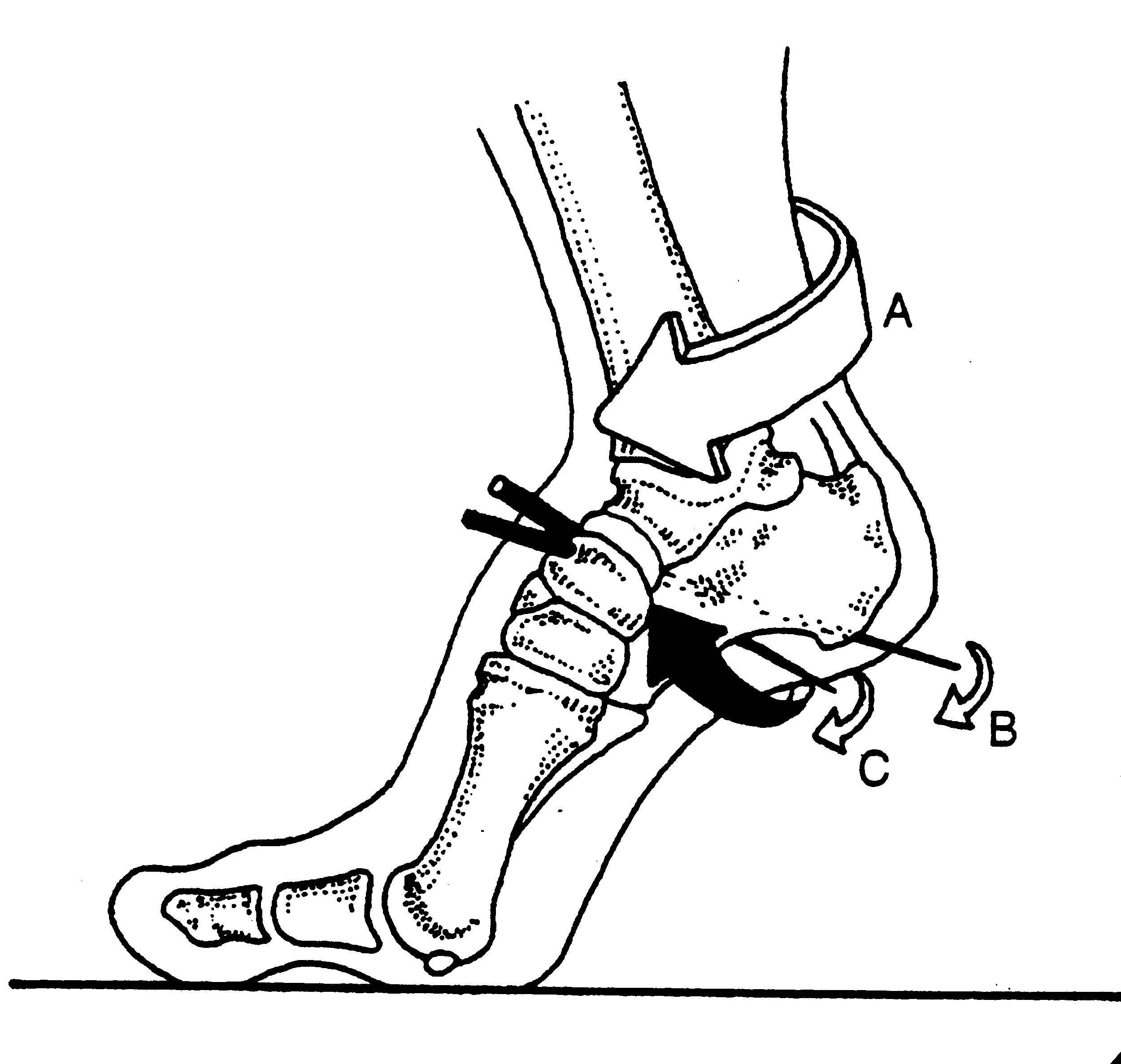
image source: Foot Orthoses and Other Conservative Forms of Foot Care. Michaud 1997; Williams an Wilkins: with permission

image source: Foot Orthoses and Other Conservative Forms of Foot Care. Michaud 1997; Williams an Wilkins: with permission

When it comes to gait, getting the 1st ray to the ground is the name of the game. When weight travels through the medial forefoot and we are able to push off the 1st ray complex, that is called "high gear push off". This was 1st discussed F Bojsen-Møller in this excellent paper (1), that just happens to be a free full text! Craig Payne, The Running Research Junkie has offered and excellent commentary on the topic here as well.

High Gear Push Off can happen when 3 conditions are met: 

  1. we have a intact visual, vestibular and kinesthetic systems that ensure we can remain upright in the gravitational plane.
  2. we have an intact calcanocuboid locking mechanism
  3. we have adequate skill, endurance and strength of our extensor hallucis brevis

The 1st condition is more global and ensures that our cerebellum and vestibular apparatus are playing nice together to create balance, of the literal sort, We seek to keep our visual axes parallel and our center of gravity over our foot. Remember that the BODY will move itself AROUND the visual axes. If the axes are off, the brain will tilt the head and the body will move AROUND the head to accommodate. We have talked about that in these posts here on the blog. 

The 2nd condition, the calcaneo-cuboid locking mechanism, works in the coronal plane and relies on a functioning peroneal group, where the peroneus longus and brevis wrap around the lateral malleolus, cuboid and tail of the 5th metatarsal, crossing the foot diagonally to insert on the base of the 1st metatarsal. When working properly, its actions will be to plantar flex and everting the forefoot, lowering the 1st ray complex down and assisting the shift of the center of gravity more medial for the weight to pass through the medial foot and out through the hallux (ideally). 

The 3rd condition, the ability to descend the 1st ray, relies on the actions of the peroneus, appropriate supination of the forefoot and ability of the extensor hallucis brevis to do its job.  Because the tendon travels behind the axis of rotation of the 1st metatarsal phalangeal joint, in addition to providing extension of the proximal phalynx of the hallux, it can also provide a downward moment on the distal 1st metatarsal (when properly coupled to and temporally sequenced with the flexor hallicus brevis and longus). If the axis of motion for the 1st metatarsal phalangeal joint moves posteriorly, to behind (rather than under) the joint, the plantar pressures increase at MTP’s 4-5 and decrease at the medial mid foot, moving you into low gear push off.  If moved even further posteriorly, the plantar pressures, and contact time in the mid foot and hind foot (2). For more on the extensor hallucis brevis, see our post here.

As you can see, high gear is desirable over low gear push off, but sometimes circumstances or biomechanics do not permit. High gear push off ensures the forefoot is dorsiflexed and everted with respect to the rearfoot and the calcaneocuboid and talonavicular joint axes are perpendicular to one another, giving us a rigid lever to push off of as the center of gravity moves medially across the foot. In low gear push off, the foot is inverted and plantarflexed and the stress falls on the lesser metatarsals and lateral stabilizing complex of the ankle, moving the center of gravity laterally, in addition to the calcaneocuboid and subtalar joint axes being more parallel,  creating a less rigid lever for push off and decreased mechanical efficiency.


1. Calcaneocuboid joint and stability of the longitudinal arch of the foot at high and low gear push off. J Anat. 1979 Aug; 129(Pt 1): 165–176.  link to free full text: https://www.ncbi.nlm.nih.gov/pmc/articles/PMC1233091/

2. van der Zwaard BC1, Vanwanseele B, Holtkamp F, van der Horst HE, Elders PJ, Menz HB Variation in the location of the shoe sole flexion point influences plantar loading patterns during gait. J Foot Ankle Res. 2014 Mar 19;7(1):20.


The Calcaneo Cuboid Locking Mechanism...Revisited...

Do you know what this is? You should if you treat folks who walk or run!

It is the mechanism by which the tendon of the peroneus longus travels behind the lateral malleolus of the ankle, travels underfoot, around the cuboid to insert into the lateral aspect of the base of the 1st metatarsal and adjacent 1st cunieform. Remember the peroneus longus?

The peroneus (or fibularis) longus arises from the head and upper two-thirds of the lateral surface of the fibula, from the deep surface of the fascia, and from the intermuscular septa between it and the muscles on the front and back of the leg; occasionally also by a few fibers from the lateral condyle of the tibia.  You can see from it attachments that it can influence the entire upper lateral leg.

It’s tendon runs down the fibular shaft, wraps around the lateral malleolus, travels obliquely under the foot, crossing the lateral cuboid (which it everts after midstance to help with supination) crosses the sole of the foot obliquely, and inserts into the lateral side of the base of the first metatarsal and lateral aspect of the 1st cunieform.  

It acts from just prior to heel strike to limit excessive rearfoot inversion, through midstance to decelerate subtalar pronation and assists in stabilization of the midfoot articulations, and into terminal stance and pre swing to lock the lateral column of the foot for toe off and plantar flex the 1st ray (creating a good foot tripod), allowing dorsal posterior shift of the 1st metatarsal-phalangeal joint axis (necessary for dorsiflexion of the hallux (big toe)).

When the peroneus longus contracts, in addition to plantar flexing the 1st ray, it everts the cuboid and locks the lateral column of the foot, minimizing muscular strain required to maintain the foot in supination (the locked position for propulsion). Normally, muscle strength alone is insufficient to perform this job and it requires some help from the adjacent articulations.

In addition, the soleus maintains spuination during propulsion by plantar flexing and inverting rear foot via the subtalar joint. This is assisted by the peroneus brevis and tertius which also dorsflex and evert the lateral column, helping keep it locked. Can you see why the peroneii are so important?

signs of a faulty calcaneo cuboid locking mechanism

  • weak peroneus longus, brevis and or tertius
  • excessive rear or midfoot pronation
  • low arch during ambulation
  • poor or low gear “push off”
  • subluxated cuboid


The calcaneo cuboid locking mechanism. Essential for appropriate supination and ambulation. Insufficiency, coming to a foot you will soon examine.

Supination, anyone?

Pronation gets all the press; but what about its counterpart, supination? There could not be one without the other. If anything, supination is at least as, if not more important to create propulsion.

Pronation is dorsiflexion, eversion and abduction of the foot. It provides shock absorption. Supination is plantar flexion, inversion and adduction. It helps the foot become a rigid lever so we can GO (Like in Theo Selig’s “Go Dig Go” ).

external leg rotation supination.gif

Locking of the lateral column of the foot (4th and 5th metatarsal, cuboid and calcaneus) is a necessary prerequisite for normal force transmission through the foot and ultimately placing weight on the head of the 1st metatarsal for proper (high gear) toe off . Locking of the lateral column minimizes muscular strain as the musculature (soleus, peroneus longus and brevis, EHL, EDL, FDL and FHL) is usually not strong enough to perform the job on its own.

external rotary moment.gif

This process is initiated by the opposite leg going into swing phase, which initiates dorsiflexion, inversion and abduction of the talus

The peroneus longus tendon aids this process by wrapping around the cuboid (the brevis attaches to the base of the 1st metatarsal) on its way to insert onto the base of the 1st metatarsal. When the peoneus longus contracts, it dorsiflexes and everts the cuboid, which, along with the soleus (which plantar flexes and inverts the subtalar joint) allows dorsiflexion of 4th and 5th metatarsals and “locks” the lateral column. Without this mechanism, there is no locking. Without locking, there is no supination. Without supination, there is little rigidity and inefficient propulsion.

The calcaneo cuboid locking mechanism. Another cool thing you learned about gait today from The Gait Guys.

Something a little different for a change: Case Management of a Post Surgical Foot

In this series, we will follow the progress of a post surgical, post rehab foot. These are the actual case notes and you can follow our thought process as we move along. 

History:

JM presented with left-sided content foot pain. On July 24 she broke her left navicular and cuboid (pretty unusual, as these fractures are rare. Navicular fractures are usually stress fractures (1), occurring in about .6% of fractures in one study (2).  Cuboid fractures are also rare and occur in less than 1.8 per 100,000 (3) ) She also tore the reticular ligaments. this happened when she fell down the stairs, inverting and plantar flexing the foot.

She has had extensive physical therapy as well as plate fixation of the navicular but is still having constant discomfort; she feels a pinching and shock like sensation in the right arch, particularly when loading the foot (whenever you hear about a "shock like pain, begin thinking about nerve related pain). She has been on gabapentin in the past which helped but she stopped it 3 weeks ago with no regression of her symptoms. She feels frustrated.

She was in physical therapy until the end of December. She has continued with exercises consisting of plantar flexion/toe raises, mobilization and inversion/eversion, squats/lunges as well as massage. She has improved but not completely better. She is able to hike 4-5 miles with little pain (boy, those Colorado women are tough!). The foot generally feels better with non weight bearing and rest as well as avoiding impact. The foot feels stiff in the morning and  she limps for the first 10 minutes after getting out of bed. Most recently she has had x-rays at VSO with Dr. X.

What did we find?

There was swelling noted over the extensor digitorum brevis with significant weakness of it as well as the extensor longus. No sensory deficits, reflexes intact. She had an anatomically short left short leg which appeared to be functional. There was a scar visible over the dorsum of the foot approximately 2 inches in length (see photos) and some discoloration lateral just anterior to the lateral malleolus. palpation along the medial plantar nerve revealed increased sensitivity below the navicular and into the medial heel. 

She has external tibial torsion bilaterally and limited eversion of the forefoot on the right. Her cuboid was moving appropriately but talonavicular articulation was not. She has adequate hip extension, 15+ degrees and ankle dorsiflexion bilaterally in excess of 15 degrees.

One leg standing with eyes open was less than 10 seconds. Loss of flexion and extension about the L SI joint. 

no x rays available for this visit

What we think was wrong:

Left foot pathomechanics (talonavicular) secondary to surgery and fracture. She has significant weakness of the short and long extensors as well as limited eversion and proprioceptive difficulties.

Discussion:

The talonavicular articulation is one of the key joints in mid foot pronation. Pathomechanics appear to be compromising the medial plantar nerve. This is exacerbated by her inability to fully evert the forefoot and pronate through the mid and forefoot (pronation is dorsiflexion, eversion and abduction). 

What We did:

We manipulated the sacroiliac joint and metatarsophalangeal articulations. We held off on any mobilization of the foot until we see her x-rays, hopefully available next visit.

We treated with neuromuscular acupuncture at the origin/insertion of the long extensors as well about the short extensor mass where the swelling was located. She was given the tripod standing, lift spread reach, toes up walking, and tiptoe waiting exercises to perform 2-3 times daily. She is scheduled for followup next week with x-ray review.


1. http://emedicine.medscape.com/article/85973-overview

2. http://journals.sagepub.com/doi/pdf/10.1177/2473011416S00299

3. http://www.uptodate.com/contents/cuboid-and-cuneiform-fractures