The top 6 reasons we like hills for training ankle rocker and hip extension

image source: https://commons.wikimedia.org/wiki/File:Caer_Caradoc_hill.jpg

image source: https://commons.wikimedia.org/wiki/File:Caer_Caradoc_hill.jpg

1. Hills do not cost money and are almost always readily available : )

2. Being outside is good for your health

3. Hills do not pull the hip into extension and place a stretch (pull) on the anterior hip musculature including the rectus femoris, iliopsoas and iliacus. This causes a slow stretch of the muscle, activating the muscle spindles (Ia afferents) and causing a muscle contraction via the stretch reflex. This will inhibit the posterior compartment of hip extensors (especially the glute max) through reciprocal inhibition, making it difficult to fire them.

4. A hill does not force your knee into extension, eliciting a stretch reflex in the hamstrings like a treadmill does

5. A hill naturally puts the ankle into dorsiflexion, and, along with active pulling up of the toes, helps you to get more into your anterior compartment and eliminates the tendency of the ankle being pulled into dorsiflexion (like with a treadmill) which would initiate a stretch reflex in the gastroc/soleus and long flexors.

6. The increased hip flexor requirement of going uphill gives you more opportunity to engage the abs before the psoas and rectus femoris/TFL and on the stance phase leg, you can get an increased stretch of those muscles

Tips for picking the right hill and using it to your advantage

  • When just starting out, try and pick an incline that does not exceed the ankle dorsiflexion available to the patient/client

  • It’s OK if it’s uncomfortable, but not if its painful

  • Concentrate on pulling up the toes and dorsiflexing the ankle

  • Squeeze your glute at heel strike and toe off

  • leave your stance phase heel on the ground as long as possible

  • Place your hands on your abs and concentrate on activating them PRIOR to flexing your hip

Dr Ivo Waerlop, one of The Gait Guys

#walkinghills #traininganklerocker #thegaitguys # increasinghipextension


The muscle they named wrong?

Why would you name a muscle after its supposed function when its function is actually something totally different? Probably due to what made sense from how it looked, not by how it acted. Of course, we are talking about the abductor hallucis.

tumblr_nn1ca5fx0C1qhko2so1_r1_1280.jpg

Think about all the anatomy you have learned over the years. Think about all the taxonomy and how it was done: sometimes by thename of the discoverer and more often by its anatomical location. The abductor hallucis seems to be the latter. 

The abductor and adductor hallicus function from approximately midstance to pre swing (1-4) (toe off), applying equal and opposite rotational vectors of force (in an ideal world) of the proximal phalynx of the hallux. This should resolve into a purely compressive force (5). In a closed chain environment, the transverse head of the adductor hallicus should act to prevent “splay” of metatarsals, along with the lumbricals and interossei (6), providing stabilzation of the forefoot (7) and rearfoot (8) during preswing, while the oblique head serves to help maintain the medial longitudinal arch. 

The abductor hallicus is actually a misnomer, as it most cases it is not an abductor but rather a plantar flexor of the 1st ray, particularly the proximal hallux, (assisting the peroneus longus) and supinator about the oblique midtarsal joint axis (5).  In the majority of cases, there doesn’t appear to be a separate, distinct insertion of the adductor hallicus to the base of the proximal phalynx, but rather a conjoint insertion with the lateral head of the flexor hallicus bevis into the lateral sesamoid and base of the proximal phalynx (9-11), emphasizing more of its plantar flexion function and stabilizing actions, rather than abduction. 

In one EMG study of 20 people with valgus (12) they looked at activity of adductor and abductor hallucis, as well as flexor hallucis brevis and extensor hallucis longus. They found that the abductor hallucis had less activity than the adductor. No surprise here; think about reciprocal inhibition and increased activity of the adductor when the 1st ray cannot be anchoroed. They also found EMG amplitude greater in the abductor hallucis by nearly two fold in flexion. 

So, the abductor hallucis seems to be important in abduction but more important in flexion. Either way, it is a stance phase stabilizer that we are beginning to know a lot more about. As for the name? You decide...


Dr Ivo Waerlop, one of The Gait Guys


1. Basmajian JV, Deluca CJ . Muscle Alive. Their Functions Revealed by Electromyography Williams and Wilkins. Baltimore, MD 1985, 377

2. Root MC, Orien WP, Weed JH. Normal and Abnormal Function of the Foot. Clinical Biomechanics, Los Angeles, CA 1977

3. Mann RA. Biomechanics of Running. In Pack RP. d. Symposium on the foot and leg in running sports. Mosby. St Louis, MO 1982:26

4. Lyons K, Perry J, Gronley JK. Timing and relative intensity of the hip extensor and abductor muscle action during level and stair ambulation. Phys Ther 1983: 63: 1597-1605

5. Michaud T. Foot Orthoses and Other Forms of Conservative Foot Care. Newton MA 1993: 50-55

6. Fiolkowski P, Brunt D, Bishop et al. Intrinsic pedal musculature support of the medial longitudinal arch: an electromyography study. J Foot & Ankle Surg 42(6) 327-333, 2003

7. Travell JG, Simons DG. Myofascial Pain and Dysfunction: The Trigger Point Manual. Williams and Wilkins, Baltimore 1992; 529

8. Kalin PJ, Hirsch BE. The origin and function of the interosseous muscles of the foot. J Anat 152, 83-91; 1987

9. Owens S, Thordarson DB. The adductor hallucis revisited. Foot Ankle Int. 2001 Mar;22(3):186-91. Am J Phys Med Rehabil. 2003 May;82(5):345-9.

10. Brenner E.Insertion of the abductor hallucis muscle in feet with and without hallux valgus. Anat Rec. 1999 Mar;254(3):429-34.

11. Appel M, Gradinger R. [Morphology of the adductor hallux muscle and its significance for the surgical treatment of hallux valgus][Article in German] Orthop Ihre Grenzgeb. 1989 May-Jun;127(3):326-30.

12. Arinci I, Geng H, Erdem HR, Yorgancioglu ZR Muscle imbalance in hallux valgus: an electromyographic study. Am J Phys Med Rehabil. 2003 May;82(5):345-9.


#halluxvalgus #halluxabductovalgus #bunion #footmuscleactivity #gait #thegaitguys


The Sartorius: insertional tendinitis and medial knee pain?

We all see folks with medial knee pain, many times women, with the pain located just below the medial tibial plateau. It often results from running, but sometimes with jumping sports like basketball as well. It has been our experience that these people are often diagnosed with an MCL type injury, but when you examine them further, they do not really fit the bill. All the ligaments are stable and there is no tenderness at the joint line. The is often tenderness at the pes anserine, but who is driving the bus here?

image source: https://commons.wikimedia.org/wiki/File:Muscles_and_tendons_of_the_legs_and_feet;_écorch_́figur_Wellcome_V0008276.jpg

image source: https://commons.wikimedia.org/wiki/File:Muscles_and_tendons_of_the_legs_and_feet;_écorch_́figur_Wellcome_V0008276.jpg

The sartorius originates from the anterior compartment of the thigh. During an ideal gait cycle, the sartorius fires from toe off through nearly terminal swing (1)

We remember that the abdominals should initiate thigh flexion with the iliopsoas, rectus femoris, tensor fascia lata and sartorius perpetuating the motion. Sometimes, when the abdominals are insufficient, we will substitute other thigh flexors, often the psoas and/or rectus femoris, but sometimes sartorius, especially in people with excessive midfoot pronation. Think about all of the medial rotation occurring at the knee during excessive midfoot pronation and when overpronation occurs, the extra compensatory external rotation that must occur to try and bring the knee back into the sagittal plane. The sartorius is positioned perfectly for this function, along with the semitendinosus which assists and external rotation in closed chain. This is why it is often implicated as the culprit in many cases of pes anserine bursitis (or as we like to say “sartorius insertional tendinitis” (2-3)

Some other things you may find interesting is that it is utilized more in crossing or cutting maneuvers while changing directions while running (4). This makes sense, given its anatomical course and origin/insertion. It can often be overlooked in adductor strains. It can also be avulsed during sprints, particularly in adolescents (5) and because of the course of the lateral femoral cutaneus nerve beneath it, can be the cause of meralgia paresthetica (6). It is proprotionally smaller in females (along with the gracilis and short head of the biceps femoris) (7). And during vertical jumping, is considered an internal rotator, along with the semimembranosis, semitendinosis, gracilis, and popliteus (8).

The sartorius is superficial in the anterior thigh, just under the skin, running from the ASIS, coursing lateral to medial and inserting at the pes anserine at its most superior aspect, just overlying the gracilis. Since it is an external rotator, knee flexor and assists in thigh abduction, you can easliy locate it by placing the patient in a "figure 4" position and having them resist as you pull downward on the leg. Be careful if you are needling this muscle because of the subsartorial canal (ie Hunters canal) lying just beneath it in the middle 1/3 of the thigh, from the apex of the femoral triangle to the adductor hiatus in the adductor magnus. It houses the femoral artery and vein, as well as the saphenous nerve and nerve to the vastus medialis.


  1. Michaud T: in Human Locomotion: The Conservative Management of Gait-Related Disorders 2011

  2. Imani F, Rahimzadeh P, Abolhasan Gharehdag F, Faiz SH. Sonoanatomic variation of pes anserine bursa. Korean J Pain. 2013;26(3):249-54. 

  3. Gupta, Aman & Saraf, Abhinesh & Yadav, Chandrajeet. (2013). ISSN 2347-954X (Print) High-Resolution Ultrasonography in PesAnserinus Bursitis: Case Report and Literature Review. 1. 753-757. 

  4. Rand MK, Ohtsuki T. EMG analysis of lower limb muscles in humans during quick change in running directions. Gait Posture. 2000 Oct;12(2):169-83.

  5. Manning CJ, Singhai S, Marshall P. Synchronised sartorius avulsions in adolescent sprinter. BMJ Case Rep. 2016 Jul 13;2016.

  6. Hsu CY, Wu CM, Lin SW, Cheng KL. Anterior superior iliac spine avulsion fracture presenting as meralgia paraesthetica in an adolescent sprinter. J Rehabil Med. 2014 Feb;46(2):188-90. doi: 10.2340/16501977-1247.

  7. Behan FP, Maden-Wilkinson TM, Pain MTG, Folland JP. Sex differences in muscle morphology of the knee flexors and knee extensors. PLoS One. 2018 Jan 23;13(1):e0190903.

  8. Cleather DJ. An important role of the biarticular hamstrings is to exert internal/external rotation moments on the tibia during vertical jumping. J Theor Biol. 2018 Oct 14;455:101-108

Subtle clues to flexor dominance

IMG_5515.jpg

Take a close look at these photographs. Compare the prominence of the extensor tendon‘s left to right. What do you see? Do you notice the deeper furrowing of the extensor tendons on the left? Do you see the subtle increased extension of the metatarsophalangeal and requisite increased flexion of the inter-phalangeal articulations, left versus right? What about the height of the arches?

Keep a keen eye out for subtle signs. They can make a real difference in your clinical diagnosis and results…

What did you notice? The Devil is in the details...

 Cavus foot? Loss of the transverse arch? Prominence of extensor tendons?

The question is: Why?

It’s about reciprocal inhibition. The concept, though observed in the 19th century, was not fully understood and accepted until it earned a Nobel prize for its creditor, Sir Charles Sherrington, in 1932. Simply put, when a muscle contracts, its antagonist is neurologically inhibited, So when your bicep contracts, your tricep is inhibited. This holds true whether you actively contract the muscle or if the muscle is irritated (causing contraction).

So how does this apply to this foot?

We see prominence of the extensor tendons (particularly the extensor digitorum brevis EDB; the longus would have caused extension at the distal interphalangeal joint). The belly of the muscle is visible, telling us that it is active. It is neurologically linked to the flexor digitorum brevis (FDB). This muscle, in turn, has slips which attach it to the abductor hallucis brevis (AHB) medially and the abductor digiti minimi (ADM) laterally. These muscles together form 2 triangles (to be discussed in another post) on the bottom of the foot, which lend to the stability of the foot and the arches, especially the transverse.

When the EDB fires, it inhibits the FDB, (which, in addition to flexing the MTP’s, assists in maintaining the arch). The EDB has an effect which drops the distal heads of the metatarsals as well (Hmm, think about all the people with met head pain) Now, look at the course of the tendons of the EDB. In a cavus foot, there is also a mild abductory moment, which flattens the arch. Conversely, the FDB in a cavus foot would serve to actually increase the arch, and would have a ,mild adductory moment. Net result? A flattened transverse arch.

Now look at the Flexor digitorum longus, overactive in tbis foot (as evidenced by the flexion of the distal interphalangeal joints, mild adduction of the toes (due to the change of direction of pull in a cavus foot) and lowering of the met heads due to hyperextesnion at the MTP joints ). This mm is reciprocally linked with the extensor digitorum longus. The prominence of the extensor tendons is do to increased activity of the EDB (go ahead, extend all your fingers and look at the tendons in your hand. Now flex the  DIP and IP joints and extend the MTP; see how they become more prominent?).

Reciprocal inhibition. It’s not just for dinner anymore…

We are and remain; The Gait Guys

About Toe Walkers...

Photo courtesy of Surestep

Photo courtesy of Surestep

Idiopathic Toe Walking in kids..Is it flexor dominance?

You see this at times in the office. Kiddos (or adults) who walk on their toes for no apparent reason. many have shortened heel cords with limited ankle dorsiflexion (1,2). Some studies report an incidence of 7-24% in pediatric populations (3) with an average of about 5% in children that are 5.5 years old (4). It seems to occur in about 2% of normally developing kids aged 5.5 years and 40% of those that have some sort of neuropsychiatric diagnosis or missed a developmental window (5-7), with an increased incidence familialy (8). The question here is why, not what.

We have discussed our opinions of flexor dominance here many times and suffice it to say that increased corticospinal activity seems to have the double whammy effect of increased firing of the distal flexors due to a lack of input to the axial extensors in the rostral and caudal reticular formations respectively(possibly from decreased spindle and /or GTO input and/or mechanoreceptor dysafferentation?) and lack of reciprocal inhibition of the extensors from the increased firing of the flexors segmentally. Is it the cortical abnormailities and missed developmental windows seen in so many of these folks that drives this? These are the sorts of things that keep us up at night....

Physical treatment modalities (2) seem to help, we think most likely to plastic changes in the connective tissue. Orthotics may prove useful due to similar mechanisms, especially if there is an equinus deformity or forefoot to rearfoot abnormaility (9). More agressive (and invasive) measures like Botox, seem to not. An interesting study using whole body vibration (10) produced some immediate but short lived positive results. This really gets you thinking about joint and muscle mechanoreceptors and the cerebellum, and makes us think that perhaps we also should be looking (and treating) north of the foot. We could not find any studies looking at the effects of proprioceptive or vestibular exercises effects on this, but think it could be promising area of therapy and we will continue to employ them until our clinical results tell us otherwise. 


1. Barrow WJ, Jaworski M, Accardo PJ. Persistent toe walking in autism. J Child Neurol 2011;26(5):619-621

2. Harris NM. Multidisciplinary approach led to positive results for pediatric patient with idiopathic toe walking. Presented at the Association of Children’s Prosthetic-Orthotic Clinics Annual Meeting, Broomfield, CO, April 15, 2016.

3. Engelbert R, Gorter JW, Uiterwaal C, et al. Idiopathic toe-walking in children, adolescents and young adults: a matter of local or generalised stiffness? BMC Musculoskelet Disord. 2011;12:61.

4. Engström P, Tedroff K. The prevalence and course of idiopathic toe-walking in 5-year-old children. Pediatrics 2012;130(2):279-284.

5. https://tmblr.co/ZrRYjx1VV59rl

6. Williams, C. , Curtin, Wakefield and Nielsen Is idiopathic toe walking really idiopathic ? The motor skills and sensory processing abilities associated with idiopathic toe walking gait.  J Child Neurol 2014, 29:71

7. https://tmblr.co/ZrRYjx1WTNcdK

3. Is idiopathic toe walking really idiopathic ? The motor skills and sensory processing abilities associated with idiopathic toe walking gait.  J Child Neurol 2014, 29:71 Williams, C. , Curtin, Wakefield and Nielsen

8. Pomarino D, Ramirez Llamas J, Pomarino A. Idiopathic toe walking: tests and family predisposition. Foot Ankle Spec 2016;9(4):301-306

9. Herrin K, Geil M. A comparison of orthoses in the treatment of idiopathic toe walking: a randomized controlled trial. Prosthet Orthot Int 2016;40(2):262-269.

10. Williams CM, Michalitsis J, Murphy AT, et al. Whole-body vibration results in short-term improvement in the gait of children with idiopathic toe walking. J Child Neurol 2016;31(9):1143-1149.


Gaining Anterior Length, Through Posterior Strength. A Lesson in Reciprocal Inhibition

tumblr_oco89rcuI61qhko2so2_1280.png
tumblr_oco89rcuI61qhko2so1_1280.png

Gaining Anterior Length, Through Posterior Strength and vice versa….A Lesson in Reciprocal Inhibition

I found a really cool article, quite by accident. I was leafing through an older copy of one of, if not my favorite Journals “Lower Extremity Review” and there it was. An article entitled “Athletes with hip flexor tightness have reduced gluteus maximus activation”. Wow, I thought! Now there is a great article on reciprocal inhibition! This reminded me of a piece we wrote some time ago

What is reciprocal inhibition, also called “reciprocal innervation” you ask? The concept, was 1st observed as early as 1626 by Rene Descartes though observed in the 19th century, was not fully understood and accepted until it earned a Nobel prize for its creditor, Sir Charles Sherrington, in 1932.

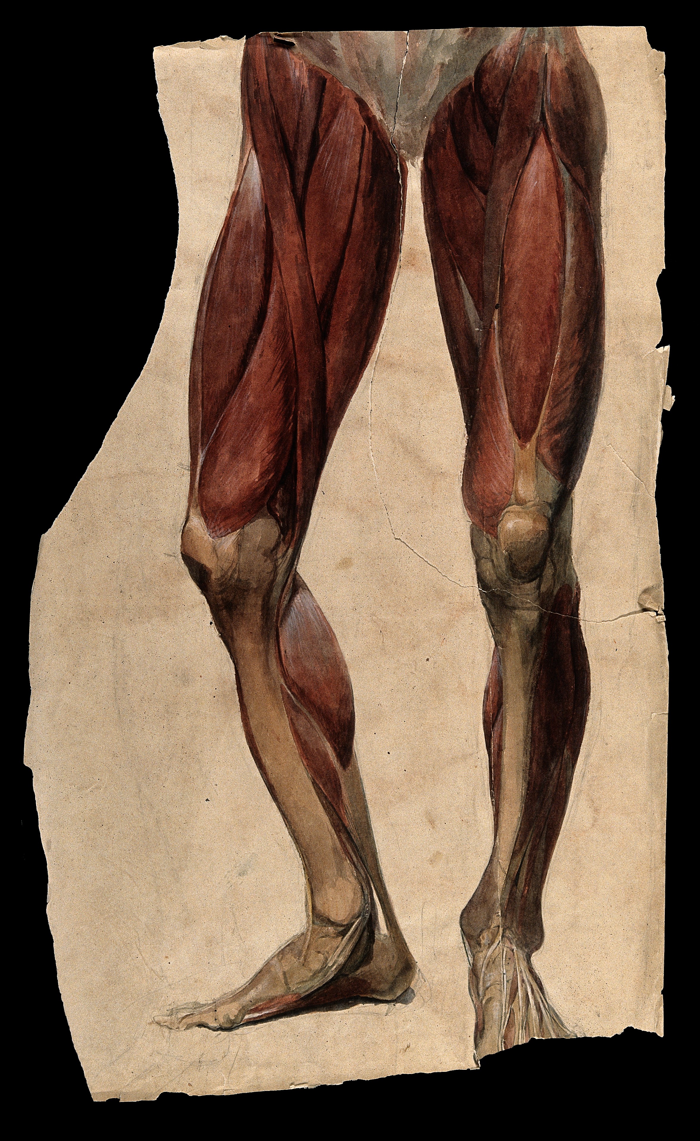
Simply put, when a muscle contracts, its antagonist is neurologically inhibited (see the diagram above) When your hip flexors contract, your hip extensors are inhibited. This holds true whether you actively contract the muscle or if the muscle is irritated in some manner, causing contraction. The reflex has to do with muscle spindles and Type I and Type II afferents which I have covered in an article I wrote some time ago.

We can (and often do) take advantage of this concept with treating the bellies of hip flexors (iliopsoas, tensor fascia lata, rectus femoris, iliacus, iliocapsularis) and extensors (gluteus maximus, posterior fibers of gluteus medius). This is especially important in folks with low back pain, as they often have increased psoas activity and cross sectional area, especially in the presence of degenerative changes.

There also appears to be a correlation between decreased hip extension and low back pain, with a difference of as little as 10 degrees being significant. Take the time to do a thorough history and exam and pay attention to hip extension and ankle dorsiflexion as they should be the same, with at least 10 degrees seeming to be the “clinical” minimum. Since the psoas should only fire at the end of terminal stance/preswing and into early swing, problems begin to arise when it fires for longer periods.

Can you see now how taking advantage of reciprocal inhibition can improve your outcomes? Even something as simple as taping the gluteus can have a positive effect! Try this today or this week in the clinic, not only with your patients hip flexors, but with all muscle groups, always thinking about agonist/antagonist relationships.


In the moment: Sports medicine  Jordana Bieze Foster: Athletes with hip flexor tightness have reduced gluteus maximus activation  Lower Extremity review Vol 6, Number 7 2014

https://tmblr.co/ZrRYjx1VG3KYy

Mills M, Frank B, Blackburn T, et al. Effect of limited hip flexor length on gluteal activation during an overhead squat in female soccer players. J Athl Train 2014;49(3 Suppl):S-83.

Ciuffreda KJ, Stark L.  Descartes’ law of reciprocal innervation. Am J Optom Physiol Opt. 1975 Oct;52(10):663-73.
Jacobson M Foundations of Neuroscience Springer Science and Business Media, Plenum Press, NY 1993 p 277

http://www.nobelprize.org/nobel_prizes/medicine/laureates/1932/sherrington-bio.html

https://thegaitguys.tumblr.com/post/9708399904/ah-yes-the-ia-and-type-ii-afferents-one-of-our

Arbanas J, Pavlovic I, Marijancic V, et al MRI features of the psoas major muscle in patients with low back pain. Eur Spine J. 2013 Sep;22(9):1965-71. doi: 10.1007/s00586-013-2749-x. Epub 2013 Mar 31.

Roach SM, San Juan JG, Suprak DN, Lyda M, Bies AJ, Boydston CR. Passive hip range of motion is reduced in active subjects with chronic low back pain compared to controls. Int J Sports Phys Ther. 2015 Feb;10(1):13-20. Erratum in: Int J Sports Phys Ther. 2015 Aug;10(4):572.

Paatelma M Karvonen E Heiskanen J Clinical perspective: how do clinical test results differentiate chronic and subacute low back pain patients from “non‐patients”? J Man Manip Ther. 2009;17(1):11‐19.[PMC free article] [PubMed]

Evans K Refshauge KM Adams R Aliprandi L Predictors of low back pain in young adult golfers: a preliminary study. Phys Ther Sports. 2005;6:122‐130.

Mellin G Correlations of hip mobility with degree of back pain and lumbar spinal mobility in chronic low‐back pain patients. Spine. June 1988;13(6):668‐670. [PubMed]

Lewis CL, Ferris DP. Walking with Increased Ankle Pushoff Decreases Hip Muscle Moments. Journal of biomechanics. 2008;41(10):2082-2089. doi:10.1016/j.jbiomech.2008.05.013.

Nodehi-Moghadam A, Taghipour M, Goghatin Alibazi R, Baharlouei H. The comparison of spinal curves and hip and ankle range of motions between old and young persons. Medical Journal of the Islamic Republic of Iran. 2014;28:74.

Daniel Moon , MD, MS; Alberto Esquenazi , MD Instrumented Gait Analysis: A Tool in the Treatment of Spastic Gait Dysfunction JBJS Reviews, 2016 Jun; 4 (6): e1. http://dx.doi.org/10.2106/JBJS.RVW.15.00076

Kilbreath SL, Perkins S, Crosbie J, McConnell J. Gluteal taping improves hip extension during stance phase of walking following stroke. Aust J Physiother. 2006;52(1):53-6.

tumblr_nozmmaOxAI1qhko2so1_500.png
tumblr_nozmmaOxAI1qhko2so2_540.png

Ahh yes, the lumbricals. 

One of our favorite muscles. And here it is in a recent paper! This one is for all you fellow foot geeks : )

Perhaps the FDL (which fires slightly earlier than the FHL) and FHL (which fires slightly later and longer) at loading response, slowing pronation and setting the stage for lumbrical function from midstance to terminal stance/preswing (flexion at the metatarsal phalangeal joint (it would have to be eccentric, if you think about this from a closed chain perspective) and extension (actually compression) of the proximal interphalangeal joints.

“The first lumbrical arose as two muscle bellies from both the tendon of the FDL and the tendinous slip of the FHL in 83.3 %, and as one muscle belly from the tendon of the FDL or the tendinous slip of the FHL in 16.7 %. These two muscle bellies subsequently merged to form the muscle belly of the first lumbrical. The second lumbrical arose from the tendinous slips of the FHL for the second and third toes as well as the tendon of the FDL in all specimens. The third lumbrical arose from the tendinous slips of the FHL for the third and fourth toes in 69.7 %, and the fourth lumbrical arose from the tendinous slip of the FHL for the fourth toe in 18.2 %. Some deep muscle fibers of the fourth lumbrical arose from the tendinous slip of the FHL for the second toe in 4.5 %, for the third toe in 28.8 %, and for the fourth toe in 15.2 %.”

Hur MS1, Kim JH, Gil YC, Kim HJ, Lee KS. New insights into the origin of the lumbrical muscles of the foot: tendinous slip of the flexor hallucis longus muscle. Surg Radiol Anat. 2015 May 12. [Epub ahead of print]

tumblr_nfzfwqcZBr1qhko2so1_1280.jpg
tumblr_nfzfwqcZBr1qhko2so2_1280.jpg
tumblr_nfzfwqcZBr1qhko2so3_r1_400.jpg
tumblr_nfzfwqcZBr1qhko2so4_r1_400.jpg
tumblr_nfzfwqcZBr1qhko2so6_r1_250.jpg

The case of the dropped (plantarflexed) metatarsal head. Or, “How metatarsalgia can happen”.

This gentleman came in with fore foot pain (3rd metatarsal head specifically), worse in the AM upon awakening, with first weight bearing that would improve somewhat during the day, but would again get worse at the end of the day and with increased activity. The began insidiously a few months ago (like so many problems do) and is getting progressively worse.

Rest and ice offer mild respite, as does ibuprofen. You can see his foot above. please note the “dropped” 3rd metatarsal head (or as we prefer to more accurately say, “plantarflexed 3rd metatarsal head”) and puffiness and prominence in that area on the plantar surface of the foot. 

To fully appreciate what is going on, we need to look at the anatomy of the short flexors of the foot. 

The flexor digitorum brevis (FDB) is innervated by the medial plantar nerve and arises from the medial aspect of the calcaneal tuberosity, the plantar aponeurosis (ie: plantar fascia) and the areas bewteen the plantar muscles. It travels distally, splitting at the metatarsal phalangeal articulation (this allows the long flexors to travel forward and insert on the distal phalanges); the ends come together to divide yet another time (see detail in picture above, yes, we are aware it is the hand, but the tendon structure in the foot is remarkably similar)) and each of the 2 portions of that tendon insert onto the middle of the middle phalanyx (1) 

As a result, in conjunction with the lumbricals, the FDB is a flexor of the metatarsal phalangeal joint, and proximal interphalangeal joint (although this second action is difficult to isolate. try it and you will see what we mean). In addition, it moves the axis of rotation of the metatasal phalangeal joint dorsally, to counter act the function of the long flexors, which, when tight or overactive, have a tendency to drive this articulation anteriorly (much like the function of the extensor hallucis brevis above in the drawing from Dr Michauds book, yes, we are aware this is a picture of the 1st MTP).

Can you see the subtle extension of the metatarsal phalangeal joint and flexion of the proximal interphalangeal joint in the picture?

We know that the FDB contracts faster than the other intrinsic muscles (2), playing a tole in postural stability (3) and that the flexors temporally should contract earlier than the extensors (4), assumedly to move this joint axis posteriorly and allow proper joint centration. When this DOES NOT occur, especially if there is a concomitant loss of ankle rocker, the metatarsal heads are driven into the ground (plantarflexion), causing irritation and pain. Metatarsalgia is born….

So what is the fix? Getting the FDB back on line for one. 

  • How about the toe waving exercise? 
  • How about the lift spread reach exercise? 
  • How about retraining ankle rocker and improving hip extension?
  • How about an orthotic with a metatarsal pad in the short term? 
  • How about some inflammation reducing modalities, like ice and pulsed ultrasound. Maybe some herbal or enzymatic anti inflammatories?

The Gait Guys. Increasing your gait and foot literacy with each and every post. 

1. http://en.wikipedia.org/wiki/Flexor_digitorum_brevis_muscle

2. Tosovic D1, Ghebremedhin E, Glen C, Gorelick M, Mark Brown J.The architecture and contraction time of intrinsic foot muscles.J Electromyogr Kinesiol. 2012 Dec;22(6):930-8. doi: 10.1016/j.jelekin.2012.05.002. Epub 2012 Jun 27.

3.Okai LA1, Kohn AF. Quantifying the Contributions of a Flexor Digitorum Brevis Muscle on Postural Stability.Motor Control. 2014 Jul 15. [Epub ahead of print]

4. Zelik KE1, La Scaleia V, Ivanenko YP, Lacquaniti F.Coordination of intrinsic and extrinsic foot muscles during walking.Eur J Appl Physiol. 2014 Nov 25. [Epub ahead of print]

tumblr_m1h0nfVgBC1qhko2so1_1280.jpg
tumblr_m1h0nfVgBC1qhko2so2_1280.jpg

Neuromechanics Weekly

Bad posture is ubiquitous:Our Flexor Driven Society

We know you visit here often and read the blog. You do it because of your desire to learn and possibly because of curiosity and wondering what exactly it is that we are saying, or not believing that we actually said it…and can back it up!  Do you remember that we said most joint and muscle receptor activity goes to the cerebellum? Do you remember that the cerebellum is NECESSARY for learning? Not just motor learning but ALL learning…

Curious? Read on…

If you think the economy is our problem, maybe you need to look a little closer. These  pictures are from a few days ago while at the playground with my kids (yes, the gait cam again…yes, we are watching YOU).

Many human ailments can be linked to imbalance. The Chinese recognized this a few thousand years ago with the concepts of Yin and Yang (pronounced Yin and Yawn; I can still hear Dr Springfield saying “Anyone who says Yang (rhymes with Tang) fails the course). Equal opposing forces are meant to remain in balance to create homeostasis.

This picture is a perfect example of flexor/extensor imbalance (stooped posture creating slow stretch of the extensors of the spine/back and gluteal muscles), which, over time (30 mins or  more) increases muscle length.

We are a flexor driven society. Think about all of the voluntary, fine manipulative movements you can make consciously (buttoning a shirt, typing, pushing off with gait). These are all corticospinal (long neurologic tracts from brain to spine) driven phenomenon (ie under conscious control) which are ultimately driving the flexors (go ahead, try and button your shirt with the backs of your fingers (ie. extensors); if you can do it, please send us the video, so we can post it).

Now think about the activities, like standing upright, that are dependent on our extensors, which are largely under the purview of the vestibulospinal system (driven from the cerebellum and vestibular nucleii in the brainstem). Try and contract your L2-L3 lumbar multifidus on the right: you can’t, because it is not under conscious control.

Let’s extrapolate further…We tend to use that which we can control, thus we use our calves and deep flexors of the posterior compartment of the leg. These cause knee flexion and plantar flexion of the foot (and dorsiflexion of the 1st MTP, provided there is adequate range of motion available), which reduce ankle rocker. They will reciprocally inhibit the anterior compartment muscles (like TA and EDL, EHL). Your center of gravity shifts and now you need to engage the quads to right it, rather than the more efficient gluteals. Now the pelvis tips forward due to action of the rectus femoris, shortening the glutes and putting them at a mechanical disadvantage (in addition to functionally weakening the lower abdominal compartment, making athletes more susceptable to hernias), so now we use the hamstrings (but these are reciprocally inhibited by the quads), so we default to the lumbar erectors and so on up the chain.

Flexor dominance leading to extensor inhibition. Not to mention that the cortico spinal pathway (flexors) inhibits the vestibulo spinal and retculospinal pathways (which both drive the extensors) in the brain stem. So, there is a local, segmental effect and cortical loop. When we continue to utilize certain pathways in the brain, they become ingrained (collateralization and facilitation) and that’s how bad motor patterns begin.

Look at the picture above again. Flexed lumbar and thoracic spines, flexed arms, flexed wrists, flexed hips, flexed knees. Not too much extensor activity going on, eh? Thus, More inhibition of the extensors.

We know you visit here often and read the blog, because of your desire to learn. Do you remember that most joint and muscle receptor activity goes to the cerebellum? Do you remember that the cerebellum is NECESSARY for learning? Not just motor learning but ALL learning…

So, sit up straight. Eat your vegetables and work your extensors. Your intelligence depends on it!

Ivo and Shawn. The Gait Guys. Telling you what you need to hear, but don’t necessarily want to hear. Yes, we are watching and yes, our glutes are engaged.