Two things we hope you see right away when viewing this video.

.

We talked about this woman in yesterday‘s post and, when viewing from the front, we wanted to bring out a few salient points

Notice how her foot progression angle is diminished bilaterally. Normally the foot will “toe out“ somewhere between 12 and 20° when the foot hits the ground and hers are nearly straightahead. This can be due to numerous things such as femoral retro torsion, internal tibial torsion, or subtalar version. All of these things will often have the foot point medially when examining a patient on the table and placing the knee in the sagittal plane. In this particular case, she has internal tibial torsion.

The next thing we would like you to look at is noting how her knee falls “outside“ the sagittal plane. In other words, instead of the knees pointing straight ahead, the point slightly out laterally. This is a cardinal sign of internal tibial torsion, especially in a gait analysis.

So what’s a clinician to do?

In this particular case, there’s nothing really to “fix“ as these are hard deformities that are often congenital. Our job is to help the patient to compensate and the best way possible so that they can remain pain and as injury free as possible. We discussed remediation in the post yesterday, So please refer back to that for what we did


Dr Ivo Waerlop, one of The Gait Guys

#invertedforefoot #forefootsupinatus #forefootvarus #pronation #forefoot #gaitanalysis #decreasedprogressionangle #toeingin

The consequences of an inverted forefoot

A forefoot that is inverted with respect to the rearfoot. Whether it is a forefoot varus, forefoot supinatus or an everted rearfoot ( because the forefoot is still inverted with respect to the rearfoot), what are the biomechanical sequelae?

If we accept the premise that the foot is basically a tripod between the calcaneus, base of the first and base of the fifth metatarsal‘s, we know that all of these parts needs to be on the ground at certain points in the gait cycle. Forces should travel from the calcaneus, up the lateral aspect of the foot, across the metatarsal heads to the first metatarsal head and hopefully out through the hallux.

The foot should hit the ground in slight inversion of the entire foot at initial contact and pronate through the middle of mid stance and then supinate through the remainder of the gait cycle. There’s an intricate balance of biomechanical events that must occur, especially in the latter half of the gait cycle when the rear foot is inverting where the forefoot is everting, so that we can have high gear push off through the distal first ray.

If the forefoot remains inverted then somehow the head of the first metatarsal needs to be brought down to the ground. If there’s not adequate range of motion in the foot, particularly the first ray, then you may pronate through the midfoot, rearfoot or in cases where this is insufficient, bring them immediately over the foot to get it down. This of course shifts center of gravity to midline and the body above must compensate in someway.

Take a look at this video footage and what do you see? She strikes on the outside of her foot but does not have adequate motion in her forefoot and therefore “crashes“ down on the forefoot, forcing a valgus moment into the ankle and the need to shift immediately by the pelvis attempts to dampen it. Notice how this is worse on the right side with more medial knee shift, pelvic shift as well as a lateral bending of the body to the right. Notice also how the upper body twists more to the left than to the right.

So what’s the fix? Well the answer is, “what’s bothering the patient?” We don’t necessarily fix what we see; we correlate what we see with what the patient’s symptoms are because that’s usually why they show up in your office. Yes, we do get people from time to time that come in strictly for “performance enhancement“ but this is pretty rare.

This woman has very little motion and plantar flexion of the first Ray complex so our primary goal was to get her to descend the first ray. We accomplished this by the following:

1. Manipulation in plantar and dorsiflexion of the first ray complex
2. Soft tissue work in the first intermetatarsal interval
3. Exercises of muscles to assist in descending the first ray including the following: extensor hallucis brevis, peroneus longus, flexor digitorum brevis
4. Pelvic stability work to improve the skill, endurance and strength of the gluteus medius complex as well as abdominal endurance work.

Your rehab program should change as the patient has more functional gains, tailoring it to the patient’s deficiencies.


Dr Ivo Waerlop, one of The Gait Guys

#invertedforefoot #forefootsupinatus #forefootvarus #pronation #forefoot #gaitanalysis

Asymmetries can make all the difference…

Take a good look at these pictures of this gentleman’s feet. Can you see any differences from side to side?

If you look closely, you’ll notice that his right foot is in and abducted position (4 foot adductus) and relatively normal. Asymmetries can make all the difference…

IMG_7024.jpg
IMG_7023.jpg


Take a good look at these pictures of this gentleman‘s feet. Can you see any differences from side to side?

If you look closely, you will notice that his right foot is in an adducted position (forefoot adductus) and the left one relatively normal. If you bisect the heel, it should pass through the second or between the second and third metatarsal in his clearly falls laterally.

So what you say?

Well, putting a foot in relative supination with respect to the other causes certain biomechanical sequela. This forefoot adductus often leads to a forefoot supinatus, so he’s unable to get the head of his first ray down to the ground. Think that might make a difference in his gait cycle?

Think about all the extra internal rotation that will have to occur in that lower extremity during a normal gait cycle. Now combine that with something like external tibial torsion or a leg length discrepancy and things can really stack up and make a big difference.

Lastly, think about the asymmetrical mechanoreceptor input from the joint mechanoreceptors and muscle spindles traveling up the neuraxis. Do you think over time that that may cause some cortical remodeling and ultimately change the way he activates muscles?

Look for asymmetries, they really do matter

Dr Ivo Waerlop, one of The Gait Guys

#asymmetriesmatter #gaitanalysis #thegaitguys #forefootadductus

Obligate Pathomechanics

Much of what we see in gait analysis is secondary to the anatomical and physiological constraints exhibited by a patient. Take a look at this gentleman running. At first glance, you may be saying “yup, crossover gait, strengthen the gluteus medius complex“.

Now let’s talk about his physical exam. He has “windswept biomechanics“, With external tibial torsion on the right and internal table torsion on the left. There is no significant difference or increase in his Q angles bilaterally. He has a forefoot supinatus on the right side (I.e his forefoot is inverted with respect to his rear foot). He has limited plantar flexion of the first Ray complex on the right.

Now watch the video again with this in mind. Can you understand that if he’s unable to get his first ray to the ground he’s going to have any sort of hike your push off, in order to get it to the Ground he’ll need to mediately rotate his lower extremity and increase the valgus angle on that side. External tibial torsion (when you drop a plum line from the tibial tuberosity, it passes medial to a line passing to the long axis of the second metatarsal) compounds this. Stand up, rotate your right foot to the outside, keep it there and walk forward. Do you see how your knee has to go to the inside to push off your big toe?

Yes, he has a crossover gait but it is obligate and a direct function of his inability to descend the first ray, at least partly due to his forefoot supinatus and his external tibial torsion on the right.

Obligate pathomechanics. Coming to a patient in your office or one of the folks you are coaching soon.

We will be talking about foot types and pathomechanics tonite, October 16th, 2019, on our 3rd Wednesday’s teleseminar on onlinece.com: Biomechanics 314

5 pacific, 6 mountain, 7 central, 8 eastern

Dr Ivo Waerlop, one of The Gait Guys

3 clues that someone has internal tibial torsion

Watch this video a few times through and see what you notice. There are three clues that this patient has internal tibial torsion, can you find them?

He presented with right sided knee pain, medial aspect of the patella and medial joint line as well as tenderness over the medial joint line and pes anserine. Lower extremity musculature test strong and 5/5 save for his semi tendinosis on the right which tested 4/5.He has diminished endurance bilaterally in the external obliques

1. Note how his knees, right greater than left, fall outside the sagittal plane

2. Note the decreased progression angle of both feet during forward motion

3. Note how he toes off in supination, right greater than left.

This patient’s knee pain is coming from irritation of the pes anserine, particularly semitendinosus and his inability to recruit his abdominals sufficiently so, instead of the usual pattern of recruiting iliopsoas or rectus femoris, he chooses his sartorius, gracious and semi tendinosis.

Pay attention to how the new tracks, the progression angle as well as if they tow off in pronation, neutral, or supination in that can offer subtle clues to internal tibial torsion.

Dr Ivo Waerlop, one of The Gait Guys

#internaltibialtorsion #gaitanalysis #thegaitguys

https://vimeo.com/365342814

What a difference a few months makes

Take a look at the pre-and post videos of this gal with a forefoot supinatus and impaired motor control of her feet and core. Shuffle walks, foot intrinsic exercises, core work and gait retraining can go a long way! The important thing to remember here is that the patient was very motivated and did what was required to make things happen. A testament to tenacity and dedication

Dr Ivo Waerlop, one of The Gait Guys

#beforeandafter #gaitretraining #gaitanalysis #forefootsupinatus

When you see this, you should be thinking one of 3 possible etiologies...

Cardinal sign of either a forefoot supinatus/forefoot varus or collapsing midfoot

I was hiking behind this young chap over the weekend along with my son and friends. Note the amount of calcaneal eversion present on the right side that is not present on the left. Also note the increased progression angle of the right foot and subtle circumduction of the extremity.

In my experience, you would generally see this much calcaneal diversion and one of three scenarios:

1. Moderate leg length discrepancy with the increased calcaneovalgus occurring on the longer leg side. This would support the amount of circumduction were seeing on the right side.

2. When there is a forefoot supinatus present and and inadequate range of motion available in the midfoot and/or forefoot. This is most likely the case here.

3. In moderate To severe midfoot collapse. This is clearly not the case as the medial aspect of the shoe is usually “blown out”.

Next time you see an everting rearfoot, think about these three possible etiologies.

Dr Ivo Waerlop, on of The Gait Guys

#evertedrrarfoot #calcanealvalgus #shortleg #forefootsupinatus #forefootvarus #gaitanalysis #thegaitguys

Holy Forefoot Flare, Batman!


Some sources say foot strike pattern does not matter. We disagree.

Look at this gal who midfoot/forefoot strikes. She also has a forefoot supinatus, a plastic condition where the forefoot is inverted with respect to the rearfoot. Take that combination and put it in a shoe with a forefoot flare and what do you get? Can you say AMPLIFICATION?

We’re not saying this is a bad shoe or even the wrong shoe. But, if she is going to run in this shoe, we will need to help her gain more ROM in her forefoot ( and some pelvic and hip stability) dodge doesn’t have to crash into eversion on each landing.

Help your patients with shoe selection. Something with less of a lateral flare in the forefoot would certainly make her life easier.

Need to know more? Consider taking our National Shoe Fit Program: link here:

Dr Ivo Waerlop, one of The Gait Guys

#badshoes #forefootflare #thegaitguys #forefootsupinatus #lateralflare #inversion

Metatarsalgia happens...

So a patient presents with forefoot pain, worse in the am upon awakening, with 1st weight bearing that would improve somewhat during the day, but would again get worse toward the end of the day and with increased activity. It began insidiously a few months ago (like so many problems do) and is getting progressively worse. Rest, ice and ibuprofen can offer some relief. You may see a dropped metatarsal head and puffiness and prominence in that area on the plantar surface of the foot, maybe not. Maybe you do a diagnostic ultrasound and see a lesion of the plantar plate as well? How did it get there? 

image courtesy of Tom Michaud: with permission

image courtesy of Tom Michaud: with permission

Lets look at the anatomy of the short flexors of the foot, as well as some biomechanics of the foot, ankle and hip. 

The flexor digitorum brevis (FDB) is innervated by the medial plantar nerve and arises from the medial aspect of the calcaneal tuberosity, the plantar aponeurosis (ie: plantar fascia) and the areas bewteen the plantar muscles. It travels distally, splitting at the metatarsal phalangeal articulation (this allows the long flexors to travel forward and insert on the distal phalanges); the ends come together to divide yet another time and each of the 2 portions of that tendon insert onto the middle of the middle phalanyx (1) 

As a result, in conjunction with the lumbricals, the FDB is a flexor of the metatarsophalangeal and proximal interphalangeal joints. In addition, it moves the axis of rotation of the metatasophalangeal joints dorsally, to counter act the function of the long flexors, which, when tight or overactive, have a tendency to drive this articulation anteriorly .Do you see any subtle extension of the metatarsophalangeal joint and flexion of the proximal interphalangeal joints on your exam?

We know that the FDB contracts faster than the other intrinsic muscles (2), playing a role in postural stability (3) and that the flexors temporally should contract earlier than the extensors (4), assumedly to move this joint axis posteriorly and allow proper joint centration. When this DOES NOT occur, the metatarsal heads are driven into the ground, causing irritation and pain.

If there is also a loss of ankle rocker this problem is made (much) worse. Why? Because, with the loss of one rocker, another must make up for the loss: ankle rocker decreases, forefoot rocker has to increase; this equals increased metatarsal head pressure. 

If you have been with us for any length of time, you know that ankle rocker and hip extension are intimately related, as one should equal the other, something we call “The “Z” angle”, that you have probably (hopefully?) read about here before. 

So what is the fix? Getting the FDB back on line for one. 

  • How about the toe waving exercise? 

  • How about the lift spread reach exercise? 

  • How about retraining ankle rocker and improving hip extension?

  • How about an orthotic with a metatarsal pad in the short term? 

  • How about some inflammation reducing modalities, like acupuncture, ice laser and pulsed ultrasound. 

  • Maybe some herbal or enzymatic anti inflammatories?


Dr Ivo Waerlop, one of The Gait Guys.

#gait #footpain #metatarsalgia #metatarsalpain #anklerocker #hipextension #thegaitguys


1. http://en.wikipedia.org/wiki/Flexor_digitorum_brevis_muscle

2. Tosovic D1, Ghebremedhin E, Glen C, Gorelick M, Mark Brown J.The architecture and contraction time of intrinsic foot muscles.J Electromyogr Kinesiol. 2012 Dec;22(6):930-8. doi: 10.1016/j.jelekin.2012.05.002. Epub 2012 Jun 27

3.Okai LA1, Kohn AF. Quantifying the Contributions of a Flexor Digitorum Brevis Muscle on Postural Stability.Motor Control. 2014 Jul 15. [Epub ahead of print]

4. Zelik KE1, La Scaleia V, Ivanenko YP, Lacquaniti F.Coordination of intrinsic and extrinsic foot muscles during walking.Eur J Appl Physiol. 2014 Nov 25. [Epub ahead of print]


The muscle they named wrong?

Why would you name a muscle after its supposed function when its function is actually something totally different? Probably due to what made sense from how it looked, not by how it acted. Of course, we are talking about the abductor hallucis.

tumblr_nn1ca5fx0C1qhko2so1_r1_1280.jpg

Think about all the anatomy you have learned over the years. Think about all the taxonomy and how it was done: sometimes by thename of the discoverer and more often by its anatomical location. The abductor hallucis seems to be the latter. 

The abductor and adductor hallicus function from approximately midstance to pre swing (1-4) (toe off), applying equal and opposite rotational vectors of force (in an ideal world) of the proximal phalynx of the hallux. This should resolve into a purely compressive force (5). In a closed chain environment, the transverse head of the adductor hallicus should act to prevent “splay” of metatarsals, along with the lumbricals and interossei (6), providing stabilzation of the forefoot (7) and rearfoot (8) during preswing, while the oblique head serves to help maintain the medial longitudinal arch. 

The abductor hallicus is actually a misnomer, as it most cases it is not an abductor but rather a plantar flexor of the 1st ray, particularly the proximal hallux, (assisting the peroneus longus) and supinator about the oblique midtarsal joint axis (5).  In the majority of cases, there doesn’t appear to be a separate, distinct insertion of the adductor hallicus to the base of the proximal phalynx, but rather a conjoint insertion with the lateral head of the flexor hallicus bevis into the lateral sesamoid and base of the proximal phalynx (9-11), emphasizing more of its plantar flexion function and stabilizing actions, rather than abduction. 

In one EMG study of 20 people with valgus (12) they looked at activity of adductor and abductor hallucis, as well as flexor hallucis brevis and extensor hallucis longus. They found that the abductor hallucis had less activity than the adductor. No surprise here; think about reciprocal inhibition and increased activity of the adductor when the 1st ray cannot be anchoroed. They also found EMG amplitude greater in the abductor hallucis by nearly two fold in flexion. 

So, the abductor hallucis seems to be important in abduction but more important in flexion. Either way, it is a stance phase stabilizer that we are beginning to know a lot more about. As for the name? You decide...


Dr Ivo Waerlop, one of The Gait Guys


1. Basmajian JV, Deluca CJ . Muscle Alive. Their Functions Revealed by Electromyography Williams and Wilkins. Baltimore, MD 1985, 377

2. Root MC, Orien WP, Weed JH. Normal and Abnormal Function of the Foot. Clinical Biomechanics, Los Angeles, CA 1977

3. Mann RA. Biomechanics of Running. In Pack RP. d. Symposium on the foot and leg in running sports. Mosby. St Louis, MO 1982:26

4. Lyons K, Perry J, Gronley JK. Timing and relative intensity of the hip extensor and abductor muscle action during level and stair ambulation. Phys Ther 1983: 63: 1597-1605

5. Michaud T. Foot Orthoses and Other Forms of Conservative Foot Care. Newton MA 1993: 50-55

6. Fiolkowski P, Brunt D, Bishop et al. Intrinsic pedal musculature support of the medial longitudinal arch: an electromyography study. J Foot & Ankle Surg 42(6) 327-333, 2003

7. Travell JG, Simons DG. Myofascial Pain and Dysfunction: The Trigger Point Manual. Williams and Wilkins, Baltimore 1992; 529

8. Kalin PJ, Hirsch BE. The origin and function of the interosseous muscles of the foot. J Anat 152, 83-91; 1987

9. Owens S, Thordarson DB. The adductor hallucis revisited. Foot Ankle Int. 2001 Mar;22(3):186-91. Am J Phys Med Rehabil. 2003 May;82(5):345-9.

10. Brenner E.Insertion of the abductor hallucis muscle in feet with and without hallux valgus. Anat Rec. 1999 Mar;254(3):429-34.

11. Appel M, Gradinger R. [Morphology of the adductor hallux muscle and its significance for the surgical treatment of hallux valgus][Article in German] Orthop Ihre Grenzgeb. 1989 May-Jun;127(3):326-30.

12. Arinci I, Geng H, Erdem HR, Yorgancioglu ZR Muscle imbalance in hallux valgus: an electromyographic study. Am J Phys Med Rehabil. 2003 May;82(5):345-9.


#halluxvalgus #halluxabductovalgus #bunion #footmuscleactivity #gait #thegaitguys


Sometimes too much shoe is too much...

Minimalist. Maximalist. Neutral. Sometimes you need to earn your way into a shoe. After all, a shoe is supposed to direct and guide your foot to better (more optimal?) mechanics, not necessarily create more work for it. The literature seems to point to maximalist shoes changing lower extremity kinematics and increasing impact forces. The body needs to have the ability to “attenuate“ these impact forces, otherwise problems could potentially arise.

Take a good look at this gal. She is having a heck of a time trying to control what her mechanics are doing in this maximalist shoe. She demonstrates poor control of the foot, as well as the knee and hip.

By design, the shoe has a thicker outsole and forefoot flare (ie: The front of the shoe is wider at the sole than it is at the interface of the foot with it). This can create accelerated forefoot pronation as you see here with the medial aspect of the foot “slapping“ down on the ground. This creates a large valgus moment at the knee, which is further accentuated by her external tibial torsion, greater on the left. Also notice the pelvic dip on the left on the right foot hits the ground; less so on the right side when the left foot strikes. Looking up the chain and as a whole, you can see that this is poor control and could potentially contribute to at the mechanics at the ankle, knee and hip. Not sure if you can see it but she also has an increase in her lumbar lordosis, diminishing her ability to be able to use her abdominal core to help to stabilize.

If she were to continue to want to utilize the shoe, we would need to work on core strength, hip stability and most likely, forefoot motion (so that she can get her first ray complex to the ground at the first metatarsal phalangeal joint), before she “earns her way” into this shoe.

Dr Ivo Waerlop, one of The Gait Guys

Kulmala JP, Kosonen J, Nurminen J, Avela J.Running in highly cushioned shoes increases leg stiffness and amplifies impact loading. Sci Rep. 2018 Nov 30;8(1):17496. FREE FULL TEXT

Law MHC, Choi EMF, Law SHY, Chan SSC, Wong SMS, Ching ECK, Chan ZYS, Zhang JH, Lam GWK, Lau FOY, Cheung RTH. Effects of footwear midsole thickness on running biomechanics. J Sports Sci. 2019 May;37(9):1004-1010

Chan ZYS, Au IPH, Lau FOY, Ching ECK, Zhang JH, Cheung RTH. Does maximalist footwear lower impact loading during level ground and downhill running? Eur J Sport Sci. 2018 Sep;18(8):1083-1089.

Sinclair J, Richards J, Selfe J, Fau-Goodwin J, Shore H.The Influence of Minimalist and Maximalist Footwear on Patellofemoral Kinetics During Running.J Appl Biomech. 2016 Aug;32(4):359-64. 

Chambon N, Delattre N, Guéguen N, Berton E, Rao G. Is midsole thickness a key parameter for the running pattern? Gait Posture. 2014;40(1):58-63

#runnning #gait #biomechanics #maximalistshoes #midsolethickness #gaitanalysis #thegaitguys

Holy twisted tibias Batman! What is going here in this R sided knee pain patient?

Screen Shot 2019-06-10 at 12.28.38 PM.png

In the 1st picture note this patient is in a neutral posture. Note how far externally rotated her right foot is compared to the left. Note that when you drop a plumbline down from the tibial tuberosity it does not pass-through or between the second and third metatarsals. Also note the incident left short leg

Screen Shot 2019-06-10 at 12.28.56 PM.png

In the next picture both of the patients legs are fully externally rotated. Note the large disparity from right to left. Because of the limited extra rotation of the right hip this patient most likely has femoral retro torsion. This means that the angle of her femoral head is at a greater than 12° angle. We would normally expect approximately 40° of external Rotation. 4 to 6° is requisite for normal gait and supination.

Screen Shot 2019-06-10 at 12.28.47 PM.png

In the next picture the patients knees are fully internally rotated you can see that she has an excessive amount of internal rotation on the right compare to left, confirming her femoral antetorsion.

Screen Shot 2019-06-10 at 12.28.38 PM.png

When this patient puts her feet straight (last picture), her knees point to the inside causing the patello femoral dysfunction right greater than left. No wonder she has right-sided knee pain!

Because of the degree of external tibial torsion (14 to 21° considered normal), activity modification is imperative. A foot leveling orthotic with a modified UCB, also inverting the orthotic is helpful to bring her foot somewhat more to the midline (the orthotic pushes the knee further outside the sagittal plane and the patient internally rotate the need to compensate, thus giving a better alignment).


a note on tibial torsion. As the fetus matures, The tibia then rotates externally, and most newborns have an average of 0- 4° of internal tibial torsion. At birth, there should be little to no torsion of the tibia; the proximal and distal portions of the bone have little angular difference (see above: top). Postnatally, the tibia should twist outward (externally) a total of 15 degrees until adult values are reached between ages 8 and 10 years of 23° of external tibial torsion (range, 0° to 40°).

Wow, cool stuff, eh? Dr Ivo Waerlop, one of The Gait Guys

#tibialtorsion #tibialversion #kneepain #thegaitguys #gaitanalysis

Ode to the Popliteus

Remember the popliteus? To recap, it contracts at the initial contact phase of the walking gait cycle, to act as an accessory PCL (look HERE

https://www.thegaitguys.com/search?q=popliteus&f_collectionId=57d4982c91b18610c6ee3e0f

to read about that), then contracts eccentrically to slow the rate of internal rotation of the femur on the tibia until midstance, so as not to macerate the meniscus; It then contracts concentrically to accelerate the external rotation of the femur on the tibial plateau so it rotates faster then the tibia, to protect the meniscus as well. So, internal rotation of the femorotibial complex from initial contact to midstance and external rotation of the complex from midstance to preswing. Got it?

Now look at the video of this gal with L sided medial knee pain and past history of a left tibial plateau fracture in her youth. Do you see it? Hmmmm; doesn’t look like internal rotation does it? Don’t see it? Remember that the whole complex SHOULD be internally rotating until the swing phase leg passes the stance pase leg. See it now? Considering that the popliteus tested weak on the clinical exam, does this surprise you?

Agreed that there are many factors initiating internal rotation (and thus pronation) of the stance phase leg from initial contact to midstance, like plantar flexion, adduction and eversion of the talus, contraction of the lower leg anterior compartment muscles, eccentric contraction of the quads and hamstrings, just to name a few, can you see how (a least theoretically) one bad player can ruin the team?

Yes, popliteus rehab, along with abdominal core and foot core endurance exercises are in her future.

Dr Ivo Waerlop, one of The Gait Guys

#popliteus #kneepain #kneeproblem #thegaitguys #gaitanalysis

Right-sided knee pain in a cyclist...due to his hip?

This 54-year-old pilot presented to our office with pain on the outside of his right knee while cycling with his wife who is currently training for the triple bypass. The discomfort comes on later in the ride and is largely lateral. He thought it may be due to a seat position so he raised his seat up but then shortly developed lower back discomfort. Lowered the seat back down and presents to the office today. He is currently on a 54 cm Pierello road bike with a straight top tube.

Physical exam revealed him to have moderately limited internal rotation of the right hip which was approximately 5 degrees external rotation; left side had approximately 5 degrees of internal rotation. There was no significant leg length discrepancy or internal tibial torsion. Musculature, save for the long extensors the toes tests 5/5 and strong. Hip extension is 0 degrees bilaterally 5 flexion approximately 120 degrees with tightness mostly in the iliopsoas and some in the rectus femoris. Knee stability tests are unremarkable. Some patellofemoral discomfort with compression on the right. Palpable tightness in the right IT band.

X-rays revealed degenerative changes at the inferior aspect of the right acetabulum with a small spur an osteophyte formation.

His seat height was set so that at bottom dead center with the seat tube he had a 30 degree bend in his knee. Seat fore and aft position placed the knee over pedal spindle behind central axis of the pedal. His pedal stroke, seen on the video, reveals moderate internal rotation and medial displacement of the knee on the right side.

So what is going on?

It’s all about how folks compensate. This gent has very limited internal rotation of the right hip. Due to the nature of cycling, he is REALLY TRYING to get his 1st MTP down to the pedal to generate power. This is not unusual among cyclists, which is why what you think should be happening in gait does not always transfer over to cycling. in doing so, he MUST rotate SOMETHING forward (in this case his pelvis) medially to create the internal rotation needed. From this scenario, you can see how the posturing would increase knee valve and offer a mechanical advantage to the vastus lateralis, causing patello femoral dysfunction and knee pain.

So we did we do?

  • Moved his seat forward so that a line drawn from between the patella and tibial tuberosity fell through the center axis of the pedal

  • Angled his cleat so that he is able to have a greater progression angle moving forward, bringing his knee more into the sagittal plane

  • Began working on the hip to increase internal rotation working on the gluteus minimus, vastus lateralis and biceps femoris as well as hip capsule and ilio/ischio/pubofemoral ligaments

Dr Ivo Waerlop, one of The Gait Guys

#kneepain #cycling #hipproblem #femoralretrotorsion #thegaitguys #torsion

The problems with some cleats....

Spring is here and Dr Ivo Waerlop of The Gait Guys talks about some common problems seen due to manufacturers defects in cleats and how they can affect athletes. From uppers put on the outsole incorrectly and contributing to and potentiating rearfoot varus and valgus to poor cleat placement affecting the 1st mtp mechanics; they all contribute to athlete performance.

Support for visual gait analysis... with respect to leg length discrepancies


image credit: https://pixabay.com/photos/eye-blue-eyelashes-vision-make-up-691269/

image credit: https://pixabay.com/photos/eye-blue-eyelashes-vision-make-up-691269/

We talk about leg length discrepancies all the time here on the blog and sometimes, how small discrepancies cause changes in peoples biomechanics. The gold standard for measuring a leg length difference is full length lower extremity X ray, but this presents a problem due to the ionizing radiation, accessibility as well as impracticality of X rays every person with a suspected difference.

We have talked about different compensations as to how to get around a leg length discrepancies. Last week we actually did a tell a seminar on this entire subject. Your patient or client needs to “create clearance” for the longer leg side. This can be accomplished in many ways.

The 5 most common strategies (keep in mind there are many more) are:

  • lean the torso to the short leg side (essentially hip adduction of the longer side)

  • hike the torso on the long leg side

  • circumduct the longer lower extremity

  • increase plantar flexion of the calf of the short leg side

  • increase hip and knee flexion on the longer leg side

And that is exactly what this study found. They looked at kinematics in people with anatomical leg length discrepancies and found that hip adduction as well as increased hip and knee flexion were 2 variables that were consistent in folks with anatomical differences and suggest these variables are a useful screening tool.

Paying attention to how people move and looking for asymmetries. In our opinion, that’s the name of the game : )

Dr Ivo Waerlop, on of The Gait Guys

Zeitoune GNadal JBatista LAMetsavaht LMoraes APLeporace G.Prediction of mild anatomical leg length discrepancy based on gait kinematics and linear regression model. Gait Posture. 2019 Jan;67:117-121. doi: 10.1016/j.gaitpost.2018.09.027. Epub 2018 Sep 29.

#LLD #leglengthdifference #leglengthinequality #visualgaitanalysis #thegaitguys #gaitanalysis

Pronation anyone? Coupling? Walking Speed? How do they relate?

We have talked many times here on TGG about pronation, supination, overpronation, asymmetrical pronation, and more.

When most people think of pronation, they think of midfoot pronation, or pronation about the subtalar or transverse tarsal joints. Pronation can actually occur about any articulation or bone, but with respect to the foot, we like to think of rearfoot (ie. talo-calcaneal), midfoot (talo-navicular) and forefoot (transverse tarsal). The question is why does this matter?

Pronation, with respect to the foot, is defined as a combination of eversion, abduction and dorsiflexion (see picture attached) which results in flattening of the planter vault encompassing the medial and lateral longitudinal arches. In a normal gait cycle, this begins at initial contact (heel strike) and terminates at midstance, lasting no more than 25% of the gait cycle.

In a perfect biomechanical world, shortly following initial contact with the ground, the calcaneus should evert 4-8 degrees, largely because the body of the calcaneus is lateral to the longitudinal axis of the tibia. This results in plantar flexion, adduction and eversion of the talus on the calcaneus, as it slides anteriorly. At this point, there should be dorsiflexion of the transverse tarsal (calcaneo-cuboid and talo-navicular joints). Due to the tight fit of the ankle mortise and its unique shape, the tibial rotates internally (medially). This translates up the kinetic chain and causes internal rotation of the femur, which causes subsequent nutation of the pelvis and extension of the lumbar spine. This should occur in the lower kinetic chain through the 1st half of stance phase. The sequence should reverse after the midpoint of midstance, causing supination and creating a rigid lever for forward propulsion.

Pronation, along with knee and hip flexion, allow for shock absorption during throughout the 1st half of stance phase. Pronation allows for the calcaneo-cuboid and talo-navicular joint axes to be parallel making the foot into a mobile adaptor so it can contour to irregular surfaces, like our hunter gatherer forefathers used to walk on before we paved the planet. Problems arise when the foot either under pronates (7 degrees valgus results in internal tibial rotation), resulting in poor shock absorption or over pronates (> 8 degrees or remains in pronation for greater than 50% of stance phase).

This paper talks about how foot and ankle pathologies have effects on other articulations in the foot. They looked at stance phase of gait in 14 people without pathology at 3 different walking speeds. they found:

coupling relationships between rear foot inversion and hallux plantar flexion and rear foot eversion with hallux dorsiflexion.... When the rear foot everts (as it does as discussed above) during pronation from initial contact to mid stance , the hallux should be extending AND when the rear foot everts, as it should from mid stance to terminal stance/pre swing, the hallux should be plantar flexing to get the 1st ray down to the ground

medial (internal) rotation of there leg was accompanied by mid foot collapse (read pronation) and lateral (external) rotation with mid foot elevation (read supination).... Because of the shape of the talar dome and shape of the talo calcaneal facet joints, the talus plantar flexes, everts and adducts from initial contact to mid stance, and dorsiflexes, inverts and adducts from mid stance to terminal stance/ pre swing

walking speed significantly influenced these coupling relationships....meaning that the faster we go, the faster these things must happen and the greater degree that the surrounding musculature and associated cortical control mechanisms must act

So, when these relationships are compromised, problems (or more often, compensations) ensue. Think about these relationships and the kinetics and kinematics the next time you are studying someones gait.

Dr Ivo Waerlop, one of The Gait Guys

Dubbeldam R1, Nester C, Nene AV, Hermens HJ, Buurke JH. Kinematic coupling relationships exist between non-adjacent segments of the foot and ankle of healthy subjects.Kinematic coupling relationships exist between non-adjacent segments of the foot and ankle of healthy subjects.Gait Posture. 2013 Feb;37(2):159-64. doi: 10.1016/j.gaitpost.2012.06.033. Epub 2012 Aug 27

cool video on our blog to go with this post:

https://www.thegaitguys.com/thedailyblog/2018/8/7/what-do-you-know-about-pronation-and-supination

#gaitanalysis #thegaitguys #pronation #couplingrelationshipsandgait #pronation

You are wondering: "Does the distance between footfalls make a difference?"

1running-iStock_000017285887Large-copy.jpg

In short, when it comes to stress fractures, IT band syndrome and patellofemoral pain, the literature says yes…

"In conclusion, decreasing stride length has been proposed as a method to treat and prevent running-related musculoskeletal injuries. While not directly examining the effect of stride length, research examining the effect of barefoot running and minimalist shoes indirectly evaluates stride length, as barefoot/minimalist runners tend to adopt a reduced stride length. Evidence suggests that decreasing stride length results in biomechanical changes, including reduced GRFs and joint moments, that can contribute to reduced injury risk. Clinical studies indicate that reducing stride length may help decrease the likelihood of stress fractures, iliotibial band syndrome, and patellofemoral pain."


a good read: https://lermagazine.com/article/implications-of-reduced-stride-length-in-running. ALSO the photo credit


#gait, #thegaitguys, #gaitanalysis, #running, #stridelength


Dr Ivo, one of The Gait Guys

Keep your eyes up and your toes up...,And it doesn’t hurt to use your abs

IMG_5822.jpg
IMG_5820.jpg
IMG_5818.jpg
IMG_5817.jpg


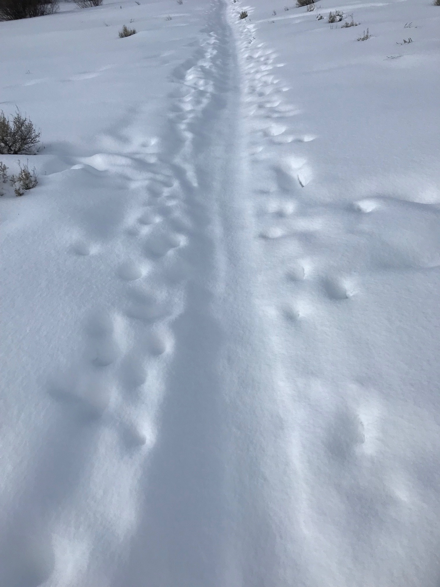
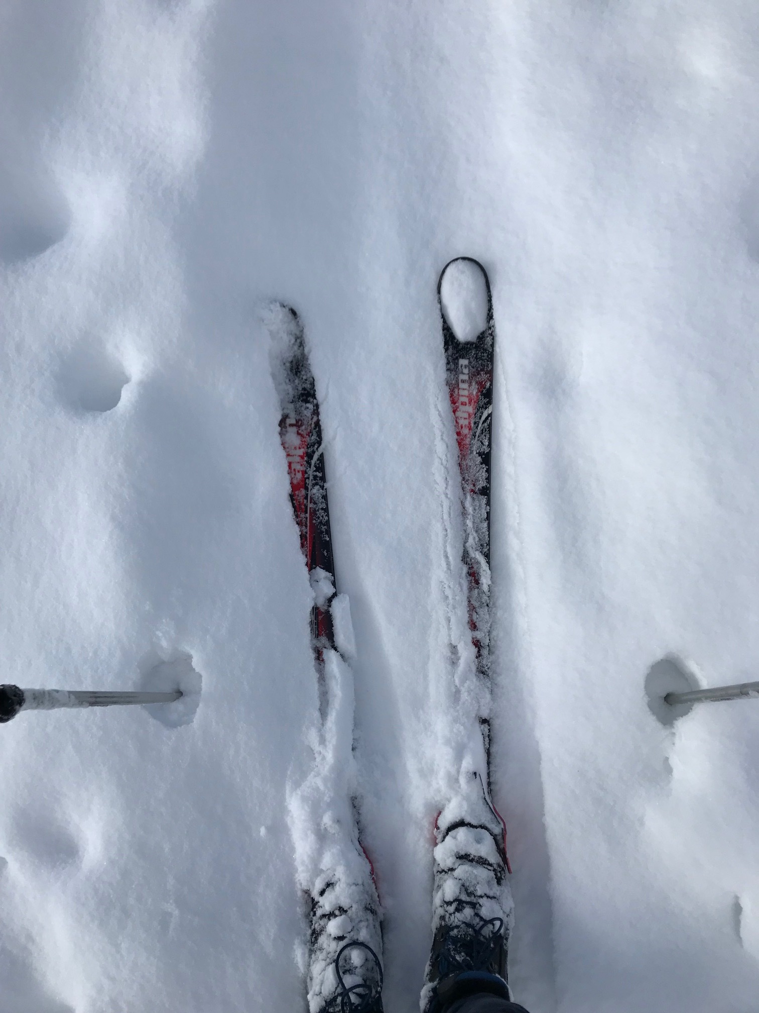
While out cross country skiing after a few inches of fresh fallen snow it dawned on me, especially when going uphill on my cross-country skis, lifting your toes up definitely pushes the head of the first metatarsal down and helps you to gain more purchase with the scales on the bottom of the skis. It also helps to press the center portion of the camber of the ski downward so that you can get better traction. Thinking about this further, lifting your toes up also helps you to engage your glutes to a greater degree.

Try this: stand comfortably with your knees slightly flexed. Lift up your toes leaving the balls of your feet on the ground. Do you feel the first metatarsal head going down and making better contact with the ground? Can you feel your foot tripod between the head of the first metatarsal, head of the fifth metatarsal and the calcaneus? Now let your toes go down. Squeeze your glute max muscles. You should still be able to fart so don’t squeeze the sphincter. You can palpate these muscles to see if you’re actually getting to them. You can do this by placing your hands on top of your hips with your fingers calling around forward like when your mom used to put her hands on her hips and yell at you. Now relax with your toes up again leaving the balls of your feet on the ground. Now engage your glutes. See how much easier it is?

Now stand with your feet flat on the ground and put your hands on your abs, specifically your external obliques. Now raise your right leg. Do you feel your external oblique engage? Now, lift your toes up leaving the balls of your feet on the ground. Now lift your leg. Do you feel how much more your abs engage?

Little tricks of the trade. That’s why you listen here and why your patients/clients come to see you. Now go out and do it!

Dr. Ivo, one of The Gait Guys

#gaitanalysis, #crosscountryskiing, #skiing, hallux, #engage, #abs