The Glute Max does that?

8.24.20.png

-There’s lots of talks about glutes and glute function. When was the last time you thought about the gluteus maximus has an internal rotator of the hip? We did a recent podcast and talked about this during climbing. 

-As the thigh flexes to 90° or greater, the internal rotational power of both the gluteus maximus and medius increases exponentially. Think about this when prescribing your rehabilitation exercises and get creative!

8.24.20 2.png

For a lot more information on this as well as other fun things with the glutes join us tomorrow evening for our monthly third Wednesdays Teleseminar on onlinece.com biomechanics 328 tomorrow night, 7 central 

Come join us for an hour of talking about peoples butts and how to make them work better :-)

#gluteus #gluteusmaximus #glute #glutes #muscleaction #muscleactions #rehab #rehabilitation #exercise #gluteexercise #gluteexercises 

Hills for training

The top five reasons we like Hills for training ankle rocker and hip extension

1. Hills do not cost money and are almost always readily available

2. Hills do not pull the hip into extension and place of stretch on the anterior hip musculature including the rectus femoris, iliopsoas and iliacus. This causes a slow stretch of the muscle activating the muscle spindles and causing muscle contraction via the stretch reflex. This will inhibit the posterior compartment of hip extensors through reciprocal inhibition, making it difficult to fire them.

3. A hill does not force your knee into extension, eliciting a stretch reflex in the hamstrings like a treadmill does

4. A hill naturally puts the ankle into dorsiflexion, And, along with active pulling up of the toes, helps you to get more into your anterior compartment and limits the tendency of the ankle being pulled into dorsiflexion (like a treadmill) which would initiate a stretch reflex in the gastroc/soleus and long flexors for the toes

5. The increased hip flexor recruitment of going uphill gives you more opportunity to engage your abs before the psoas and rectus femoris/TFL and, on the stance phase leg, you can get an increased stretch of those muscles

#hill #hills #rehab #rehabilitation#runningupthathill #runninghills#ankledorsiflexion #anklerocker#hipextension

Valgus Posts

A valgus post on an orthotic is a useful tool when you are trying to get weight off of the lateral and onto the medial column of the foot. It works best with people that have adequate range of motion in the first ray complex (they can get the head of the big toe to the ground) because if you don’t have adequate range of motion, you will only force the knee to the midline which sometimes can cause patellofemoral problems.

A valgus post as a post which begins wider laterally and narrows medially. It usually begins near the tubercle of the fifth metatarsal and can extend as far up as the end of the fifth toe. The idea is as you walk across the post, it forces the foot from lateral to medial helping to sink the first ray down to the ground. It functions similar to a first ray cut out (A notch cut out of the orthotic at the head of the first metatarsal) however is generally more gradual and sometimes better tolerated.

This type of posting is often used in people with internal tibial torsion who often having a difficult time getting weight onto the first right and keeping the knee in the sagittal plane.

In this video we briefly describe how the posting works. 

A simple test for motor programming


Marching, a rudimentary motion, is a great screening for people with motor programming difficulties. This gal has left lower extremity dystonia and we were looking to see if it was more peripheral or central in origin.


You can see how her movement breaks down after a few simple steps of attempted coordinated movement. Use this simple marching screen next time you suspect a central programming issue in your patients :-) 

3 clues that someone has internal tibial torsion

Watch this video a few times through and see what you notice. There are three clues that this patient has internal tibial torsion, can you find them?

He presented with right sided knee pain, medial aspect of the patella and medial joint line as well as tenderness over the medial joint line and pes anserine. Lower extremity musculature test strong and 5/5 save for his semi tendinosis on the right which tested 4/5.He has diminished endurance bilaterally in the external obliques

1. Note how his knees, right greater than left, fall outside the sagittal plane

2. Note the decreased progression angle of both feet during forward motion

3. Note how he toes off in supination, right greater than left.

This patient’s knee pain is coming from irritation of the pes anserine, particularly semitendinosus and his inability to recruit his abdominals sufficiently so, instead of the usual pattern of recruiting iliopsoas or rectus femoris, he chooses his sartorius, gracious and semi tendinosis.

Pay attention to how the new tracks, the progression angle as well as if they tow off in pronation, neutral, or supination in that can offer subtle clues to internal tibial torsion.

Dr Ivo Waerlop, one of The Gait Guys

#internaltibialtorsion #gaitanalysis #thegaitguys

https://vimeo.com/365342814

What a difference a few months makes

Take a look at the pre-and post videos of this gal with a forefoot supinatus and impaired motor control of her feet and core. Shuffle walks, foot intrinsic exercises, core work and gait retraining can go a long way! The important thing to remember here is that the patient was very motivated and did what was required to make things happen. A testament to tenacity and dedication

Dr Ivo Waerlop, one of The Gait Guys

#beforeandafter #gaitretraining #gaitanalysis #forefootsupinatus

Ode to the Popliteus

Remember the popliteus? To recap, it contracts at the initial contact phase of the walking gait cycle, to act as an accessory PCL (look HERE

https://www.thegaitguys.com/search?q=popliteus&f_collectionId=57d4982c91b18610c6ee3e0f

to read about that), then contracts eccentrically to slow the rate of internal rotation of the femur on the tibia until midstance, so as not to macerate the meniscus; It then contracts concentrically to accelerate the external rotation of the femur on the tibial plateau so it rotates faster then the tibia, to protect the meniscus as well. So, internal rotation of the femorotibial complex from initial contact to midstance and external rotation of the complex from midstance to preswing. Got it?

Now look at the video of this gal with L sided medial knee pain and past history of a left tibial plateau fracture in her youth. Do you see it? Hmmmm; doesn’t look like internal rotation does it? Don’t see it? Remember that the whole complex SHOULD be internally rotating until the swing phase leg passes the stance pase leg. See it now? Considering that the popliteus tested weak on the clinical exam, does this surprise you?

Agreed that there are many factors initiating internal rotation (and thus pronation) of the stance phase leg from initial contact to midstance, like plantar flexion, adduction and eversion of the talus, contraction of the lower leg anterior compartment muscles, eccentric contraction of the quads and hamstrings, just to name a few, can you see how (a least theoretically) one bad player can ruin the team?

Yes, popliteus rehab, along with abdominal core and foot core endurance exercises are in her future.

Dr Ivo Waerlop, one of The Gait Guys

#popliteus #kneepain #kneeproblem #thegaitguys #gaitanalysis

Tricks of the trade: Backward walking

image credit: https://pixabay.com/vectors/slide-sliding-falling-stickman-151861/

image credit: https://pixabay.com/vectors/slide-sliding-falling-stickman-151861/

A single event can generate asynchronous sensory cues due to variable encoding, transmission, and processing delays. Robert Peterka talks about this, along with posture compensation and system apportionment when it comes to balance and coordination of the visual, vestibular and proprioceptive systems. We have talked about that here on the blog in the past.

We are often looking for ways to “highlight” pathology and make it more visible in the clinical exam. Having your patient/client walk backwards is one of those tools.

Walking and remaining upright in the gravitational plane requires 3 integrated systems to work in concert with one another: the visual, vestibular and proprioceptive systems. Backwards walking requires a more coordinated effort AND IF there is a “hiccup” or extra demand on the system (the proprioceptive in this case), neurological processing can take a little longer, efforts can be delayed and the end result is a greater compensation is needed; this often makes pathology more evident.

Try having your client walk backwards when you are doing your exam and see what we mean. We think you will be surprised with the results : )

Dr Ivo Waerlop, one of The Gait Guys

Peterka RJStatler KDWrisley DMHorak FB. Postural compensation for unilateral vestibular loss. Front Neurol. 2011 Sep 6;2:57. doi: 10.3389/fneur.2011.00057. eCollection 2011.

temporal Shayman CSSeo JHOh YLewis RFPeterka RJHullar TE.Relationship between vestibular sensitivity and multisensory temporal integration. J Neurophysiol. 2018 Oct 1;120(4):1572-1577. doi: 10.1152/jn.00379.2018. Epub 2018 Jul 18.

Hawkins KABalasubramanian CKVistamehr AConroy CRose DKClark DJFox EJ. Assessment of backward walking unmasks mobility impairments in post-stroke community ambulators. Top Stroke Rehabil. 2019 May 12:1-7. doi: 10.1080/10749357.2019.1609182. [Epub ahead of print]

#backwardwalking #clinicalexam #thegaitguys #gaitpathology #clinicaltricksofthetrade

On the subject of manual muscle work…

image credit: https://commons.wikimedia.org/wiki/File:Muscle_spindle_model.jpg

image credit: https://commons.wikimedia.org/wiki/File:Muscle_spindle_model.jpg

Here is an older article that may seem verbose, but has interesting implications for practitioners who do manual muscle work with their clients. We would invite you to work your way through the entire article, a little at a time, to fully grasp it’s implications.

Plowing through the neurophysiology, here is a synopsis for you:

Tactile and muscle afferent (or sensory) information travels into the dorsal (or posterior) part of the spinal cord called the “dorsal horn”. This “dorsal horn” is divided into 4 layers; 2 superficial and 2 deep. The superficial layers get their info from the A delta and C fibers (cold, warm, light touch and pain) and the deeper layers get their info from the A alpha and A beta fibers (ie: joint, skin and muscle mechanoreceptors).

So what you may say

The superficial layers are involved with pain and tissue damage modulation, both at the spinal cord level and from descending inhibition from the brain. The deeper layers are involved with apprising the central nervous system about information relating directly to movement (of the skin, joints and muscles).

Information in this deeper layer is much more specific that that entering the more superficial layers. This happens because of 3 reasons:

  1. there are more one to one connections of neurons (30% as opposed to 10%) with the information distributed to many pathways in the CNS, instead of just a dedicated few in the more superficial layers

  2. the connections in the deeper layers are largely unidirectional and 69% are inhibitory connections (ie they modulate output, rather than input)

  3. the connections in the deeper layers use both GABA and Glycine as neurotransmitters (Glycine is a more specific neurotransmitter).

Ok, this is getting long and complex, tell me something useful...

This supports that much of what we do when we do manual therapy on a patient or client is we stimulate inhibitory neurons or interneurons which can either (directly or indirectly)

  • inhibit a muscle

  • excite a muscle because we inhibited the inhibitory neuron or interneuron acting on it (you see, 2 negatives can be positive)

So, much of what we do is inhibit muscle function, even though the muscle may be testing stronger. Are we inhibiting the antagonist and thus strengthening the agonist? Are we removing the inhibition of the agonist by inhibiting the inhibitory action on it? Whichever it may be, keep in mind we are probably modulating inhibition, rather than creating excitation.


Semantics? Maybe…But we constantly talk about being specific for a fix, not just cover up the compensation. Is it easier to keep filling up the tire (facilitating) or patching the hole (inhibiting). It’s your call


Yan Lu Synaptic Wiring in the Deep Dorsal Horn. Focus on Local Circuit Connections Between Hamster Laminae III and IV Dorsal Horn Neurons J Neurophys Volume 99 Issue 3

March 2008 Pages 1051-1052 link: http://jn.physiology.org/content/99/3/1051

Music to my ears. Movement to my steps...

https://en.wikipedia.org/wiki/Music

https://en.wikipedia.org/wiki/Music

"The applicable contribution of these novel findings is that music tempo could serve as an unprompted means to impact running cadence. As increases in step rate may prove beneficial in the prevention and treatment of common running-related injuries, this finding could be especially relevant for treatment purposes, such as exercise prescription and gait retraining."

Van Dyck E, Moens B, Buhmann J, Demey M, Coorevits E, Dalla Bella S, Leman M. Spontaneous Entrainment of Running Cadence to Music Tempo. Sports Med Open. 2015;1(1):15. Epub 2015 Jul 14.

link to free full text: https://www.ncbi.nlm.nih.gov/pmc/articles/PMC4526248/

image credit: http://pressplay.pbworks.com/w/page/82954552/Loebner%20Keith%20HW%203

Perhaps we need to change how we are are rehabbing X (insert your favorite weight bearing joint)

image credit: https://en.wikipedia.org/wiki/StrongBoard_balance

image credit: https://en.wikipedia.org/wiki/StrongBoard_balance

We have recently run across some research that has changed the way we look at some of the rehab we do, especially proprioceptive rehab. Perhaps it will do the same for you.

Traditionally, we present increasing balance requirements to the weight bearing structure by changing one or more of the three parameters that keep us upright in the gravitational plane: vision, the proprioceptive system (which include the muscles, joints and ligaments) and the vestibular system (the utricle, saccule and semicircular canals). We have discussed them extensively in multiple articles here on the blog. We generally would make the rehab task more difficult by removing a stimulus (closing your eyes, having someone stand on foam) or challenging (standing on one leg, putting someone on a wobble board, BOSU, extending the head, etc) the to make it more durable and "educated". More difficult task + better balance = more stable joint and better outcomes. 

The importaat thing is to think about how much of each system is apportioned; we often (wrongly) assume it is pretty equally divided between the three. It turns out, that it really depends on the surface you are standing on and the circumstances.

On flat planar surfaces, the division of labor looks something like this:

  • proprioceptive system 70%
  • vestibular system 20%
  • visual system 10 %

On uneven or unstable surfaces (like a BOSU, dynadisc, foam, Swiss ball, etc), it looks like this:

  • vestibular system 70%
  • visual system 20%
  • proprioceptive system 10%

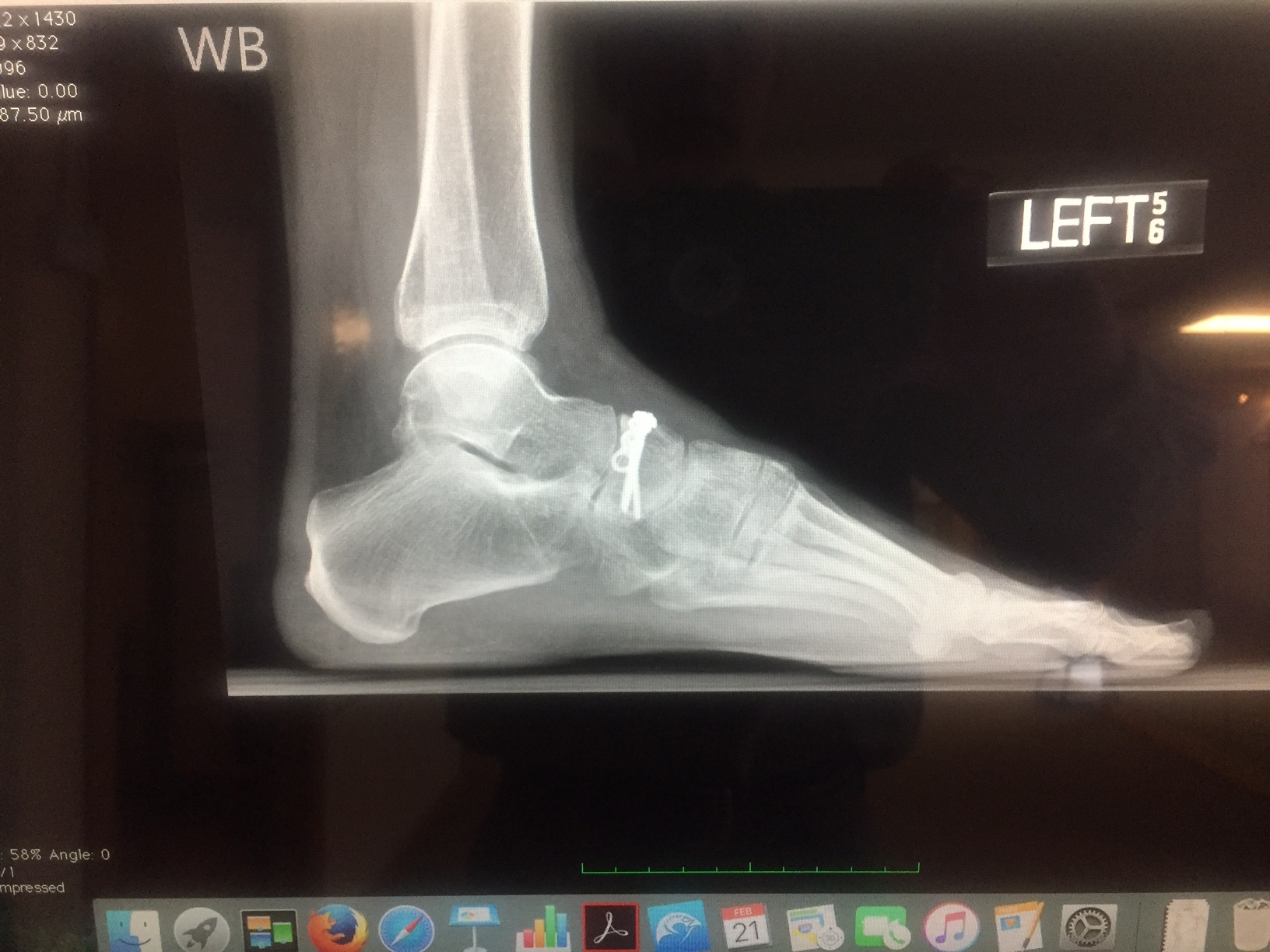
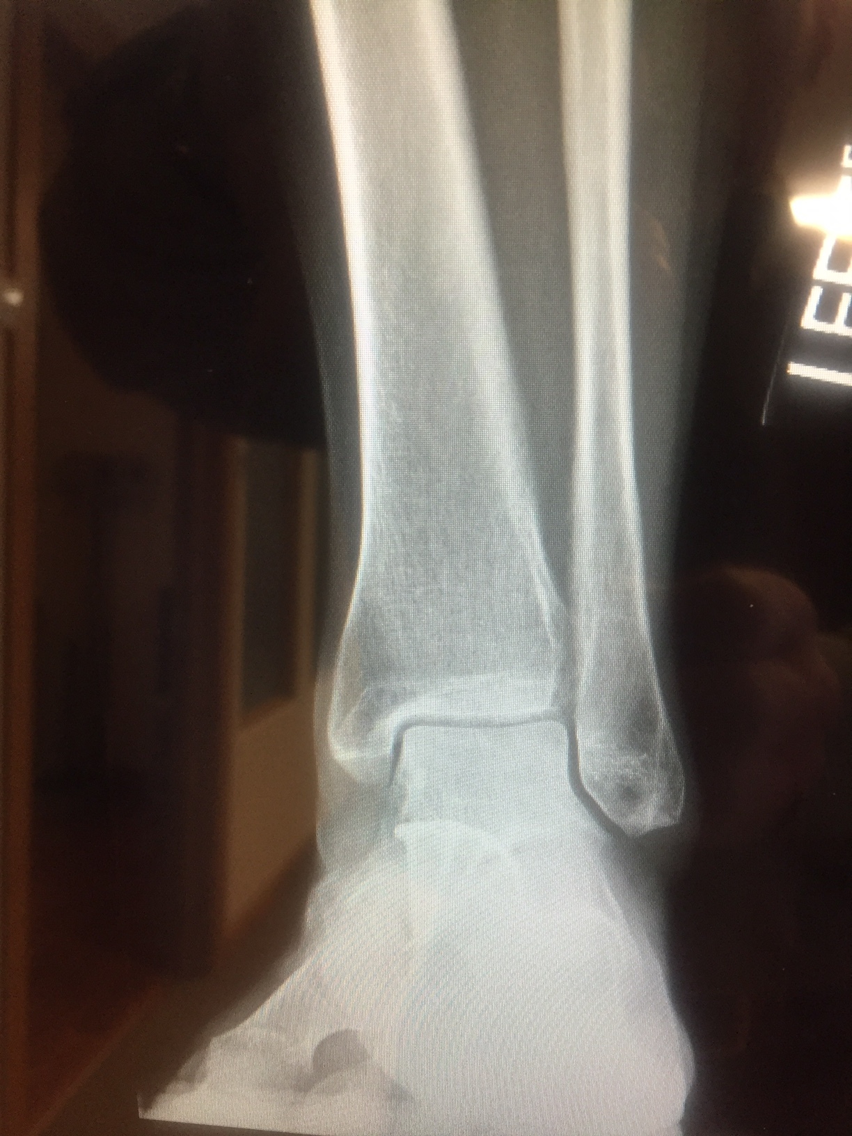
So, if we are rehabbing an ankle, it would make the most sense to do most of the rehab (and additional challenges) on a flat planar surface, perhaps incorporating things like forward, backward and side lean, toe and heel work and closed chain strengthening. WE could also close the eyes to make them more dependent on the proprio system, or extend the head 60 degrees to dampen the influence the lateral semicircular canals. We can put them on a BOSU or unstable surface but we need to remember that in that case, we will be rehabbing the vestibular system AND PERHAPS teaching THAT SYSTEM to compensate more, than the "broken" system. Yes, they get better BUT we are not fixing the system that is injured. 

You could make the argument, that your athletes/clients run/walk/exercise on uneven surfaces and use their vestibular system more.Maybe so, but is the actual injury to the vestibular system or to the musculoskeletal one?

Armed with this information, try and think of the system that is compromised and focus your efforts on that system, rather than the other two. Yes, people have vestibular dysfunction and refractive errors and need therapy, exercises and/or corrective lenses, but many of us are not vestibular or opticokinetic therapists (kudos to those of you who are!)


Peterka RJ, Statler KD, Wrisley DM, Horak FB. Postural Compensation for Unilateral Vestibular Loss. Frontiers in Neurology. 2011;2:57. doi:10.3389/fneur.2011.00057.

Horak FB. Postural Compensation for Vestibular Loss. Restorative neurology and neuroscience. 2010;28(1):57-68. doi:10.3233/RNN-2010-0515.

This simple screening test becomes a form of exercise.

Today we look at a simple CNS screen for your “central pattern generators” or “CPG’s”. If you do not pass, then the exercise becomes the rehab exercise. If you (or your client) does not have good coordination between the upper and lower extremity, then they will not be that efficient, physiologically or metabolically. 

The “cross crawl” or “step test” looks at upper and lower extremity coordination, rather than muscular strength. If performed for a few minutes, it becomes a test that can look at endurance as well. 

It is based on the “crossed extensor” response, we looked at last week. That is, when one lower limb flexes, the other extends; the contralateral upper limb also flexes and the ipsilateral upper limb extends. It mimics the way things should move when walking or running. 
 

  • Stand (or have your client stand) in a place where you will not run into anything.
  • Begin marching in place.
  • Observe for a few seconds. When you (or your client) are flexing the right thigh, the left arm should flex as well; then the left thigh and right arm. Are your (their) arms moving? Are they coordinated with the lower extremity?
  • What happens after a few minutes? Is motion good at 1st and then breaks down?
  • Now speed up. What happens? Is the movement smooth and coordinated? Choppy? Discoordinated?
  • now slow back down and try it with your (their) eyes closed


If  movement is smooth and coordinated, you (they) pass

If movement is choppy or discoordinated, there can be many causes, from simple (muscle not firing, injury) to complex (physical or physiological lesion in the CNS).

  • If movement is not smooth and coordinated, try doing the exercise for a few minutes a day. You can even start sitting down, if you (they) cannot perform it standing. If it improves, great; you were able to help “reprogram” the system. If not, then you (they) should seek out a qualified individual for some assistance and to get to the root of the problem.

Arm swing and instability. To train or not to train... Should we do it?

We have long talked about arm swing and whether to change it, encourage it or just observe it. It appears to be an indicator of potential instability as well as a portent for more dire neurological problems (Alzheimers, Parkinson's)

This study looks at altered arm swing in kids with CP; how it is an indicator that there is a problem and how it can profoundly effect their gait and stability. Cerebral palsy may be an extreme case, but how does it differ REALLY (other than severity) from someone who has a mild neurological impairment, such as movement patterning disorders, that we see each and every day in our friends, family, clients and patients? Try and think out of the box and investigate the implications.

"Observational research suggests that in children with cerebral palsy, the altered arm swing is linked to instability during walking. Therefore, the current study investigates whether children with cerebral palsy use their arms more than typically developing children, to enhance gait stability. Evidence also suggests an influence of walking speed on gait stability. Moreover, previous research highlighted a link between walking speed and arm swing. Hence, the experiment aimed to explore differences between typically developing children and children with cerebral palsy taking into account the combined influence of restricting arm swing and increasing walking speed on gait stability. Spatiotemporal gait characteristics, trunk movement parameters and margins of stability were obtained using three dimensional gait analysis to assess gait stability of 26 children with cerebral palsy and 24 typically developing children. Four walking conditions were evaluated: (i) free arm swing and preferred walking speed; (ii) restricted arm swing and preferred walking speed; (iii) free arm swing and high walking speed; and (iv) restricted arm swing and high walking speed. Double support time and trunk acceleration variability increased more when arm swing was restricted in children with bilateral cerebral palsy compared to typically developing children and children with unilateral cerebral palsy. Trunk sway velocity increased more when walking speed was increased in children with unilateral cerebral palsy compared to children with bilateral cerebral palsy and typically developing children and in children with bilateral cerebral palsy compared to typically developing children. Trunk sway velocity increased more when both arm swing was restricted and walking speed was increased in children with bilateral cerebral palsy compared to typically developing children. It is proposed that facilitating arm swing during gait rehabilitation can improve gait stability and decrease trunk movements in children with cerebral palsy. The current results thereby partly support the suggestion that facilitating arm swing in specific situations possibly enhances safety and reduces the risk of falling in children with cerebral palsy."

Front Hum Neurosci. 2016 Jul 15;10:354. doi: 10.3389/fnhum.2016.00354. eCollection 2016.
Restricted Arm Swing Affects Gait Stability and Increased Walking Speed Alters Trunk Movements in Children with Cerebral Palsy.
Delabastita T, Desloovere K, Meyns P.

link to FREE FULL TEXT: https://www.ncbi.nlm.nih.gov/pmc/articles/PMC4945643/

The Beef on the EDL.....

We have long been promoting appropriate function of the long extensors of the toes  here, in our practices, our lectures, on Youtube, in our book......You get the idea. Lets take a closer look at this often weakened and overlooked muscle.

We remember that the EDL lies mostly in the superior and somewhat lateral part of the anterior compartment of the lower leg, comprising approximately the upper 2/3 from under the lateral tibial plateau and fibula, and from the interosseus membrane. It lies under the tibialis anterior, and the extensor hallucis longus lies below it. Its tendons pass inferiorly and travel under the extensor retinaculum and attaches to the base of the distal phalanges of toes 2-4. These muscles act from initial contact to loading response to help eccentrically lower the foot to the ground and ensure smooth heel rocker and most likely attenuate the speed of initial pronation as the talus glides anteriorly on the calcaneal facets and again from terminal stance through initial swing to provide compression of the metatarsal phalangeal and interphalangeal joints, to offset the long flexors (which are often overactive) and create clearance for the toes during swing.  

jhowardL.gif
jhowardR.gif

What does it look like when the long extensors don’t work so well? Have a look at the pedograph on the right (pair J howard r). what do we see? First we notice the lack of printing under the head of the 1st metatarsal and increased printing of the second metatrsal head. Looks like this individual has a forefoot supinatus, or possibly a forefoot varus (cannot get the head of the 1st metatarsal to the ground, and thus a weak medial tripod, possibly insufficient extensor hallucis brevis, peroneus longus, flexor digitorum brevis, or all of the above). Next we see increased printing of the distal phalanges of digits 2-4. Looks like the long flexors are dominant, which means the long extensors are inhibited. What about the lack of printing of the 5th toe? I thought the flexors were overactive? They are, but due to the supinatus, the foot is tipped to the inside and the 5th barely contacts the ground!

How do you fix this?

  • Help make a better foot tripod using the toe wave, tripod standing and extensor hallucis brevis exercises.
  • Make sure the articulations are mobile with joint mobilization, manipulation and massage.
  • How about dry needling and acupuncture to improve function?
  • Make sure the knee and hip are functioning appropriately.
  • Put them in footwear that will allow the foot to function better (a less rigid, less ramp delta shoe).
  • As a last resort, if they cannot make an adequate tripod because of lack of motivation, anatomical constraints or both, use a foot leveling orthotic.


Want to be faster?

superhero.jpg

Take this simple test. 

If you want to be faster, you had better incorporate some proprioceptive training into your plan. It is the 1st part of our mantra: Skill, Endurance, and Strength (in that order). Proprioceptive training appears to be more important that strength or endurance training from an injury rehabilitation perspective injury rehabilitation perspective as well part of an injury prevention program

 What is proprioception? It is body position awareness; ie: knowing what your limbs are doing without having to look at them.

Take this simple test:

  • Stand in a doorway with your shoes off. Keep your arms up at your sides so that you can brace yourself in case you start to fall. Lift your toes slightly so that only your foot tripod remains on the ground (ie the base of the big toe, the base of the little toe and the center of the heel.). Are you able to balance without difficulty? Good, all 3 systems (vision, vestibular and proprioceptive) are go.
  • Now close your eyes, taking away vision from the 3 systems that keep us upright in the gravitational plane. Are you able to balance for 30 seconds? If so, your vestibular and proprioceptive systems are intact.
  • Now open your eyes and look up at the ceiling. Provided you can balance without falling, now close your eyes. Extending your neck 60 degrees just took out the lateral semicircular canals of the vestibular system (see here for more info). Are you still able to balance for 30 seconds? If so, congrats; your proprioceptive system (the receptors in the joints, ligaments and muscles) is working great. If not, looks like you have some work to do. You can begin with exercises we use every day by clicking here.

Proprioception should be the 1st part of any training and/or rehabilitation program. If you don’t have a good framework to hang the rest of your training on, then you are asking for trouble. 


Timothy E. Hewett, PhD, , Kevin R. Ford, MS, Gregory D. Myer, MS, CSCS Anterior Cruciate Ligament Injuries in Female Athletes: Part 2, A Meta-analysis of Neuromuscular Interventions Aimed at Injury Prevention The American Journal of Sports Medicine Vol 34, Issue 3, pp. 490 - 498   link to free full text: http://journals.sagepub.com/doi/abs/10.1177/0363546505282619

Lephart SM1, Pincivero DM, Giraldo JL, Fu FH. The role of proprioception in the management and rehabilitation of athletic injuries. Am J Sports Med. 1997 Jan-Feb;25(1):130-7.


You can only take so many whacks to the head before it starts to show...

In addition to vision and the vestibular system, proprioceptive information is gathered by primarily 2 sources: The peripheral joint mechanoreceptors left (type 1, type 2, type 3, and type 4 ), as well as the muscle mechanoreceptors: Muscle spindles and golgi tendon organs. This information is transmitted to the cortex via the dorsal column and spinocerebellar pathways. The information is then integrated in the parietal lobe (for information in the dorsal columns) and cerebellum. The information is then relayed to the motor cortex, basal ganglia, and vestibular system. The interplay of these 3 systems, vision, the vestibular system, and the joint/muscle mechanoreceptors is what allows us to keep our bodies up right and functioning in the gravitational field. When integration is compromised, at either a peripheral or cortical level, proprioception suffers.

" Measures of cumulative subconcussive head impacts during a men’s lacrosse season are associated with decreases in balance scores from pre- to postseason, according to findings from Sacred Heart University in Fairfield, CT, that could have implications for lower extremity injury risk. The findings suggest that, even in the absence of a concussion, repetitive subconcussive impacts can negatively affect an athlete’s balance, which in turn can increase the risk of lower extremity injury"

http://lermagazine.com/news/in-the-moment-sports-medicine/subconcussive-subtleties-lacrosse-study-links-balance-impacts

Miyashita TL, Diakogeorgiou E, Marrie K. Correlation of head impacts to change in balance error scoring system scores in Division I Men’s lacrosse players. Sports Health 2017 Jan 1. [Epub ahead of print]

Plisky PJ, Rauh MJ, Kaminski TW, Underwood FB. Star Excursion Balance Test as a predictor of lower extremity injury in high school basketball players. J Orthop Sports Phys Ther 2006;36(12):911-919.

Where do you do YOUR gait retraining?

It may seem like we are stating the obvious, but visual contrast seems to matter more than visual acuity. When we look at light contrast in lower frequencies (red ranges), we start to see some decomposition of gait with regards to step length, but not necessarily cadence or speed in this study of over 4,000 folks over 50 (hey, that’s us!). What are your patients wearing on their feet? What color is your floor? How about the walls?

Do your gait retraining in a well lit area with lots of contrast between the floor and your patients shoes, as well as the surroundings.


Duggan E, Donoghue O, Kenny RA, Cronin H, Loughman J, Finucane C. Time to Refocus Assessment of Vision in Older Adults? Contrast Sensitivity but Not Visual Acuity Is Associated With Gait in Older Adults. J Gerontol A Biol Sci Med Sci. 2017 Feb 28. doi: 10.1093/gerona/glx021. [Epub ahead of print]


More on that post operative foot

This is part 2 of a series following a case. If you missed part 1, please go back here and read what we found.

The patient returns 1 week later and reports being approximately 25% improved. She has been performing her "toes up" exercises while walking all the time. She is having some difficulty still with balance. She has been performing her toe waving exercises a few times daily.

X-rays performed 2/17 reveal screw fixation of the navicular. I cannot find evidence of a previous cuboid fracture. The ankle mortise is clear.

She still has 4/5 weakness of the long and short toe extensors; long greater than short. She has tenderness to palpation along the anterior aspect of the deltoid ligament on the left hand side which is made worse with eversion of the ankle. There is a loss of long axis extension at the talocrural and talonavicular articulations. Less tenderness is noted in the inter metatarsal intervals and the interossei musculature.

There is significant improvement over last time. Lack of fixation of the navicular to other articulations will allow us to perform manipulation/mobilization of the foot.

We treated with diagnostic manipulation and mobilization of the foot. I reviewed exercises to date and added the shuffle walk exercise. Since acupunture and needling can influence blodd flow (1-4) We utilized acupuncture points stomach 36, spleen 6, gallbladder 41, liver 3, points in the inter metatarsal intervals, bladder 67 and liver 1.Neelding has been shown to improve muscke activation (4-7) so I did origin/insertion stimulation of the long extensors with 3 sets of 10 repetition cocontraction along with origin/insertion stimulation of the short extensors with 3 sets, 10 repetitions cocontraction was performed. She will follow back in approximately 2 weeks because of travel.

So far, so good. We will keep you posted : )


1. Sandberg, M., Larsson, B., Lindberg, L.-G. and Gerdle, B. (2005), Different patterns of blood flow response in the trapezius muscle following needle stimulation (acupuncture) between healthy subjects and patients with fibromyalgia and work-related trapezius myalgia. European Journal of Pain, 9: 497. doi:10.1016/j.ejpain.2004.11.002

2.  Cagnie, Barbara et al. The Influence of Dry Needling of the Trapezius Muscle on Muscle Blood Flow and Oxygenation Journal of Manipulative & Physiological Therapeutics , Volume 35 , Issue 9 , 685 - 691

3. Tsuchiya, Masahiko; Sato, Eisuke F.; Inoue, Masayasu; Asada, Akira† Acupuncture Enhances Generation of Nitric Oxide and Increases Local Circulation  Anesthesia & Analgesia: February 2007 - Volume 104 - Issue 2 - pp 301-307

4. Jan Dommerholt Dry needling — peripheral and central considerations Journal Of Manual & Manipulative Therapy Vol. 19 , Iss. 4,2011

5. Zanin, Marília Silva et al. Electromyographic and Strength Analyses of Activation Patterns of the Wrist Flexor Muscles after Acupuncture Journal of Acupuncture and Meridian Studies , Volume 7 , Issue 5 , 231 - 237

6. https://youtu.be/02-M0i6AKAk

7. Fragoso APS, Ferreira AS. Immediate effects of acupuncture on biceps brachii muscle function in healthy and post-stroke subjects. Chinese Medicine. 2012;7:7. doi:10.1186/1749-8546-7-7.


Cerebellar impairment = Gait Changes = Happy Patient

This is a fairly info dense post with many links. please take the time to explore each one to get the most out of it. 

If you have been with us here on TGG long enough, you know the importance of the cerebellum and gait. Mechanoreceptor information travels north to the cortex via the dorsal (and ventral) spinocerebellar pathways to be interpreted (and interpolated, in the case of the ventral pathway), with the information relaying back to the motor cortex and vestibular nucleii and eventually back down to the alpha (and gamma) motor neurons that proved the thing you call movement and thus gait. (Cool video on spinocerebellar pathways here and here).

This FREE FULL TEXT paper has some cool charts, like this one, that show the parameters of gait that change with cerebellar dysfunction (in this case, disease, although idiopathic means they really don't know. Anatomical or physiological lesions will behave the same, no? Doesn't the end result of a functional short leg look the same as an anatomical one?)

Looking tat this chart, what do we really see? People with cerebellar dysfunction:

  • a shorter step length
  • a wider base of gait
  • decreased velocity
  • increased lateral sway
  • slower overall gait cycle

Hmmmm...Beginning to sound like a move toward more primitive gait. Just like we talked about in this post on the 5 factors and proprioception here several years ago. We like to call this decomposition of gait. 

They go on to talk about specific anatomic regions of the cerebellum and potential correlation to specific gait abnormalities, like the intermediate zone and interposed nucleii controlling limb dynamics and rhythmic coordination like hypermetria (overshooting a target), especially when walking in uneven surfaces or when gait is perturbed, like walking into something or changes in surface topography, or the lateral zone of the cerebellum, for voluntary limb control, such as where you place your foot. Definitely gait nerd material.

There aren't any direct tips on rehab, but it would stand to reason that activities that activate the cerebellum and collateral pathways would give you the most clinical gains. Lots of propriosensory exercises like here, here, here and here for a start.

Happy cerebellum = Happy patient

The Gait Guys


Winfried Ilg, Heidrun Golla, Peter Thier, Martin A. Giese; Specific influences of cerebellar dysfunctions on gait. Brain 2007; 130 (3): 786-798. doi: 10.1093/brain/awl376  FREE FULL TEXT

One of life's great mysteries....Some folks will do what they want anyway....

The origins of the species, gravity and women...Just a few of life mysteries. Reading this article (1) made us sad in many ways. It's like smoking. You know it's bad for you but you keep doing it. Why? The mystery remains to us.

Vanity seems to often trump biomechanics, as we see in pencil skirts (see our post here), droopy pants (see here)  and high heels (here).

Yet, here is yet another study about women, heels and bunion surgery. 

"Almost two thirds (31) of the 50 patients who said they wanted to go back to wearing heels after surgery did so, and 24 of these women said their postoperative use equaled or exceeded the frequency of their preoperative wear. There were no differences between pre- and postoperative heel heights.

In the study, women older than 65 years were more likely than younger women to report high-heel use prior to hallux valgus surgery.

However, 58.5% of study participants reported difficulty with heel wear, and 13.9% said they had significant restriction, and couldn’t wear anything without pain but custom orthopedic shoes or slippers. Most women (86%) were able to return to comfortable shoes after surgery with minimal or no discomfort; 27.7% said their footwear choice was unrestricted, meaning they could wear both comfortable shoes and heels with minimal discomfort. The 23 women older than 65 years were twice as likely to report significant restriction as those in the younger cohort; compared by operative type, patients who had the most extensive procedures had the highest rates of restriction. The findings were published in June by the World Journal of Methodology. (2)"

Bunions are believed to be caused by an inability to anchor the 1st ray and the untoward action of the adductor hallucis, acting from the transverse and oblique insertions more proximally on the foot, make the hallux head west. This is under the purview of the peroneus longus, extensor hallucis brevis as well as the short flexors of the lesser toes (see here).

The components of supination are plantar flexion, inversion and adduction. Why would you continue to wear a shoe with a narrow toe box that forces the big toe medially and that puts you in plantar flexion? We won't even begin to talk about the loss of ankle rocker.....

We guess folks will continue to do what they will do....


1. Robinson C, Bhosale A, Pillai A. Footwear modification following hallux valgus surgery: The all-or-none phenomenon. World J Methodol 2016;6(2):171-180.

2. http://lerfoothealth.com/archives/2016/most-women-who-want-to-wear-heels-after-bunion-surgery-do-so/