The EHB....In all its glory...

The extensor hallucis brevis : An overlooked "miracle worker"

tumblr_n3vbw7hW5t1qhko2so2_400.jpg

The Extensor Hallicus Brevis, or EHB as we fondly call it is an important muscle for descending the distal aspect of the 1st ray complex (1st metatarsal and medial cunieform) as well as extending the 1st metatarsophalangeal joint. It is in part responsible for affixing the medial tripod of the foot to the ground.  Its motion is generally triplanar, with the position being 45 degrees from the saggital (midline) plane and 45 degrees from the frontal (coronal) plane, angled medially, which places it almost parallel with the transverse plane. With pronation, it is believed to favor adduction (1).

It arises from the anterior calcaneus and inserts on the dorsal aspect of the proximal phalynx. It is that quarter dollar sized fleshy protruding, mass on the lateral aspect of the dorsal foot.  The EHB is the upper part of that mass. It is innervated by the lateral portion of one of the terminal branches of the deep peronel nerve (S1, S2), which happens to be the same as the extensor digitorum brevis (EDB), which is why some sources believe it is actually the medial part of that muscle. It appears to fire from loading response to nearly toe off, just like the EDB; another reason it may phylogenetically represent an extension of the same muscle (2-4).

Because the tendon travels behind the axis of rotation of the 1st metatarsal phalangeal joint, in addition to providing extension of the proximal phalynx of the hallux (as seen in the child above), it can also provide a downward moment on the distal 1st metatarsal (when properly coupled to and temporally sequenced with the flexor hallicus brevis and longus), assisting in formation of the foot tripod we have all come to love (the head of the 1st met, the head of the 5th met and the calcaneus).

Why is this so important?

The central axis of a joint (sometimes called the instantaneous axis of motion) is the center of movement of that articulation. It is the location where the motion will occur around, much like the center of a wheel, where the axle attaches. In an articulation, it usually involves one bone moving around another. Lets look at an example with a door hinge.

A hinge is similar to a joint, in that it has parts with is joining together (the door and the jamb), with a “joint” in between, The axis of rotation of the hinge is at the pivot rod. When the door, hinge and jamb are all aligned, it functions smoothly. Now imagine that the hinge was attached to the jamb 1/4” off center. What would happen? The hinge would bind and the door would not operate smoothly.

Now let’s think about the 1st metatarsal phalangeal joint. It exists between the head of the 1st metatarsal and the proximal part of the proximal part of the proximal phalanyx. Normally, because the head of the 1st metatarsal is larger than the heads of the lesser ones, the center of the joint is higher (actually,almost 2X as high; 8mm as opposed to 15mm). We also remember that the 1st metatarsal is usually shorter then the 2nd, meaning during a gait cycle, it bears the brunt of the weight and hits the ground earlier than the head of the 2nd.

tumblr_lij2n4n1mK1qggnse.jpg

The head of the 1st metatarsal should slide (or should we say glide?) posteriorly on the sesamoids during dorsiflexion of the hallux at pre swing (toe off). It is able to do this because of the descent of the head of the 1st metatarsal, which causes a dorsal posterior shift of the axis of rotation of the joint. We remember that the head of the 1st descends through the conjoined efforts of supination and the coordinated efforts of the peroneus longus, extensor hallucis brevis, extensor hallucis longus, dorsal and plantar interossei and flexor hallucis brevis (which nicely moves the sesamoids and keeps the process going smoothly)(1, 5).

Suffice it to say, if things go awry, the axis does not shift, the sesamoids do not move, and the phalanyx crashes into the 1st metatarsal, causing pain and if it continues, a nice spur you can write home about!

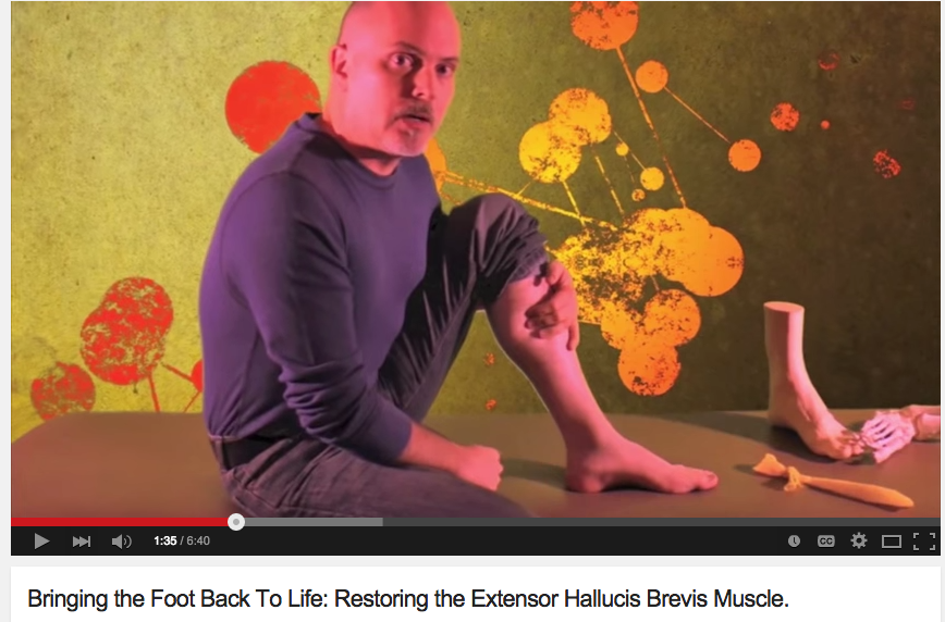
Treating and needling this muscle is easy, as it is very accessible on the dorsum of the foot and due to the decreased receptor density, is not too uncomfortable. We like to needle the peroneus longus and short flexors as well, as they all have the function of lowering the head of the 1st ray. Check it out in this quick how to video.

1. Michaud T: Human Locomotion: The Conservative Management of Gait Related DisordersNewton Biomechanics; First Edition 2011

2. https://www.physio-pedia.com/Extensor_Hallucis_brevis

3. http://www.wheelessonline.com/ortho/extensor_hallucis_brevis

4. Becerro de Bengoa Vallejo R., Losa Iglesias M.E., Jules K.T.  Tendon Insertion at the Base of the Proximal Phalanx of the Hallux: Surgical Implications (2012)  Journal of Foot and Ankle Surgery,  51  (6) , pp. 729-733.

5. Zelik, K.E., La Scaleia, V., Ivanenko, Y.P. et al. Eur J Appl Physiol (2015) 115: 691. https://doi.org/10.1007/s00421-014-3056-x

tumblr_nj93ad9yEM1qhko2so1_1280.jpg
tumblr_nj93ad9yEM1qhko2so2_r1_1280.png
tumblr_nj93ad9yEM1qhko2so3_r1_1280.png
tumblr_nj93ad9yEM1qhko2so4_r1_1280.png

Pain at toe-off; Stopping Big Toe Impingement with the extensor hallucis capsularis.

Photo: note the AET coming off the EHL tendon in the diagram

What if there was a mechanism in place by which to pull structures out of the way of a joint moving to end range ? If you know your biomechanics, you know this is a true phenomenon on several levels. We know of one at the knee, the articularis genu has been written about having function of drawing the suprapatellar bursa and joint capsule/synovial tissue cephalad (upward) during knee extension preventing an impingement phenomenon during full quadriceps contraction in knee extension loading. 

What if there were a similar mechanism in the big toe ? When teaching we are sometimes asked what joint, that when it goes sour, creates more devastation to the entire biomechanical chain than any other joint. I like to choose the big toe/1st metatarsophalangeal joint because failure to fully push off the big toe at full joint range impairs hip extension, stride and step lengths, and creates compensations far and wide ipsilaterally and contralaterally in the body. Most everyone knows about bunions, turf toe, hallux valgus, sesamoiditis and the like, but there are many other things that can make this joint painful. Today we bring you another “clearing mechanism” that acts to pull synovial and capsular tissues out of a joint that is nearing end range.
As seen in the anatomy dissection photo above, the extensor hallucis capsularis (EHC) is an accessory tendon slip off of the extensor hallucis longus (EHL). Interestingly, one study found that 8% of the dissections showed the EHC came off of the tibialis anterior tendon slip. This EHC accessory slip typically originates off the long extensor tendon (EHL) and traverses medially to the dorsomedial joint capsule region. Some studies suggest it is found in 80-98% of people. We propose it is most likely present in everyone because of the critical nature of its function. We propose that perhaps it may be missed on traditional dissections because of its blending with fascial tissues and because of its sometimes trivial size and girth. Just like when we fully extend our knee we want to be sure the articularis genu will draw the synovial capsular tissue up and out of the patellar/femoral approximation, the EHC has been shown on intra-operative testing to exert a pretension on the metatarsophalangeal (MTP) joint capsule similarly pulling the synovial-capsular tissue free from the end range dorsiflexing toe. Without this function, synovial-capsular impingement can occur and create pain and an inhibitory arthrogenic reflex to the EHL, tibialis anterior or any other muscles around the joint for that matter. This can act and feel like an acute “turf toe” (hyper-dorsiflexion event) and yet, not be true turf toe osseous impingement.
So if your client has pain at the dorsal joint on end range extension of the great toe, meaning things like toe-off, doing push ups from the ball of the foot, jumping, kneeling or squatting with the hallux in forced dorsiflexion etc, this tendon slip (and its origin, the EHL muscle) should be on your mind and assessment of the anterior compartment for S.E.S. must commence (S.E.S.= skill, endurance and strength, our Gait Guys mantra). This is why you need to intimately understand this important video (link) and need to know how to do this exercise, the shuffle walks (video link) and build clean ankle rocker ranges of motion via S.E.S. of the anterior compartment.  Pulling on the great toe, twisting it like a radio knob, and forcing end range shouldn’t be the biggest guns in your arsenal, logically restoring all the dysfunctional components should be.

We wonder how many of the videos online of people demonstrating big toe mobilizations, toe distractions, fancy exercises and various toe circus tricks to regain motion and function and reduce pain actually truly know about the anatomy and function of the big toe and how ankle rocker and other things can impair its function.  We wonder about these kinds of things.  

Please just remember, the average uneducated viewer is merely looking for solutions to their painful parts. Those in the know have a responsibility to deliver as complete a package as possible, within reason. 

“With great powers (and knowledge) there must also come, great responsibility.”-Stan Lee  

Dr. Shawn Allen

the gait guys

Photo credit link: http://www.wisconsinfootandankleinstitute.com

www.wisconsinfootandankleinstitute.com/img/research/The-Accessory-Extensor-Tendon_fig1.jpg

references:

Foot Ankle Surg. 2014 Sep;20(3):192-4. doi: 10.1016/j.fas.2014.04.001. Epub 2014 Apr 16.
The extensor hallucis capsularis tendon–a prospective study of its occurrence and function.Bayer T1, Kolodziejski N2, Flueckiger G2.

Foot Ankle Int. 2006 Mar;27(3):181-4.
Extensor hallucis capsularis: frequency and identification on MRI.
Boyd N1, Brock H, Meier A, Miller R, Mlady G, Firoozbakhsh K.

Foot Ankle Int. 2004 Jun;25(6):387-90.
The accessory extensor tendon of the first metatarsophalangeal joint.
Bibbo C1, Arangio G, Patel DV.

A year ago we produced this short video (link) on how to bring back the EHB (extensor hallucis Brevis muscle). Well, it continues to do its magic on a regular basis. Here is a patient’s foot with clear demonstration of unassisted success of isolated…

A year ago we produced this short video (link) on how to bring back the EHB (extensor hallucis Brevis muscle). Well, it continues to do its magic on a regular basis. Here is a patient’s foot with clear demonstration of unassisted success of isolated engagement of the EHB while simultaneous release of the EHL (ext. Hallucis long us) while engaging the FHL (flexor hallucis long us).  This patient could not isolate any of the long or short hallux muscles on his own. “I can’t find it, my brain doesn’t know what it is supposed to do or how to do it ! (paraphrased)  But after just 24 hours consisting of a few sessions of the exercise here is the result in the photo above.  Success !  And here were his comments: 

Doc, you were right - the brain is an amazingly plastic thing!

I’ll keep working on it, but happy to see such quick progress!

The client’s problem was some medial mid-rear foot pain from the resultant excessive increased pronation because of a forefoot varus.  Well, it is a bit more complicated than that to be precise. There was some true clinical ankle and rearfoot instability because of a lifetime of ankle sprains as well as some highly suspect lower syndesmosis hypermobility from probable distal anterior tib-femoral ligamentous attenuation/tears but the main point is that these were clinically manifesting themselves because of the apparent forefoot varus and the resultant pronatory foot mechanics to get the 1st metatarsal head (medial tripod) to the ground; a typical phenomenon .  Here is the kicker, he did  not have a fixed forefoot varus, it was a mirage, it was functional. What he had was an inability to descend the first metatarsal (plantarflex the Metatarsal) / medial tripod of the foot.  He could not do this because he could not separate ankle dorsiflexion and hallux dorsiflexion.  There was essentially no hallux dorsiflexion at all because he could not descend the 1st MET (head).  So, we knew it was time to break out the nuclear EBH exercise in the video above !  Big problems require big guns !

The rest is history. We fully expect to see a virtual disappearance of the “so called” forefoot varus (because it was never present in the first place). 

“If you have never seen the beast, you will not recognize it when you see it.”-unknown

Did you know that the EHB (extensor hallucis brevis) the topic of today’s video tutorial, originates off of the forepart of the medial aspect of calcaneus & lateral talocalcaneal ligament. It is just above the bulk origin of the EDB (exten…

Did you know that the EHB (extensor hallucis brevis) the topic of today’s video tutorial, originates off of the forepart of the medial aspect of calcaneus & lateral talocalcaneal ligament. It is just above the bulk origin of the EDB (extens

or digitorum brevis). It is frequently torn/strained in ankle inversion sprains and frequently goes undiagnosed. It can be torn/avulsed from the bone if the inversion sprain is focused below the lateral ankle joint. This occurs mostly when the foot is more plantarflexed before the inversion event. A foot cannot afford to have an impaired big toe ! Don’t miss this one !

Part 2 of the EHB: Bringing the Extensor Hallucis Brevis of the Foot Back to Life.

Today we show you a proprietary exercise we developed here at The Gait Guys. It was developed out of necessity for those clients who are too EHL dominant (long big toe extensor muscle) and big toe short flexor dominant (FHB). These two muscles are what we call a foot functional pair.  Big toes like these will be dysfunctional and will not be able to gain sufficient purchase on the ground to produce stability and power without impacting the joint (1st metatarsophalangeal joint).  Imbalances like these lead to altered joint loading responses and can be a possible predictor for premature damage to the joint over time. These imbalances are also what lead to injuries to the big toe, the arch and other areas of the foot. After all, when the big is weak or dysfunctional gait will be compensated.  When imbalance at this joint occurs because of EHB weakness the medial tripod anchor (the head of the 1st metatarsal) is compromised possibly transmitting stress into the foot, arch and medial stabilizers such as the tibialis posterior for example.

This exercise is to be weaned back to less and less yellow band resistance until the EHB can be engaged on its own. Then the gait retraining must begin. Simply reactivating and strengthening the skill and muscle is not enough. The pattern must be then taken to the floor and learned how to be used in the gait cycle.

Do we need to mention the critical function this muscle plays in extension of the 1st MPJ, of its importance in hallux rigidus/limitus, in bunions, hallux valgus, toe off function, arch height and function ? We hope not.

It is a process restoring gait. All too often the “Devil is in the Details”.
If you liked this video, visit our daily blog: www.thegaitguys.tumblr.com
or our website: www.thegaitguys.com
See our other free videos here on youtube on our “The Gait Guys Channel”.
Or our other videos here: http://store.payloadz.com/results/results.asp?m=80204

Shawn and Ivo
The Gait Guys

all material copyright 2012 The Gait Guys/ The Homunculus Group: all rights reserved.