The Latissimus in Gait

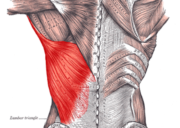
Affording itself a large attachment centrally from the T7 to L5 spinouses, laterally to the iliac crest and thoracolumbar fascia, rostrally to the lower 3 or 4 ribs and inferior angle of the scapula, to travel superiorly and laterally to the attach to the medial lip of the intertubercular sulcus, it is perfectly situated to effect both the upper and lower extremities in a large variety of movements.

It is one of the quintessential and often overlooked muscles in gait. It is generally quiet electrophysiologically during walking gait (1,2) until speed increases or you are running (2-4).  The latissimus dorsi is THE functional link between the upper and lower extremity, particularly though its connections with the thoracolumbar fascia (5,6). Latissimus activity, through gait and arm swing seems to profoundly influence and be influenced by gluteal activity, through the posterior oblique sling system (3,4), consisting of the latissimus dorsi, thoracolumbar fascia and contralateral gluteus maximus (7). The posterior oblique sling system provides trunk stability and power delivery to the upper extremity from the contralateral lower extremity and promotes mutual gait patterns between the upper and lower extremities (8), while creating joint contraction in running, turning and walking (9,10). Recent studies conversely show that arm swing can, in turn, effect lower extremity movement as well (3,4). 

Given the importance of the latissimus, it would stand to reason we would want it to function at its best. Dry needling is one modality we seem to be able to use to change its function,not only reducing central sensitization, but reducing local and referred pain, improves range of motion and muscle activation patterns (11-13). There are other modalities, including supportive exercises, that can be used to both activate and rehabilitate the lat as well (14-17)

Here is one method we like to use to needle the latissimus dorsi. Consider adding this to your clinical toolbag. 


references: 

1. Houglum P, Bertoti D in: Brunstrums Clinical Kinesiology 6th Edition, FA Davis 2012 p.558

2. G. Cappellini, Y. P. Ivanenko, R. E. Poppele, F. Lacquaniti Motor Patterns in Human Walking and Running Journal of Neurophysiology Published 1 June 2006 Vol. 95 no. 6, 3426-3437 DOI: 10.1152/jn.00081.2006

3. Shin S, Kim T, Yoo W. Effects of Various Gait Speeds on the Latissimus Dorsi and Gluteus Maximus Muscles Associated with the Posterior Oblique Sling System. Journal of Physical Therapy Science. 2013;25(11):1391-1392. doi:10.1589/jpts.25.1391.

4. Kim T, Yoo W, An D, Oh J, Shin S. The Effects of Different Gait Speeds and Lower Arm Weight on the Activities of the Latissimus Dorsi, Gluteus Medius, and Gluteus Maximus Muscles. Journal of Physical Therapy Science. 2013;25(11):1483-1484. doi:10.1589/jpts.25.1483.

5. Vleeming A, Pool-Goudzwaard AL, Stoeckart R, van Wingerden JP, Snijders CJ. The posterior layer of the thoracolumbar fascia. Its function in load transfer from spine to legs. Spine (Phila Pa 1976). 1995 Apr 1;20(7):753-8.

6. Willard FH, Vleeming A, Schuenke MD, Danneels L, Schleip R. The thoracolumbar fascia:anatomy,  function and clinical considerations. Journal of Anatomy. 2012;221(6):507-536.doi:10.1111/j.1469-7580.2012.01511.x.

7. Mooney V, Pozos R, Vleeming A, Gulick J, Swenski D Exercise treatment for sacroiliac pain. Orthopedics. 2001 Jan; 24(1):29-32.

8. Page P, Frank C, Lardner R: Assessment and treatment of muscle imbalance. Champaign: Human Kinetics Pub, 2010, pp 30–37. 

9. Bergmark A Stability of the lumbar spine. A study in mechanical engineering. Acta Orthop Scand Suppl. 1989; 230():1-54.

10. Collins SH, Adamczyk PG, Ferris DP, Kuo AD A simple method for calibrating force plates and force treadmills using an instrumented pole. Gait Posture. 2009 Jan; 29(1):59-64.

11. Dar GHicks GE. The immediate effect of dry needling on multifidus muscles function in healthy individuals. J Back Musculoskelet Rehabil. 2016 Apr 27;29(2):273-278.

12. Ortega-Cebrian S, Luchini N, Whiteley R. Dry needling: Effects on activation and passive mechanical properties of the quadriceps, pain and range during late stage rehabilitation of ACL reconstructed patients.Phys Ther Sport. 2016 Sep;21:57-62. doi: 10.1016/j.ptsp.2016.02.001. Epub 2016 Feb 24.

13. Dommerholt J. Dry needling — peripheral and central considerations. The Journal of Manual & Manipulative Therapy. 2011;19(4):223-227. doi:10.1179/106698111X13129729552065.

14. Youdas JW, Coleman KC, Holstad EE, Long SD, Veldkamp NL, Hollman JH. Magnitudes of muscle activation of spine stabilizers in healthy adults during prone on elbow planking exercises with and without a fitness ball. Physiother Theory Pract. 2017 Sep 18:1-11. doi: 10.1080/09593985.2017.1377792. [Epub ahead of print]

15. Crane P, Ladden J, Monica D. Treatment of axillary web syndrome using instrument assisted soft tissue mobilization and thoracic manipulation for associated thoracic rotation dysfunction: A case report. Physiother Theory Pract. 2017 Aug 30:1-5. doi: 10.1080/09593985.2017.1368755. [Epub ahead of print]

16. Massé-Alarie H, Beaulieu LD, Preuss R, Schneider C. Influence of paravertebral muscles training on brain plasticity and postural control in chronic low backpain. Scand J Pain. 2016 Jul;12:74-83. doi: 10.1016/j.sjpain.2016.03.005. Epub 2016 May 11.

17. Snarr RL, Hallmark AV, Casey JC, Esco MR. Electromyographical Comparison of a Traditional, Suspension Device, and Towel Pull-Up. J Hum Kinet. 2017 Aug 1;58:5-13. doi: 10.1515/hukin-2017-0068. eCollection 2017 Sep.

An often overlooked culprit in hip pain...

We often find clinically that the quadratus femoris as becoming the 1st dysfunctional muscle of the deep 6 external rotators (1) and its pain referral pattern can mimic the piriformis (2) and piriformis syndrome (3) as well as hamstring insertional tendinitis.  It has also been implicated in some cases of femoroacetabular impingement (4)  as well as ishiofemoral impingement (5). It is active during walking stance phase, and moreso during stance while running as well as with a clamshell exercise with external rotation (6). It appears to be maximally lengthened with flexion and adduction or abduction, with internal rotation ( a great position of you need to stretch this muscle), and is deducted to be strongest going from a 60-90 degree flexed position into extension (ie: it has the with the largest moment arms observed for extension in the deduced force-length efficient range of 60-90° flexion)(7).

Needling this muscle can sometimes pose a challenge. Here is a demo of one way to accomplish it I often employ while needling some of the other surrounding hip musculature.

Consider the QF the next time you have someone with hamstring insertional pain, or diffuse hip pain that you are having a difficult time localizing.

  1. Personal observation
  2. Janet G. Travell , M.D., and David G. Simons, M.D., Myofascial Pain and Dysfunction: The Trigger Point Manual, The Lower Extremities vol. 2 (Baltimore: Williams & Wilkins, 1992) pp. 186-193.
  3. Dalmau-Carolà J Myofascial pain syndrome affecting the quadratus femoris Pain Pract. 2010 May-Jun;10(3):257-60. doi: 10.1111/j.1533-2500.2009.00347.x. Epub 2010 Feb 11

  4.  Diamond LEVan den Hoorn WBennell KLWrigley TVHinman RSO'Donnell JHodges PW. Coordination of deep hip muscle activity is altered in symptomatic femoroacetabular impingement.  J Orthop Res. 2016 Aug 11. doi: 10.1002/jor.23391. [Epub ahead of print]

  5. http://radsource.us/ischiofemoral-impingement-syndrome/
  6. Semciw, Adam I. et al. Quadratus femoris: An EMG investigation during walking and running Journal of Biomechanics , Volume 48 , Issue 12 , 3433 - 3439

  7. Vaarbakken KSteen HSamuelsen GDahl HALeergaard TBStuge B .Primary functions of the quadratus femoris and obturator externus muscles indicated from lengths and moment arms measured in mobilized cadavers. Clin Biomech (Bristol, Avon). 2015 Mar;30(3):231-7. doi: 10.1016/j.clinbiomech.2015.02.004. Epub 2015 Feb 11.

More on that post operative foot

This is part 2 of a series following a case. If you missed part 1, please go back here and read what we found.

The patient returns 1 week later and reports being approximately 25% improved. She has been performing her "toes up" exercises while walking all the time. She is having some difficulty still with balance. She has been performing her toe waving exercises a few times daily.

X-rays performed 2/17 reveal screw fixation of the navicular. I cannot find evidence of a previous cuboid fracture. The ankle mortise is clear.

She still has 4/5 weakness of the long and short toe extensors; long greater than short. She has tenderness to palpation along the anterior aspect of the deltoid ligament on the left hand side which is made worse with eversion of the ankle. There is a loss of long axis extension at the talocrural and talonavicular articulations. Less tenderness is noted in the inter metatarsal intervals and the interossei musculature.

There is significant improvement over last time. Lack of fixation of the navicular to other articulations will allow us to perform manipulation/mobilization of the foot.

We treated with diagnostic manipulation and mobilization of the foot. I reviewed exercises to date and added the shuffle walk exercise. Since acupunture and needling can influence blodd flow (1-4) We utilized acupuncture points stomach 36, spleen 6, gallbladder 41, liver 3, points in the inter metatarsal intervals, bladder 67 and liver 1.Neelding has been shown to improve muscke activation (4-7) so I did origin/insertion stimulation of the long extensors with 3 sets of 10 repetition cocontraction along with origin/insertion stimulation of the short extensors with 3 sets, 10 repetitions cocontraction was performed. She will follow back in approximately 2 weeks because of travel.

So far, so good. We will keep you posted : )


1. Sandberg, M., Larsson, B., Lindberg, L.-G. and Gerdle, B. (2005), Different patterns of blood flow response in the trapezius muscle following needle stimulation (acupuncture) between healthy subjects and patients with fibromyalgia and work-related trapezius myalgia. European Journal of Pain, 9: 497. doi:10.1016/j.ejpain.2004.11.002

2.  Cagnie, Barbara et al. The Influence of Dry Needling of the Trapezius Muscle on Muscle Blood Flow and Oxygenation Journal of Manipulative & Physiological Therapeutics , Volume 35 , Issue 9 , 685 - 691

3. Tsuchiya, Masahiko; Sato, Eisuke F.; Inoue, Masayasu; Asada, Akira† Acupuncture Enhances Generation of Nitric Oxide and Increases Local Circulation  Anesthesia & Analgesia: February 2007 - Volume 104 - Issue 2 - pp 301-307

4. Jan Dommerholt Dry needling — peripheral and central considerations Journal Of Manual & Manipulative Therapy Vol. 19 , Iss. 4,2011

5. Zanin, Marília Silva et al. Electromyographic and Strength Analyses of Activation Patterns of the Wrist Flexor Muscles after Acupuncture Journal of Acupuncture and Meridian Studies , Volume 7 , Issue 5 , 231 - 237

6. https://youtu.be/02-M0i6AKAk

7. Fragoso APS, Ferreira AS. Immediate effects of acupuncture on biceps brachii muscle function in healthy and post-stroke subjects. Chinese Medicine. 2012;7:7. doi:10.1186/1749-8546-7-7.


3 points to use with ankle instability

In this study they stimulated 3 points: ST41, BL60 and GB40. Take a look at their locations (above). ST41 is at the base of the long extensor tendons; gee, we never emphasize long extensor function, do we? GB 40 is at the lateral malleolus between the peroneus longus/brevis and peroneus tertius; how important are these for coronal plane stability, not to mention the ability to descend the 1st ray. BL60 is just anterior to the lateral malleolus, right by the peroneus longus and brevis (again). Could they have included K6, under the medial malleolus and near the long flexors? Sure. How about SP4 or 4, in the substance of the flexor hallucis brevis and anterior to the extensor hallucis longus. Of course. You can probably think of other points to include as well.

Do you think it was by accident that their muscle selection included dorsiflexors (excepting the peroneus longus) and everters? How about a muscle that would help descend the 1st ray and complete the medial tripod? Hmmm... There is always a reason and a rationale....


"CONCLUSION: Electroacupuncture can effectively improve the proprioception of athletes with FAI and achieves a superior efficacy as compared with the conventional physiotherapy."...or in this case, low level e stim to the medial and lateral malleolus.

How about adding these points, no matter how you would like to stimulate them, to your CAI toolkit?


Zhu Y, Qiu ML, Ding Y, Qiang Y, Qin BY. [Effects of electroacupuncture on the proprioception of athletes with functional ankle instability]. Zhongguo Zhen Jiu. 2012 Jun;32(6):503-6.


Acupuncture/Dry Needling and Proprioception. A Winning combination.


What a great combination of therapies for folks with chronic ankle instability, or almost any injury for that matter! Taking 2 modalities that emphasize afferent input from the peripheral mechanoreceptor system, which has such a large influence on the cerebellum as well as the segmental and descending pain inhibition pathways.

Did you notice they used the trigger points in the peroneus longs muscle to needle? Though they didn't say it, did you remember that that the point correlates to a great point: Gallbladder 34, which is an empirical point for musculoskeletal pain? Interesting how this muscle influences both frontal and saggital plan stability. 

Though the techniques of exercise could use some refinement (check out the gents posture in the photo, sure looks like he could use some gluteus medius work!), this is a good overview that provides evidence that utilizing spacial summation (combining multiple techniques that provide afferent input to more than one modality to cause an effect) has better outcomes than one alone. Put this one on your reading list : )

Salom-Moreno J, Ayuso-Casado B, Tamaral-Costa B, Sánchez-Milá Z, Fernández-de-Las-Peñas C, Alburquerque-Sendín F.Trigger Point Dry Needling and Proprioceptive Exercises for the Management of Chronic Ankle Instability: A Randomized Clinical Trial. Evid Based Complement Alternat Med. 2015;2015:790209. doi: 10.1155/2015/790209. Epub 2015 Apr 30.

link to FREE FULL TEXT: https://www.ncbi.nlm.nih.gov/pmc/articles/PMC4430654/

Those MultifidiThe multifidi are important proprioceptive sentinels for the low back, as well as the rest of the body, for virtually every activity you do weight bearing, including gait. They are implicated in many instances of low back pain, especi…

Those Multifidi

The multifidi are important proprioceptive sentinels for the low back, as well as the rest of the body, for virtually every activity you do weight bearing, including gait. They are implicated in many instances of low back pain, especially folks with flexion or extension intolerance, since their fiber orientation and thus mechanical advantage (or disadvantage) is dependent upon whether or not you are maintaining a normal lumbar lordosis.

Modalities which boost their function are an excellent adjunct to the rehabilitation process. Since they are not under volitional control (go ahead, try and contract your L2/L3 multifidus), they are innervated by the vestibulospinal tract and we must use proprioceptive work to engage them. Dry Needling is one modality that can help them to become functional again.

RESULTS and CONCLUSION:
“Significant difference was found in the percentage of change of muscle activation post needling between groups on the right side at level L4-5. A slight increase in the percentage of muscle activity, post procedure was observed in the dry needling group compared with the control group, although not significant in other segments examined. An improvement of back muscle function following dry needling procedure in healthy individuals was found. This implies that dry needling might stimulate motor nerve fibers and as such increase muscle activity.”

see also our post here.

J Back Musculoskelet Rehabil. 2015 Sep 6. [Epub ahead of print]
The immediate effect of dry needling on multifidus muscles’ function in healthy individuals. Dar G1,2, Hicks GE3.

tumblr_o9vc4xvrkE1qhko2so1_400.jpg
tumblr_o9vc4xvrkE1qhko2so2_400.jpg

One point and 1 treatment can profoundly influence gait

When talking about the lower extremity and gait (as I have been know to do at more that one seminar), I often talk about the “reverse engineering” principle. This is looking at a muscle or muscle group from a “ground up” perspective, as it would be functioning during the gait cycle. This, along with knowing when a muscle should be firing in the gait cycle, can provide clues to what may be going on and how you may be able to help.

When discussing the quads, we often employ this principle. It can be a little difficult to think of the vastus medialis as a lateral rotator of the thigh and the rectus femoris as a flexor (anterior nutator) of the pelvis, but if you put your foot on the ground and think about it, you will see what I mean.

The VMO is often implicated in patello femoral syndromes but cannot be selectively activated. The ratio between vastus medialis and vastus lateralis does seem to be alterable and perhaps is a siginificant factor.

How about if we look at the vastus lateralis instead?

The vastus lateralis is the largest and most powerful portion of the quadriceps. One paper reports that the muscle volume of the the vastus lateralis was 674 cm3 followed by the vastus intermedius at 580 cm3, vastus medialis 461 cm3 and lowest in the rectus femoris 339 cm3.  This makes the vastus lateralis is twice the volume of the rectus femoris!

Studies of muscle fiber orientation show that VL force component is directed approximately 12-15° laterally with respect to the longitudinal axis of the femoral shaft. This would mean it has a tremendous mechanical advantage and could (should?) pull the patella directly laterally compared to the VMO force, whose component is directed approximately 55 ° medially.   The muscle “balance” between the VMO and the VL, along with the periarticular soft tissue structures acting on the patella, is considered major component in the control of normal patellar alignment and function. The VL is often considered to be the “overactive” one by many clinicians, particularly in cases of patellofemoral dysfunction. It turns out that from an EMG standpoint, they may be correct. 

The vastus lateralis arises posteriorly from the femur along the linea aspera and circumnavigates the thigh in a counterclockwise fashion to attach laterally to the patellar tendon.   Because of its size and fiber orientation, it would stand to reason that needling it would have more cortical representation than say the vastus medialis.

There is an interesting paper where they needled a single acupuncture point: Stomach 34. For those who haven’t studied acupuncture (or don’t remember) this point is located on the thigh, in a small depression about 2.5 inches (63 mm for the metric folks) lateral to and above lateral border of the patella. In other words, it is in the vastus lateralis (see above).

The results showed statistically significant improvement in velocity, cadence, stride length, cycle time, step time and single/double leg support after treatment. The effect was small, but positive.

Think about where the trigger points are for this muscle (see above) ; fairly close to this point, sometimes (depending on the trigger point), even directly over this point. Needling has many effects on muscle and its trigger points and we like to think that needling “normalizes” function of a muscle; perhaps it influences the apparent “dominance” of this muscle and allows the patella to track more medially?

So, in this popultion of patients of elderly individuals, 1 acupuncture (needling) treatment  had a positive influence on their gait. Perhaps if the folks in the knee study were treated a few more times, we would have seen a change. Imagine what could have happened if aditional treatment modalities, like exercise, proprioceptive work and gait retraining were added! 

What a great, cost effective alternative or addition to your rehabilitation this could be. Consider adding this modality (and point!) to your current clinical toolbox, not only for older patients but for any patients that may have a gait abnormality.


https://tmblr.co/ZrRYjx24ecKUx

Boucher JP, King MA, Lefebvre R, Pépin A. “Quadriceps femoris muscle activity in patellofemoral pain syndrome.” Am J Sports Med. 1992 Sep-Oct;20(5):527-32. Web. 17 Nov 2012.

Souza DR, Gross MT. “Comparison of vastus medialis obliquus: vastus lateralis muscle integrated electromyographic ratios between healthy subjects and patients with patellofemoral pain.” Phys Ther. 1991 Apr;71(4):310-6. Web. 25 Nov 2012.

Cowan SM, Bennell KL, Crossley KM, Hodges PW, McConnell J. “Physical therapy alters recruitment of the vasti in patellofemoral pain syndrome.” Med Sci Sports Exerc. 2002 Dec;34(12):1879-85. Web. 26 Nov 2012.

Boling MC, Bolgla LA, Mattacola CG, Uhl TL, Hosey RG. “Outcomes of a weight-bearing rehabilitation program for patients diagnosed with patellofemoral pain syndrome.” Arch Phys Med Rehabil. 2006 Nov;87(11):1428-35

Kim, H. H., & Song, C. H. (2010). Effects of knee and foot position on EMGactivity and ratio of the vastus medialis oblique and vastus lateralis during squatexercise. Journal of Muscle and Joint Health, 17(2), 142-150.

Lam, P. L., & Ng, G. Y. (2001). Activation of the quadriceps muscle during semisquatting with different hip and knee positions in patients with anterior knee pain. American Journal of Physical Medicine & Rehabilitation, 80(11), 804-808.

Erskine, R. M., Jones, D. A., Maganaris, C. N., & Degens, H. (2009). In vivo specific tension of the human quadriceps muscle. European journal of applied physiology, 106(6), 827-838. [PubMed]

Grabiner MD: Current Issues in Biomechanics (9th ed). Champaign, Human Kinetics Publishers, 1993.

http://www.orthobullets.com/anatomy/10058/vastus-lateralis

Hauer K, Wendt I, Schwenk M, Rohr C, Oster P, Greten J. Stimulation of acupoint ST-34 acutely improves gait performance in geriatric patients during rehabilitation: A randomized controlled trial. Arch Phys Med Rehabil. 2011 Jan;92(1):7-14. doi: 10.1016/j.apmr.2010.09.023.

Peter Deadman, Mazin Al-Khafaji, Kevin Baker: A Manual of Acupuncture (2nd Edition) Journal of Chinese Medicine Esat Sussex, England 2007

Travell JG, Simons DG. Myofascial Pain and Dysfunction: The Trigger Point Manual: The Lower Extremities. Vol.2 . Baltimore, Md: Williams & Wilkins;1992

 http://www.medscape.org/viewarticle/521494_3

tumblr_o2uunml08W1qhko2so2_500.jpg
tumblr_o2uunml08W1qhko2so1_1280.jpg

Leg Pain? Are you SURE its a disc?

Gluteus minimus dysfunction is often present in gait disorders, including stance phase mechanical problems, since it fires from initial contact through pre swing, like it better known counterpart, the gluteus medius. It is interesting that the trigger point referral pattern of the gluteus minimus has a sciatic distribution, whereas the gluteus medius is more in the local area of the hip. 

 There are several, well known effects of dry needling:

decreased central sensitization

increased range of motion

changes in muscle activation

changes in the chemical environment surrounding a trigger point

changes in local and referred pain


and now we can add (not surprisingly), changes in autonomic function. The mechanism probably has something to do with pain and the reticular formation sending information down the cord via the lateral cell column (intermediolateral cell nucleus) or pain (nociceptive) afferents sending a collateral in the spinal cord to the dysfunctional muscle (Dr Ivo talks about these mechanisms in his dry needling and acupuncture lectures). 


Conclusions

The presence of active TrPs within the gluteus minimus muscle among subacute sciatica subjects was confirmed. Every TrPs-positive sciatica patient presented DN related vasodilatation in the area of referred pain. The presence of vasodilatation suggests the involvement of sympathetic nerve activity in myofascial pain pathomechanism.

BMC Complement Altern Med. 2015; 15: 72. Published online 2015 Mar 20. doi:  10.1186/s12906-015-0587-6PMCID: PMC4426539 Intensive vasodilatation in the sciatic pain area after dry needling

link to full text: http://www.ncbi.nlm.nih.gov/pmc/articles/PMC4426539/

One way to correct an dysfunctional Extensor Hallucis Brevis

The Extensor Hallicus Brevis, or EHB  (beautifully pictured above causing the  extension (dorsiflexion) of the proximal big to is an important muscle for descending the distal aspect of the 1st ray complex (1st metatarsal and medial cunieform) as well as extending the 1st metatarsophalangeal joint.

Since this muscle is frequently dysfunctional, and is one of THE muscles than can lower the head of the 1st metatarsal, along with the peroneus longus and most likely the tibialis posterior (through its attachment to the 1st or medial cunieform), needling can often assist in normalizing function and works especially well, when coupled with an appropriate rehab program. Here is one way to needle it effectively. 

Treat the paraspinals in addition to the peripheral muscleAs people who treat a wide variety of gait related disorders we often emphasize needling the paraspinal muscles associated with the segemental innervation of the peripheral muscle you are tre…

Treat the paraspinals in addition to the peripheral muscle

As people who treat a wide variety of gait related disorders we often emphasize needling the paraspinal muscles associated with the segemental innervation of the peripheral muscle you are treating. For example, you may facilitate or needle the L2-L4 paraspinals (ie: femoral nerve distribution) along with the quads, or perhaps the C5-C6 PPD’s along with the shoulder muscles for the deltiods or rotator cuff for arm swiing. We do this to get more temporal and spacial summation at a spinal cord level, to hopefully get better clinical results.

White and Panjabi described clinical instability as the loss of the ability of the spine, under physiologic loads, to maintain relationships between vertebrae in such a way that there is neither damage nor subsequent irritation to the spinal cord or nerve roots, and, in addition there is no development of incapacitating deformity or pain due to structural changes.

Increased movement between vertebrae (antero or retrolisthesis) of > 3.5 mm (or 25% of the saggital body diameter) during flexion and/or extension suggests clinical instability. This often leads to intersegmental dysfunction and subsequent neurological sequelae which could be explained through the following mechanisms:

Recall that the spinal nerve, formed from the union of the ventral (motor) and dorsal (sensory) rami, when exiting the IVF splits into an anterior and posterior division, supplying the structures anterior and posterior to the IVF respectively. The posterior division has 3 branches: a lateral branch that supplies the axial muscles such as the iliocostalis and quadratus; an intermediate branch, which innervates the medial muscles, such as the longissimus, spinalis and semispinalis; and a medial branch, which innervates the segmental muscles, (multifidus and rotatores) as well as the joint capsule. Inappropriate intersegmental motion has 2 probable neurological sequelae: I) alteration of afferentation from that level having segmental (reflexogenic muscle spasm or vasoconstrictive/vasodilatory changes from excitation of primary afferents and gamma motoneurons) and suprasegmental (less cerebellar afferentation, less cortical stimulation) effects and II) compression or traction of the medial branch of the PPD, causing,  over time, demyelination and resultant denervation, of the intrinsic muscles, resulting in impaired motor control both segmentally and suprasegmentally. The segmental effects are directly measurable with needle EMG. This is a form of paraspinal mapping, which has also been explored by Haig et al. So, in short, instability can lead to denervation and denervation can lead to instability.

We often see clinically that treating a trigger point (needling, dry needling, acupuncture, manual pressure) can alter the function of the associated muscle . Improvements in muscle strength and changes in proprioception are not uncommon. Needling also seems to increase fibroblastic activty through the local inflammation it causes. Wouldn’t better muscle function and some scar tissue be a beneficial thing to someone with instability?

The next time you have a patient with instability, make sure to include the paraspinals in your quest for better outcomes.

The Vasti

Do you treat runners? Do you treat folks with knee pain? Patellar tracking issues? Do you treat the quadriceps? Do you realize that the vastus lateralis, in closed chain, is actually an INTERNAL rotator of the thigh (not a typo), and many folks have a loss of internal rotation of the hip? Do you give them “IT band stretches” to perform?

In this short video, Dr Ivo demonstrates some needling techniques for the quads and offers some (entertaining) clinical commentary on the IT band. A definite view for those of you who have needling in their clinical tool box.

Dry Needling and Proprioception. What a great combination. Since dry needling and proprioception both have such profound effects on muscle tone, why not combine them to treat chronic ankle instability? We do all the time and here is a FREE FULL TEXT…

Dry Needling and Proprioception. What a great combination.

Since dry needling and proprioception both have such profound effects on muscle tone, why not combine them to treat chronic ankle instability? We do all the time and here is a FREE FULL TEXT article that ties the two together nicely!

And what better to muscle to use than the peroneii? These babies help control valgus/varus motions of the foot and influence plantar and dorsiflexion AND the longus descends the 1st ray. We call that a triple win!

“This study provides evidence that the inclusion of TrP-DN within the lateral peroneus muscle into a proprioceptive/strengthening exercise program resulted in better outcomes in pain and function 1 month after the end of the therapy in individuals with ankle instability. Our results may anticipate that the benefits of adding TrP-DN in the lateral peroneus muscle for the management of ankle instability are clinically relevant as large between-groups effect sizes were observed in all the outcomes.”

link to full text
http://www.ncbi.nlm.nih.gov/pmc/articles/PMC4430654/

photo from this past weekends Dry Needling Seminar: working on the dorsal interossei

Welcome to Monday and News You Can Use!

Any of your patients of clients taking anti inflammatories? Especially after a rehab session or dry needling/acupuncture? They may be thwarting the healing process. Excerpted from a recent lecture, Dr Ivo talks about how they can down regulate the healing process.

tumblr_m72w8bsnBi1qhko2so1_500.jpg
tumblr_m72w8bsnBi1qhko2so2_1280.gif
tumblr_m72w8bsnBi1qhko2so3_1280.jpg

A Great Reference Text:

Biomedical Acupuncture for Sports and Trauma Rehabilitation
Dry Needling
Techniques,

Yun-tao Ma, Elsevier, 2010

Both Dr Allen and Dr Waerlop have studied under Dr Ma and Dr Waerlop has had the privilege of teaching with Dr Ma (Dr Waerlop is one of the leading acupuncture instructors in the US and one of the few Dry Needling Instructors)

This book is a follow up to Dr Ma’s 1st book: Biomedical Acupuncture for Pain Management.

They are both great resources for the Western approach to needling for pain management and dry needling, explaining the physiological mechanisms behind both.

Dr Waerlop uses acupuncture extensively for muscle stimulation and facilitation in many gait related disorders, and finds it a valuable adjunct to therapeutic exercise.

The Gait Guys

You can download many lectures on acupuncture by Dr Waerlop and many videos by Drs Allen and Waerlop on rehabilitation and gait, as well as basic science here.