Better gait AFTER rhizotomies?

Nothing surprised me more than reading this paper and finding out that folks that have had rhizotomies, which removes the afferent input from the dorsal horn and sensory information from the reflex loops in the cord, actually had better gait. Of course these children had severe spastic diplegia, which means they have lost descending inhibition from higher center's and most likely had increased flexor tone in the lower extremities. 

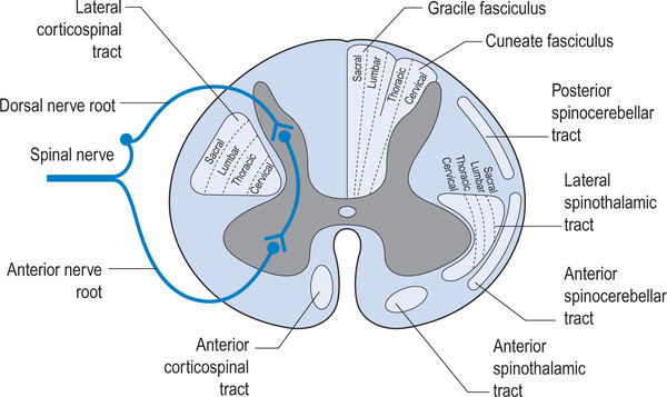
image credit: http://realtyconnect.me/spinal-cord-cross-section-tracts/background-information-musculoskeletal-key-within-spinal-cord-cross-section-tracts/

image credit: http://realtyconnect.me/spinal-cord-cross-section-tracts/background-information-musculoskeletal-key-within-spinal-cord-cross-section-tracts/

Remember that the fibers entering the dorsal horn not only go to the dorsal columns but also to the spinocerebellar pathways. When someone has spasticity, the feedback loops are skewed and flexor drive coming from the rostral reticular formation generally is increased are often kept in check by the cerebellar and vestibular feedback loops. Perhaps the interruption of this feedback loop and lack of information from type IA and II afferents of the muscle spindles as well as Ib afferents from the globe tendon organs modulated the tone sufficiently to improve gait. This study did a selective dorsal rhizotomy which means only a portion of it was ablated. 

The somatotopic organization  of the dorsal horn of the spinal cord (i.e.: certain areas of the dorsal horn correspond to certain body parts) is well documented in humans; It would make sense that the dorsal root itself (i.e.: the afferent fibers in the nerve going into the dorsal horn) would be as well, as they are that way in murines (2) and felines (3). 

So, how does this apply to gait? People with strokes, cortical lesions, diseases like cerebral palsy and even possibly increased flexor tone, may benefit from altered input into the dorsal horn. It would have been really cool to see if they increased extensor activity in this individuals, if they would be benefited further. 


Abstract

OBJECTIVE: To identify factors associated with long-term improvement in gait in children after selective dorsal rhizotomy (SDR).

DESIGN: Retrospective cohort study SETTING: University medical center PARTICIPANTS: 36 children (age 4-13y) with spastic diplegia (gross motor classification system level I (n=14), II (n=15) and III (n=7) were included retrospectively from the database of our hospital. Children underwent selective dorsal rhizotomy (SDR) between January 1999 and May 2011. Patients were included if they received clinical gait analysis before and five years post-SDR, age >4 years at time of SDR and if brain MRI-scan was available.

INTERVENTION: Selective dorsal rhizotomy MAIN OUTCOME MEASURES: Overall gait quality was assessed with Edinburgh visual gait score (EVGS), before and five years after SDR. In addition, knee and ankle angles at initial contact and midstance were evaluated. To identify predictors for gait improvement, several factors were evaluated including: functional mobility level (GMFCS), presence of white matter abnormalities on brain-MRI, and selective motor control during gait (synergy analysis).

RESULTS: Overall gait quality improved after SDR, with a large variation between patients. Multiple linear regression analysis revealed that worse score on EVGS and better GMFCS were independently related to gait improvement. Gait improved more in children with GMFCS I & II compared to III. No differences were observed between children with or without white matter abnormalities on brain MRI. Selective motor control during gait was predictive for improvement of knee angle at initial contact and midstance, but not for EVGS.

CONCLUSION: Functional mobility level and baseline gait quality are both important factors to predict gait outcomes after SDR. If candidates are well selected, SDR can be a successful intervention to improve gait both in children with brain MRI abnormalities as well as other causes of spastic diplegia.


1. Oudenhoven LM, van der Krogt MM, Romei M, van Schie PEM, van de Pol LA, van Ouwerkerk WJR, Harlaar Prof J, Buizer AI. Factors associated with long-term improvement of gait after selective dorsal rhizotomy. Arch Phys Med Rehabil. 2018 Jul 4. pii: S0003-9993(18)30442-8. doi: 10.1016/j.apmr.2018.06.016. [Epub ahead of print]

2. Wessels WJ1, Marani E. A rostrocaudal somatotopic organization in the brachial dorsal root ganglia of neonatal rats. Clin Neurol Neurosurg. 1993;95 Suppl:S3-11.

3. Koerber HRBrown PB. Somatotopic organization of hindlimb cutaneous nerve projections to cat dorsal horn. J Neurophysiol. 1982 Aug;48(2):481-9.

This simple screening test becomes a form of exercise.

Today we look at a simple CNS screen for your “central pattern generators” or “CPG’s”. If you do not pass, then the exercise becomes the rehab exercise. If you (or your client) does not have good coordination between the upper and lower extremity, then they will not be that efficient, physiologically or metabolically. 

The “cross crawl” or “step test” looks at upper and lower extremity coordination, rather than muscular strength. If performed for a few minutes, it becomes a test that can look at endurance as well. 

It is based on the “crossed extensor” response, we looked at last week. That is, when one lower limb flexes, the other extends; the contralateral upper limb also flexes and the ipsilateral upper limb extends. It mimics the way things should move when walking or running. 
 

  • Stand (or have your client stand) in a place where you will not run into anything.
  • Begin marching in place.
  • Observe for a few seconds. When you (or your client) are flexing the right thigh, the left arm should flex as well; then the left thigh and right arm. Are your (their) arms moving? Are they coordinated with the lower extremity?
  • What happens after a few minutes? Is motion good at 1st and then breaks down?
  • Now speed up. What happens? Is the movement smooth and coordinated? Choppy? Discoordinated?
  • now slow back down and try it with your (their) eyes closed


If  movement is smooth and coordinated, you (they) pass

If movement is choppy or discoordinated, there can be many causes, from simple (muscle not firing, injury) to complex (physical or physiological lesion in the CNS).

  • If movement is not smooth and coordinated, try doing the exercise for a few minutes a day. You can even start sitting down, if you (they) cannot perform it standing. If it improves, great; you were able to help “reprogram” the system. If not, then you (they) should seek out a qualified individual for some assistance and to get to the root of the problem.
tumblr_mg6a1wNO4i1qhko2so1_1280.jpg
tumblr_mg6a1wNO4i1qhko2so2_400.jpg

A little neuro, anyone?

Welcome to Monday, and yes, it is a NEURO day. In fact, if you got up this morning, you too are having a NEURO day. Dr Allen thinks it’s all about the ORTHOPEDICS, but without NEURO, there would not be any orthopedics : )

A dialogue from one of our avid readers, Dr. Ryan.

Dr. Ryan: Hey Ivo,

I just read this article on Mercola’s site which is an interview with Dr. Craig Buhler who does muscle activation techniques.  Can you check this for accuracy?  This must be a mistake b/c I always thought spindle activation will facilitate the muscle to contract.  Also, I always wondered why the O/I attachment points are tender in muscles that are inhibited.  Does his description sound right to you.  If not, do you have a better explanation?

“Your muscle system and nervous system relate to each other from within tiny muscle fibers called spindle cells, which monitor stretch. If your muscle is overloaded too rapidly, the spindle cells will temporarily inhibit the muscle. The next time you contract the muscle, it will fire again. Similarly, cells within your tendons called Golgi tendon organs also measure and monitor stretch. If your tendon is stretched too rapidly or exceeds its integrity, the Golgi tendon organs will temporarily inhibit the muscle. But the next time the muscle fires, it will again fire appropriately.

"But there’s a fail-safe system," Dr. Buhler explains. "It’s where the tendon attaches into the periosteum of the bone and the little fibers there are called Sharpey’s fibers. Those fibers are loaded with little receptors that monitor tension. And if the integrity of those fibers are exceeded, they inhibit the muscle, just like a circuit breaker would inhibit an electrical circuit.

Once that happens, the muscle will still fire under passive range of motion. But if you load the muscle, it gives way. If you continue to load the muscle, your body creates pain at the attachment points to protect you. What the central nervous system does at that point is compute an adaptive strategy by throwing stress into the muscle next to it. Other tissues begin to take on more of the load for the muscle that’s been injured.”

Here is a link to the entire article if you want to check it out:

http://fitness.mercola.com/sites/fitness/archive/2013/01/04/advanced-muscle-integration-technique.aspx?e_cid=20130104_DNL_art_1"

Dr Ivo: Thanks Dr. Ryan.

Spindles monitor length and GTO’s monitor tension. My understanding is spindles, when activated, stimulate the alpha motor neuron(at the cord) and cause contraction of that muscle or motor unit. GTO’s, when activated, cause inhibition of the muscle they are associated with. I am not aware of them being inhibitory, only GTO’s. They are believed to be GABAnergic synapses. The impulse (at least in cats) can be smaller or inhibited if the muscle is held in contraction for an extended period of time (see attached)

Perhaps he is talking about spindle dysfunction, where the intrafusal portion of the spindle (which is innervated by a gamma motor neuron) is either excited or inhibited. The gamma’s are more of a slave to the interneuronal pool (in the cord), which would be the sum total of all excitatory and inhibitory input to that area (ie the central integrated state). This not only reflects local receptor input but also cortical information descending (from areas 4s and 6 in the precentral gyrus) AND descending information from the caudal reticular formation.

Based on what you sent, I do not agree with the 1st 2 sentences. I was not aware about increased receptor density of Sharpeys fibers. I did a quick search and found this: http://www.ncbi.nlm.nih.gov/pmc/articles/PMC2100202/  , which eludes to it and here: http://www.ncbi.nlm.nih.gov/pmc/articles/PMC3098959/. I will have to dive in more when I have time.

Not sure why O/I attachments are tender in inhibited muscles. I find them tender in most folks. Maybe because inhibited muscles ave altered receptor function and that preloads the nociceptive afferent pathway or at least that neuronal pool? Are they closer to threshold for some reason? Not sure. LMK what you find.

Thanks for getting me jazzed about sharpeys fibers!

for those of you who need to know YES, there will be a forthcoming Sharpeys fibers article

Dr. Ryan: That’s what I thought.  Thanks for looking into it and I will check out those links.  You have jazzed me plenty of times over the years.  Glad I could jazz you up for a change.  Have a great weekend.


Yes, Dr Ivo is definitely an uber neuro geek, especially when he spends time on the weekend talking about spindles!


all material copyright 2013 The Homunculus Group/ The Gait Guys. All rights reserved. Please as before you lift our stuff.