Malleolar Fracture

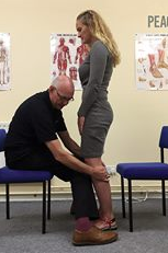
What does a post lateral malleolar fracture patient look like?

Can you say loss of proprioception? Can you say loss of triceps surae strength? Can you say loss of hip abductor strength?


Whenever we have a fracture and that segment is subsequently immobilized, we usually have damage to joint and muscle Mechanoreceptors as well as some atrophy of the receptors because of the immobilization. Lucky for us, and this patient in particular, those changes are rapidly reversed due largely to initially neural adaptation which is responsible for most of the early strength changes and then later, after a few weeks, muscular hypertrophy.

The Short Foot Exercise

Here it is, in all its glory...Our version of the short foot exercise. Love it or hate it, say it “doesn’t translate”, we find it a useful training tool for both the patient/client as well as the clinician. It awakens and creates awareness of the sometimes dormant muscles in the user and offers a window to monitor progression for them, as well as the observer.

Remember that the foot intrinsics are supposed to be active from midstance through terminal stance/pre swing. Having the person “walk with their toes up” to avoid overusing the long flexors is a cue that works well for us. This can be a useful adjunct to your other exercises on the road to better foot intrinsic function.


Dr Ivo Waerlop, one of The Gait Guys

Sulowska I, Mika A, Oleksy Ł, Stolarczyk A. The Influence of Plantar Short Foot Muscle Exercises on the Lower Extremity Muscle Strength and Power in Proximal Segments of the Kinematic Chain in Long-Distance Runners Biomed Res Int. 2019 Jan 2;2019:6947273. doi: 10.1155/2019/6947273. eCollection 2019

Okamura K, Kanai S, Hasegawa M, Otsuka A, Oki S. Effect of electromyographic biofeedback on learning the short foot exercise. J Back Musculoskelet Rehabil. 2019 Jan 4. doi: 10.3233/BMR-181155. [Epub ahead of print]

McKeon PO, Hertel J, Bramble D, et al. the foot core system: a new paradigm for understanding intrinsic foot muscle function Br J Sports Med March 2014 doi:10.1136/bjsports-2013- 092690

Dugan S, Bhat K: Biomechanics and Analysis of Running Gait Phys Med Rehabil Clin N Am 16 (2005) 603–621

Bahram J: Evaluation and Retraining of the Intrinsic Foot Muscles for Pain Syndromes Related to Abnormal Control of Pronation http://www.aptei.ca/wp-content/uploads/Intrinsic-Muscles-of-the-Foot-Retraining-Jan-29-05.pdf


#shortfootexercise #footexercises #footrehab #thegaitguys #gaitanalysis #gaitrehab #toesupwalking


https://vimeo.com/342800960

Hearing IS related to balance

Footnotes 7 - Black and Red.jpg

"Hearing had a clearly beneficial effect of auditory inputs on vestibulospinal coordination, especially for distance of displacement and angle of rotation." 

Given that these 2 systems, hearing as well as balance, are contained within the same location; “the organ of corti”, this is not surprising. The cochlea is the organ for hearing and the semicircular canals, utricle of saccule componenents of the vestibular apparatus. They share a common internal "fluid" called endolymph in the membranous labrynth of the inner ear that can flow freely from the vestibular apparatus to the cochlea. You will remember that the vestibular apparatus controls the vestibula spinal pathway which keeps us upright in the gravitational plain, along with vision and the joint mechanoreceptors. Involvement of 1 system will usually affect the other.

Dr Ivo, one of The Gait Guys

Seiwerth I, Jonen J, Rahne T, Schwesig R, Lauenroth A, Hullar TE, Plontke SK. Influence of hearing on vestibulospinal control in healthy subjects. HNO. 2018 Jul;66(Suppl 2):49-55. doi: 10.1007/s00106-018-0520-7.

#hearing, #balance, #gait, #thegaitguys, #gaitanalysis, #gaitcompensations, #vestibular

LLD's and Achilles Tendinopathy

Sometimes, it doesn't matter whether it is long or short.

 Achilles tendinopathy .. there are many factors that can contribute. Have you considered leg length inequality? Generally speaking, People have a tendency to overpronate on the longer leg side and under prone only shorter leg side with strain on the medial and lateral aspects of the Achilles tendon respectively. It would make sense that this could be a contributing factor.

 "The mean inequality in length of legs (ILL) was 5 +/- 4 mm. Among the 48 patients with ILL > or = 5 mm, the side affected with ruptured tendon was longer in 48% of cases and shorter in 52%. "

Age and pathology can play a role with younger, healthy tender and having greater compliance.

Proprioception is impaired on the affected side of folks with Achilles tendinopathy. This is a "chicken and the egg" scenario. Did impaired proprioception cause the tendinopathy or is the tendinopathy causing the impaired proprioception? Probably, a little bit of both.

Dr Ivo, one of The Gait Guys

Leppilahti J, Korpelainen R, Karpakka J, Kvist M, Orava S. Ruptures of the Achilles tendon: relationship to inequality in length of legs and to patterns in the foot and ankle. Foot Ankle Int. 1998 Oct;19(10):683-7.

Scholes M, Stadler S, Connell D, Barton C, Clarke RA, Bryant AL, Malliaras P. Men with unilateral Achilles tendinopathy have impaired balance on the symptomatic side. J Sci Med Sport. 2018 May;21(5):479-482. doi: 10.1016/j.jsams.2017.09.594. Epub 2017 Oct 6.

Intziegianni K, Cassel M, Rauf S, White S, Rector M, Kaplick H, Wahmkow G, Kratzenstein S, Mayer F. Influence of Age and Pathology on Achilles Tendon Properties During a Single-leg Jump. Int J Sports Med. 2016 Nov;37(12):973-978. Epub 2016 Aug 8.

#achilles,#tendon, #achillestendon, #tendinopathy, #proprioception

Balance..

Footnotes 7 - Black and Red.jpg

Did you know that the posterior spinocerebellar tract is essential for normal gait? It receives information from ALL muscle spindles, Golgi tendon organs and joint mechanoreceptors and coordinates them not only with the cerebellum but also with the vestibular system. Abnormalities within this system are present (but perhaps not apparent) all gait pathologies.

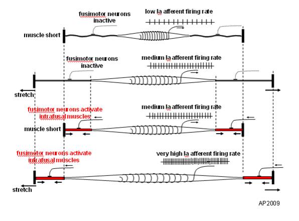
Muscle Spindles and Proprioception

image source: https://en.wikipedia.org/wiki/File:Fusimotor_action.jpg

image source: https://en.wikipedia.org/wiki/File:Fusimotor_action.jpg

And what have we been saying for the last 6 years? 

Connected to the nervous system by large diameter afferent (sensory) fibers, they are classically thought of as appraising the nervous system of vital information like length and rate of change of length of muscle fibers, so we can be coordinated. They act like volume controls for muscle sensitivity. Turn them up and the muscle becomes more sensitive to ANY input, especially stretch (so they become touchy…maybe like you get if you are hungry and tired and someone asks you to do something); turn them down and they become less or unresponsive.

Their excitability is governed by the sum total (excitatory and inhibitory) of all neurons (like interneuron’s) acting on them (their cell bodies reside in the anterior horn of the spinal cord).

Along with with Golgi tendon organs and joint mechanoreceptors, they also act as proprioceptive sentinels, telling us where our body parts are in space. We have been teaching this for years. Here is a paper that exemplifies that, identifying several proteins responsible for neurotransduction including the Piezo2 channel as a candidate for the principal mechanotransduction channel. Many neuromuscular diseases are accompanied by impaired  muscle spindle function, causing a decline of motor performance and coordination. This is yet another key finding in the kinesthetic system and its workings. 

Remember to include proprioceptive exercises and drills (on flat planar surfaces, like we talked about here) in your muscle rehab programs


Kröger S Proprioception 2.0: novel functions for muscle spindles. Curr Opin Neurol. 2018 Oct;31(5):592-598. 

Woo SH, Lukacs V, de Nooij JC, Zaytseva D, Criddle CR, Francisco A, Jessell TM, Wilkinson KA, Patapoutian A. Piezo2 is the principal mechanotransduction channel for proprioception.Nat Neurosci. 2015 Dec; 18(12):1756-62. Epub 2015 Nov 9.

Fusimotor control of proprioceptive feedback during locomotion and balancing: can simple lessons be learned for artificial control of gait?

Hulliger M. Fusimotor control of proprioceptive feedback during locomotion and balancing: can simple lessons be learned for artificial control of gait? Prog Brain Res. 1993; 97:173-80.

Perhaps we need to change how we are are rehabbing X (insert your favorite weight bearing joint)

image credit: https://en.wikipedia.org/wiki/StrongBoard_balance

image credit: https://en.wikipedia.org/wiki/StrongBoard_balance

We have recently run across some research that has changed the way we look at some of the rehab we do, especially proprioceptive rehab. Perhaps it will do the same for you.

Traditionally, we present increasing balance requirements to the weight bearing structure by changing one or more of the three parameters that keep us upright in the gravitational plane: vision, the proprioceptive system (which include the muscles, joints and ligaments) and the vestibular system (the utricle, saccule and semicircular canals). We have discussed them extensively in multiple articles here on the blog. We generally would make the rehab task more difficult by removing a stimulus (closing your eyes, having someone stand on foam) or challenging (standing on one leg, putting someone on a wobble board, BOSU, extending the head, etc) the to make it more durable and "educated". More difficult task + better balance = more stable joint and better outcomes. 

The importaat thing is to think about how much of each system is apportioned; we often (wrongly) assume it is pretty equally divided between the three. It turns out, that it really depends on the surface you are standing on and the circumstances.

On flat planar surfaces, the division of labor looks something like this:

  • proprioceptive system 70%
  • vestibular system 20%
  • visual system 10 %

On uneven or unstable surfaces (like a BOSU, dynadisc, foam, Swiss ball, etc), it looks like this:

  • vestibular system 70%
  • visual system 20%
  • proprioceptive system 10%

So, if we are rehabbing an ankle, it would make the most sense to do most of the rehab (and additional challenges) on a flat planar surface, perhaps incorporating things like forward, backward and side lean, toe and heel work and closed chain strengthening. WE could also close the eyes to make them more dependent on the proprio system, or extend the head 60 degrees to dampen the influence the lateral semicircular canals. We can put them on a BOSU or unstable surface but we need to remember that in that case, we will be rehabbing the vestibular system AND PERHAPS teaching THAT SYSTEM to compensate more, than the "broken" system. Yes, they get better BUT we are not fixing the system that is injured. 

You could make the argument, that your athletes/clients run/walk/exercise on uneven surfaces and use their vestibular system more.Maybe so, but is the actual injury to the vestibular system or to the musculoskeletal one?

Armed with this information, try and think of the system that is compromised and focus your efforts on that system, rather than the other two. Yes, people have vestibular dysfunction and refractive errors and need therapy, exercises and/or corrective lenses, but many of us are not vestibular or opticokinetic therapists (kudos to those of you who are!)


Peterka RJ, Statler KD, Wrisley DM, Horak FB. Postural Compensation for Unilateral Vestibular Loss. Frontiers in Neurology. 2011;2:57. doi:10.3389/fneur.2011.00057.

Horak FB. Postural Compensation for Vestibular Loss. Restorative neurology and neuroscience. 2010;28(1):57-68. doi:10.3233/RNN-2010-0515.

Gait: How we stay upright in the gravitational plane.

We remember that we have 3 systems that keep us upright in the gravitational plane: The visual system, The vestibular system and the proprioceptive system. As we age, we seem to become more dependent upon the visual system to maintain stability of the head (which is largely under the purview of the vestibular system). Older folks seem to be less stable than younger ones....At least when it comes to head stability.

This study looked at the neuromechanical mechanisms of head stability in younger and older women during gait initiation, a time when we employ the visual system for things like balance and foot placement. The visual system in this case is king and trumps the other systems in that it will do whatever it need to to keep the eyes level with the horizon.

They used a stereophotogrammetric system to measure angular displacement and acceleration and surface emg (no, not the best) to look at activation latencies of neck (particularly the SCM), trunk and pelvic muscles. Older women had higher variability in angular displacement of the head (possibly age related breakdown of the vestibular system?), decreased ability to attenuate accelerations in the sagittal (forward) plane, and increased SCM activation latencies.

The bottom line?

Make sure the older women you are working with have:

- better functioning joints
- better functioning muscles (appropriate skill, endurance and strength)
- better proprioception

You could help these by:

- manipulating and mobilizing joints that have pathomechanics
- improving muscular function through endurance and strength based exercises of the neck as well as core
-improving muscular function through modalities you use
-give them more proprioceptive based exercises, especially ones which incorporate the head, like head repositioning exercises,
-encourage them to engage in proprioceptive heavy activities, like rock climbing (which also works the axial extensors), cross country skiing, bike riding, etc

Maslivec A, Bampouras T, Dewhurst S, Vannozzi G, Macaluso A, Laudani L. Mechanisms of head stability during gait initiation in young and older women: A neuro-mechanical analysis.
J Electromyogr Kinesiol. 2017 Nov 23;38:103-110. doi: 10.1016/j.jelekin.2017.11.010. [Epub ahead of print]

link to free full text: https://www.sciencedirect.com/science/article/pii/S1050641116302036?via%3Dihub

Arm swing and Gait Stability

"When discussing the effects of arm swing on gait stability, it is
necessary to start with a definition of gait stability. In steady-state gait, infinitesimally small perturbations are ever present, and the system’s response to such perturbations may be called local stability. When gait is externally perturbed, global stability can be assessed by quantifying the response to such a perturbation. Following Bruijn et al. [12], in human gait, this response may be divided into two phases: an initial phase, which is dependent upon both the steady state of the system (as it was before the perturbation) and the system’s intrinsic mechanical properties (e.g. inertia, stiffness), and a second, reactive phase (‘recovery’), which is mainly dependent on active control and reflexes." - P. Meyns et al. / Gait & Posture 38 (2013) 555–562

*The how and why of arm swing during human walking
Pieter Meyns a,1 , Sjoerd M. Bruijn a,b,1, Jacques Duysens a,c,

Try THIS at home...

Screen Shot 2018-02-07 at 11.23.24 AM.png

Cool test, results you can see and some literature to back it up. If you are interested at all in proprioception, this is an interesting read.

So, the question for us is: "Does ankle dorsiflexion actually create more stability, like is purported?"

“The point is that if I make their ankle rigid this way, then they can more effectively use the balance mechanisms at their knees, hips, and proximally, because they’re on a stable base. My proposition is that their balance is actually normal, apart from that distal segment. When their ankle is stabilized, they use their knees more effectively, and they become less dependent on their eyesight to maintain their balance.”

http://lermagazine.com/article/afos-and-balance-issues-in-peripheral-neuropathy

The SCM and Gait?

We remember that we have 3 systems that keep us upright in the gravitational plane: The visual system, The vestibular system and the proprioceptive system. As we age, we seem to become more dependent upon the visual system to maintain stability of the head (which is largely under the purview of the vestibular system).  Older folks seem to be less stable than younger ones....At least when it comes to head stability.

This study looked at the neuromechanical mechanisms of head stability in younger and older women during gait initiation, a time when we employ the visual system for things like balance and foot placement. The visual system  in this case is king and trumps the other systems in that it will do whatever it need to to keep the eyes level with the horizon.

They used a  stereophotogrammetric system to measure angular displacement and acceleration and surface emg (no, not the best) to look at activation latencies of neck (particularly the SCM), trunk and pelvic muscles. Older women had higher variability in angular displacement of the head (possibly age related breakdown of the vestibular system?), decreased ability to attenuate accelerations in the saggital (forward) plane, and increased SCM activation latencies.

The bottom line?

Make sure the older women you are working with have:

- better functioning joints
- better functioning muscles (appropriate skill, endurance and strength)
- better proprioception

You could help these by:

- manipulating and mobilizing joints that have pathomechanics
- improving muscular function through endurance and strength based exercises of the neck as well as core
-improving muscular function through modalities you use
-give them more proprioceptive based exercises, especially ones which incorporate the head, like head repositioning exercises,
-encourage them to engage in proprioceptive heavy activities, like rock climbing (which also works the axial extensors), cross country skiing, bike riding, etc

Maslivec A, Bampouras T, Dewhurst S, Vannozzi G, Macaluso A, Laudani L.  Mechanisms of head stability during gait initiation in young and older women: A neuro-mechanical analysis.
J Electromyogr Kinesiol. 2017 Nov 23;38:103-110. doi: 10.1016/j.jelekin.2017.11.010. [Epub ahead of print]

link to free full text: http://www.sciencedirect.com/science/article/pii/S1050641116302036?via%3Dihub

Want more stability, NOW?

balancing-stones.jpg

Try this...

While walking or running running (or watching a client walk, amble or run) you may be thinking  “I need to do something to improve my (their) proprioception, or they are going to fall (again)” If you were to increase your (their) surface area, and make yourself (theirself)vless top heavy, I (they) would be more stable. How can we accomplish that?

Here is what you can do:

First, spread your toes.; why not maximize the real estate available to your feet?

Next,  widen your stance (or base of gait). Spreading your weight over a larger surface area would be more stable and provide stability.

Third, raise your arms out from your sides (no don't try to fly) to provide more input from your upper extremities to your proprioceptive system (more input from peripheral joint and muscle mechanoreceptors = more input to cerebellum = better balance)

Lastly, Slow down from your blistering pace. this will give your (aging) nervous system more time to react.

All these actions were all “primitive” reactions of the nervous system when learning to walk. We did a post on that when my youngest son was learning to walk.

Want to have better balance?

  • Spread your toes
  • Widen your stance
  • Raise your arms
  • Slow down

Notice I didn’t say this would make you faster. Who is more likely to fall on a corner when being chased by a predator; the tortoise or the hare?

A little practical neurology for you this morning brought to you by the geeks of gait. Ivo and Shawn.

Arm Swing and dynamic stability of the system.

Screen Shot 2017-10-16 at 1.33.03 PM.png

We have discussed the arm swing issue so many times over the years that we have lost count. By many sources, arm swing is a product of lower limb action and a product of the effective, or ineffective, relationship between the shoulder "girdle" (maybe thoracic rotation component) and the pelvic girdle (lumbopelvic rhythm) during gait.  This is the concept of phasic and anti-phasic limb swing. If you want to dive into that, and you should if you are unfamiliar with the concept, you can look it up on our blog using the search box.  We are not to forget that the arms, and thus arm swing, is a major factor in maintaining balance. We have used the term "ballast" many times to describe the effects of arm swing, rotation, abduction, circumduction etc on assisting balance maintenance of the body during various locomotion strategies. These are largely subconscious actions, hence why we agree with the research suggesting that arm swing is secondary, compensatory, and takes its queues off of the activity of the lower limb motor actions. In essence, arm swing variants are necessary compensations to assist in maintaining things like balance, center of pressure, equilibrium and the like. 

In this recent 2017 study, we have another suggesting arm swings function in assisting, even improving, dynamic stability. We are reminded of MdGill's suggestion, and the concepts of phasic and antiphasic torso-pelvis counter rotational movements, of how spinal loads can be affected by changes or differences in arm position.  Even arm position changes in sitting and standing can alter spinal loads, so during movement it is a virtual guarantee. 

This study looked at "how arm swing could influence the lumbar spine and hip joint forces and motions during walking." In this study, the researchers had each subject perform walking with different arm swing amplitudes and arm positions. Here is a comment from the researchers on what they found, it is pretty much what we have been writing about for several years based off of other research"

"The range of motion of the thorax with respect to the pelvis and of the pelvis with respect to the ground in the transversal plane were significantly associated with arm position and swing amplitude during gait. The hip external-internal rotation range of motion statistically varied only for non-dominant limb. Unlike hip joint reaction forces, predicted peak spinal loads at T12-L1 and L5-S1 showed significant differences at approximately the time of contralateral toe off and contralateral heel strike."

Thus, we find yet another study confirming what many will say is obvious, that being arm position and movements have notable effects on whole body kinetics and spinal loads. This study suggested that arm variations did not have an effect on spinal loads during walking. We find this curious; it is something we will be looking into, and pondering. We hope you do as well.

Effect of arm swinging on lumbar spine and hip joint forces. Lorenza Angelini et al. Journal of Biomechanics, Sept 2017
http://www.sciencedirect.com/science/article/pii/S0021929017304670

Vitamin D and Gait?

Vitamin-D.jpg

So, is it the effects on calcium and nerve function (neurotransmitter release), the effects on calcium and muscular contraction, the antioxidants properties, some other function? Supplementing Vitamin D and getting people more sun exposure are easy things to do...

"These findings reveal an important new relationship between parathyroid hormone and gait stability parameters and add to understanding of the role of 25-OHD in motor control of gait and dynamic balance in community-dwelling women across a wide age span."

Bird MLEl Haber NBatchelor FHill KWark JD. Vitamin D and parathyroid hormone are associated with gait instability and poor balance performance in mid-age to older aged women. Gait Posture. 2017 Sep 28;59:71-75. doi: 10.1016/j.gaitpost.2017.09.036. [Epub ahead of print]


https://www.ncbi.nlm.nih.gov/pubmed/29017107

What? Gait and hearing....

No, not how does the gait SOUND, but can they hear? We have long payed attention to the mechanics of gait, muscles and proprioception, and many times, if you have been following us for any length of time, the interplay of all three. But how often have you considered someones hearing in your evaluation?

Anatomically, the hearing organs (hair cells, otolithic apparatus, cochlea) are conveniently located right next store to the vestibular system (utricle, saccule and semicircular canals). The hearing organs start with mechanical vibrations of the tympanic membrane (ear drum) which move 3 auditory bones (or ossicles) called the incus, malleus and stapes. The vibrations move the stapes moves in and out of the fluid filled cochlea and vibrate crystals laying on a platform (otolith) in which hair cells are imbedded and if the vibration sufficient, the nerves attached to the hair cells fire and you can hear your favorite baseline by Cliff Williams (or Cliff Burton, John Myung, Bootsy Collins or whomever you like to listen to).

The vestibular system, as you probably know, monitors position and velocity of movement of the head. There are three hula hoop type structures called “semicircular canals” (see picture above) that monitor rotational and tilt position and angular acceleration, as well as two other structures, the utricle and saccule, which monitor tilt and linear acceleration.

The vestibular apparatus (the canals and the utricle and saccule) feed into a part of the brain called the floccular nodular lobe of the cerebellum, which as we are sure you can imagine, have something to do with balance and coordination. This area of the cerebellum feeds back to the vestibular system (actually the vestibular nucleii); which then feed back up to the brain as well as (you guessed it) down the spinal cord and to predominantly the extensor muscles.

Can you see how the 2 systems are “attached” to one another and could conceivably be interrelated? When you move fluid in one system (endo or peri lymph) you HAVE to move fund in the other. The fluid displaces, (like air, in air filled shoes or insoles), it does not deform (like EVA in an insole)

It is well established that hearing impaired folks (especially kids) have gait (and often balance) problems (1-3) and the worse the hearing loss, the worse the gait disturbance (1). This can occur in middle aged (4), as well as older adults as well (5-7). Perhaps this is due to viscosity changes in the endo or perilymph (8), otosclerosis of the ossicles (9,10) or other age related changes in utricle and saccule function (5-7). Also, for the 1st time, we are documenting a “sensory reweighting” which occurs when auditory cues are changed (4), with a greater reliance on visual cues. Hmmm. We wonder how this plays out with listening to music while walking or running?

So, The next time someone you see has a gait or balance problem, check their hearing and adjust your treatment program accordingly : )


References:

1. Melo RS.  Gait performance of children and adolescents with sensorineural hearing loss.
Gait Posture. 2017 Jun 3;57:109-114. doi: 10.1016/j.gaitpost.2017.05.031.

2. Jafarnezhadgero AA, Majlesi M, Azadian E. Gait ground reaction force characteristics in deaf and hearing children. Gait Posture. 2017 Mar;53:236-240. doi: 10.1016/j.gaitpost.2017.02.006. Epub 2017 Feb 14.

3. Janky KL, Givens D.Vestibular, Visual Acuity, and Balance Outcomes in Children With Cochlear Implants: A Preliminary Report. Ear Hear. 2015 Nov-Dec;36(6):e364-72. doi: 10.1097/AUD.0000000000000194.

4. Maheu M, Sharp A, Landry SP, Champoux F. Sensory reweighting after loss of auditory cues in healthy adults. Gait Posture. 2017 Mar;53:151-154. doi: 10.1016/j.gaitpost.2017.01.015. Epub 2017 Jan 24.

5. Agmon M, Lavie L, Doumas M. The Association between Hearing Loss, Postural Control, and Mobility in Older Adults: A Systematic Review. J Am Acad Audiol. 2017 Jun;28(6):575-588. doi: 10.3766/jaaa.16044. Review.

6. Layman AJ1, Li C, Simonsick E, Ferrucci L, Carey JP, Agrawal Y. Association Between Saccular Function and Gait Speed: Data From the Baltimore Longitudinal Study of Aging. Otol Neurotol. 2015 Jan 7. [Epub ahead of print]

7. Otol Neurotol. 2012 Jul;33(5):832-9. doi: 10.1097/MAO.0b013e3182545061.
Decline in semicircular canal and otolith function with age.
Agrawal Y1, Zuniga MG, Davalos-Bichara M, Schubert MC, Walston JD, Hughes J, Carey JP.

8. Wu T, Marcus DC. Age-Related Changes in Cochlear Endolymphatic Potassium and Potential in CD-1 and CBA/CaJ Mice . JARO: Journal of the Association for Research in Otolaryngology. 2003;4(3):353-362. doi:10.1007/s10162-002-3026-6.

9. Grayeli AB1, Sterkers O, Toupet M. Audiovestibular function in patients with otosclerosis and balance disorders. Otol Neurotol. 2009 Dec;30(8):1085-91. doi: 10.1097/MAO.0b013e3181b0fd5d.

10. Ozmen AO1, Aksoy S, Ozmen S, Saraç S, Sennaroğlu L, Gürsel B. Balance after stapedotomy: analysis of balance with computerized dynamic posturography. Clin Otolaryngol. 2009 Jun;34(3):212-7. doi: 10.1111/j.1749-4486.2009.01915.x.


Things often work better in pairs… Especially with Exercise

You have heard us always talk about how the lower kinetic chain is connected, how ankle rocker effects hip extension and how important hallux (great toe) extension is.

What can we conclude form this study?

  • toe spreading exercises are important for reducing navicular drop (and thus mid foot pronation, at least statically)
  • In addition to increased abductor hallucis recruitment in ascending and descending stairs, when hip external rotation exercises were added along with toe spreading exercises folks had more recruitment of the vastus medialis (a closed chain external rotator of the leg and thigh)

Keep in mind:

  • the exercises given were all non weight bearing and open chain for the external rotators. Imagine what might have happened if they were both closed chain AND weight bearing!
  • They concentrated on the effects of toe spreading (AKAlift/spread/reach) on the abductor hallucis. It also has far reaching effects on the dorsal interossei, long and short extensors of the toes.


Goo YM, Kim DY, Kim TH. The effects of hip external rotator exercises and toe-spread exercises on lower extremity muscle activities during stair-walking in subjects with pronated foot. J Phys Ther Sci. 2016 Mar;28(3):816-9. doi: 10.1589/jpts.28.816. Epub 2016 Mar 31.

link toFREE FUL TEXT: https://www.ncbi.nlm.nih.gov/pmc/articles/PMC4842445/


Cerebellar impairment = Gait Changes = Happy Patient

This is a fairly info dense post with many links. please take the time to explore each one to get the most out of it. 

If you have been with us here on TGG long enough, you know the importance of the cerebellum and gait. Mechanoreceptor information travels north to the cortex via the dorsal (and ventral) spinocerebellar pathways to be interpreted (and interpolated, in the case of the ventral pathway), with the information relaying back to the motor cortex and vestibular nucleii and eventually back down to the alpha (and gamma) motor neurons that proved the thing you call movement and thus gait. (Cool video on spinocerebellar pathways here and here).

This FREE FULL TEXT paper has some cool charts, like this one, that show the parameters of gait that change with cerebellar dysfunction (in this case, disease, although idiopathic means they really don't know. Anatomical or physiological lesions will behave the same, no? Doesn't the end result of a functional short leg look the same as an anatomical one?)

Looking tat this chart, what do we really see? People with cerebellar dysfunction:

  • a shorter step length
  • a wider base of gait
  • decreased velocity
  • increased lateral sway
  • slower overall gait cycle

Hmmmm...Beginning to sound like a move toward more primitive gait. Just like we talked about in this post on the 5 factors and proprioception here several years ago. We like to call this decomposition of gait. 

They go on to talk about specific anatomic regions of the cerebellum and potential correlation to specific gait abnormalities, like the intermediate zone and interposed nucleii controlling limb dynamics and rhythmic coordination like hypermetria (overshooting a target), especially when walking in uneven surfaces or when gait is perturbed, like walking into something or changes in surface topography, or the lateral zone of the cerebellum, for voluntary limb control, such as where you place your foot. Definitely gait nerd material.

There aren't any direct tips on rehab, but it would stand to reason that activities that activate the cerebellum and collateral pathways would give you the most clinical gains. Lots of propriosensory exercises like here, here, here and here for a start.

Happy cerebellum = Happy patient

The Gait Guys


Winfried Ilg, Heidrun Golla, Peter Thier, Martin A. Giese; Specific influences of cerebellar dysfunctions on gait. Brain 2007; 130 (3): 786-798. doi: 10.1093/brain/awl376  FREE FULL TEXT

Rock Your Rehab Process with these simple Proprioceptive Exercises

In this capsule, excerpted from a recent Dry Needling Seminar, Dr Ivo talks about one of his proprioceptive sequences and the neurological reasoning behind it

Today we give away some of the farm with a great proprioceptive exercise sequence that we use ALL THE TIME.

Skill (proprioception), Endurance, Strength. In that order.

Try incorporating this simple and effective sequence into your rehab program and watch your results get even better!


Comparative effects of proprioceptive and isometric exercises on pain and difficulty in patients with knee osteoarthritis: A randomised control study. Ojoawo AO, Matthew O, Mariam HA.Technol Health Care. 2016 Jul 8. [Epub ahead of print]

Efficacity of exercise training on multiple sclerosis patients with cognitive impairments. Chenet A, Gosseaume A, Wiertlewski S, Perrouin-Verbe B. Ann Phys Rehabil Med. 2016 Sep;59S:e42. doi: 10.1016/j.rehab.2016.07.097.

Exercise strategies to protect against the impact of short-term reduced physical activity on muscle function and markers of health in older men: study protocol for a randomised controlled trial. Perkin OJ, Travers RL, Gonzalez JT, Turner JE, Gillison F, Wilson C, McGuigan PM, Thompson D, Stokes KA. Trials. 2016 Aug 2;17:381. doi: 10.1186/s13063-016-1440-z.

Leg and trunk muscle coordination and postural sway during increasingly difficult standing balancetasks in young and older adults. Donath L, Kurz E, Roth R, Zahner L, Faude O.Maturitas. 2016 Sep;91:60-8. doi: 10.1016/j.maturitas.2016.05.010. Epub 2016 May 27.

Hip proprioceptive feedback influences the control of mediolateral stability during human walking. Roden-Reynolds DC, Walker MH, Wasserman CR, Dean JC. J Neurophysiol. 2015 Oct;114(4):2220-9. doi: 10.1152/jn.00551.2015. Epub 2015 Aug 19.

Proprioceptive Training and Injury Prevention in a Professional Men's Basketball Team: A Six-Year Prospective Study. Riva D, Bianchi R, Rocca F, Mamo C.J Strength Cond Res. 2016 Feb;30(2):461-75. doi: 10.1519/JSC.0000000000001097.

Proprioceptive feedback contributes to the adaptation toward an economical gait pattern. Hubbuch JE, Bennett BW, Dean JC. J Biomech. 2015 Aug 20;48(11):2925-31. doi: 10.1016/j.jbiomech.2015.04.024. Epub 2015 Apr 23.

Take a good look at this gals gait. In the 1st section, she is walking on relatively level ground and in the second part, the topography changes and the balance requirements become much greater. You may remember a post we did some time ago talking about proprioception and learning to walk here.

Besides the obvious gluteus medius weakness, genu valgum and pronation, R>L, can you see how when the task becomes more complex, that the system begins to break down? Did you see the increased base (wider) of gait? did you see the decreased speed of movement? Did you see the increased ancillary arm movements?

Keep your eyes open for clues like this in your clinical exam. When the going gets rough, the nervous system often reverts to what it knows best a slows down a bit.