What does progressive weakness of the posterior compartment look like?

Look at this video carefully and what do you notice? Can you see the progressive dip in the left heel as time goes on while toe walking? This is a cardinal sign of lack of endurance in the posterior compartment, in this patient’s case tibialis posterior. Your differential, in addition to lack of type one muscle fibers, insufficient vascularity or mitochondria for whatever reason would be circulatory problems as well as conditions causing progressive motor weakness like myasthenia gravis.

Fatigue testing is very important because a lot of times the problem doesn’t come out till the person reaches say a half an hour, an hour or sometimes even many miles into the run or ride. Our job as clinicians is to try to diagnose the problem to the best of our abilities. Our job also is to “exploit their weaknesses” rather than “extol their virtues”. 

If you’re getting somebody with posterior calf pain or a foot drop, or maybe somebody who gets worse over time, consider fatigue testing.

Top end heel raises. The top end might matter.

Screen Shot 2019-04-19 at 6.34.09 PM.png

Thought experiment . . .
If top end posterior compartment (loosely, the calf complex) strength is lacking, then heel rise may not be optimized to transfer body mass forward sufficiently and effectively.
This lack of forward progression, fails to move the body mass sufficiently forward enough to reduce the external moment arms and optimize the internal moment arms to take maximal advantage of the calf complex (I am talking about moment arms between the grounded 1st MTP joint and ankle mortise & ankle mortise and achilles tendon).
These are rough thoughts today gang, letting you inside our heads and how we juggle multiple parameters when we are struggling to solve a client's problems.

In the lower heel rise photo, The body mass does not progress forward enough over the grounded first MTP joint at the big toe (during gait, the heel doesn’t just rise up, the axis of the ankle joint moves both up and forward).
In this case, the foot may not be fully rigid in a supinated position to benefit from joint closed-packed positions. Thus, the foot may be more pliable and one might suppose that if not adequately supinated, they are inadequately still too much relatively pronated. This might put more load into the tibialis posterior and other soft tissue mechanical loading scenarios that are less optimally suited to do this job. Over time, might this lead to pathology? Likely.
Thus, when running on a weaker posterior mechanism (often found unilaterally) the higher up posterior chains might be overburdened, the tendon loads and loading response of the achilles, tibialis posterior, and long flexors will be most likely altered, likely negatively, the naturally occurring foot locking mechanisms might be less optimal than desired, subtalar and forefoot loading might be premature (ie. sesamoid malpositioning for one, as a simple example), etc etc. Loading a foot(the mid and forefoot) into heel rise that is still somewhat pronated creates a different moment arm around the subtalar joint axis (that moves through the 1st metatarsal), than a foot that is more supinated.

Now, put these ideas into the 2 photos from yesterday where one might be loading the forefoot laterally or more medially, and now make the top end strength more in one of those scenarios. Is it any wonder why so many struggle with posteiror mechanism tendonopathies ? There are so many parameters to consider and examine. And, if not examined in great detail, the key lacking parameter can be missed.
Hence, just forcing calf strength loading is too simple a solution, there is a needle in that haystack that upset the client's apple cart, it is the job of the clinician to find it and remedy it.

Today, looking into the research and finding some interesting things that are spurring some thoughts.

Shawn Allen, one of the gait guys

The 4 Factors of Heel Rise.

Screen Shot 2019-01-14 at 12.48.19 PM.png
Screen Shot 2019-01-14 at 12.48.08 PM.png

These SHOULD all happen to have appropriate heel rise and forward progression

1. active contraction of the posterior compartment of the calf

2. passive tension in the posterior compartment of the calf

3. knee flexion and anterior translation of the tibia ankle rocker

4. the windlass mechanism

a problem with any one of these (or more collectively) can effect heel rise, usually causing premature heel rise.

ask yourself:

  • Do you think the posterior compartment is actively contracting? not enough or too much? Remember the medial gastrocnemius adducts the heel at the end of terminal stance to assist in supination. Don't forget about the tibialis posterior as well as the flexor digitorum longs and flexor hallucinate longus.

  • Does there appear to be increased passive tension in the posterior compartment? How visible and prominent are their calf muscles?

  • Do they have forward progression of the body mass?

  • How is his windlass mechanism? Good but not good enough.

Dr Ivo Waerlop. One of The Gait Guys…

#gait, #gaitanalysis, #continuingeducation, #limp, #casestudy, #gaitparameters, #heelrise, #prematureheelrise, #windlassmechanism

Podcast 112: Strengthening the foot's arch


Interested in our stuff ? Want to buy some of our lectures or our National Shoe Fit program? Click here (thegaitguys.com or thegaitguys.tumblr.com) and you will come to our websites. In the tabs, you will find tabs for STORE, SEMINARS, BOOK etc. We also lecture every 3rd Wednesday of the month on onlineCE.com. We have an extensive catalogued library of our courses there, you can take them any time for a nominal fee (~$20).
 

Show links:
http://traffic.libsyn.com/thegaitguys/pod_112f.mp3
* and on iTunes, Soundcloud, and just about every other podcast harbor site, just google "the gait guys podcast", you will find us.
 

Show notes:

Job security, become so good and so unique that Ai cant replace your skills as a doctor
http://www.techinsider.io/age-of-ems-machines-will-take-over-all-jobs-2016-8

How prosthetics are working now, and will in the future
and why you should be scared
http://thenextweb.com/insider/2016/08/04/researches-think-we-may-have-to-protect-our-brains-from-hackers-in-a-few-years/

Open talk about how coordination is the first strength changes someone notes. It comes before true strength is achieved. It is neurologic, and its can feel decievingly safe, but it is a lie.

Foot Strengthening ?
https://drjohnrusin.com/advanced-strength-training-for-feet/

http://www.jospt.org/doi/abs/10.2519/jospt.2016.6482?platform=hootsuite&

Impaired Foot Plantar Flexor Muscle Performance in Individuals With Plantar Heel Pain and Association With Foot Orthosis Use

Tags:
foot arch, foot intrinsics, short foot, yoga toes, gastrocnemius, soleus, heel pain, hammer toes, correct toes, foot exercises, thegaitguys, squatting, gait, gait analysis, gait assessment,  orthotics, prosthetics
 

Pain on the outside of one leg, inside of the other. 

Whenever you see this pattern of discomfort, compensation is almost always at play and it is your job to sort it out. 

This patient presents with with right sided discomfort lateral aspect of the right fibula and in the left calf medially. Pain does not interfere with sleep.  He is a side sleeper 6 to 8 hours. His shoulders can become numb; left shoulder bothers him more than right.

PAST HISTORY: L shoulder surgery, rotator cuff with residual adhesive capsulitis. 

GAIT AND CLINICAL EVALUATION: see video. reveals an increased foot progression angle on the right side. Diminished arm swing from the right side. A definite body lean to the right upon weight bearing at midstance on that side.

He has external tibial torsion bi-lat., right greater than left with a right short leg which appears to be at least partially femoral. Bi-lat. femoral retrotorsion is present. Internal rotation approx. 4 to 6 degrees on each side. He has an uncompensated forefoot varus on the right hand side, partially compensated on the left. In standing, he pronates more on the left side through the midfoot. Ankle dorsiflexion is 5 degrees on each side. 

trigger points in the peroneus longus, gastroc (medial) and soles. 

Weak long toe extensors and short toe flexors; weak toe abductors. 

pathomechanics in the talk crural articulation b/l, superior tip/fib articulation on the right, SI joints b/l

WHAT WE THINK:  

1.    This patient has a leg length discrepancy right sided which is affecting his walking mechanics. He supinates this extremity as can be seen on video, especially at terminal stance/pre swing (ie toe off),  in an attempt to lengthen it; as a result, he has peroneal tendonitis on the right (peroneus is a plantar flexor supinator and dorsiflexor/supinator; see post here). The left medial gastroc is tender most likely due to trying to attenuate the midfoot pronation on the left (as it fires in an attempt to invert the calcaneus and create more supination). see here for gastroc info

2.    Left shoulder:  Frozen shoulder/injury may be playing into this as well as it is altering arm swing.

WHAT WE DID INITIALLY (key in mind, there is ALWAYS MORE we can do):    

  •  build intrinsic strength in his foot in attempt to work on getting the first ray down to the ground; EHB, the lift/spread/reach exercises to perform.
  • address the leg length discrepancy with a 3 mm sole lift
  • address pathomechanics with mobilization and manipulation. 
  • improve proprioception: one leg balancing work
  • needled the peroneus longus brevis as well as medial gastroc and soles. 
  • follow up in 1 week to 10 days.

Pretty straight forward, eh? Look for this pattern in your clients and patients

Ankle Plantarflexors as Gait compensators ?

We are always talking about compensations. We have worn out our statement “what you see in someone’s gait is not their problem, ti is their compensation stratetgy(s).”
Here is a study with an interesting thought.
Just remember, try to fix the underlying problems. But, realizing sometimes you cannot, especially in the elderly population, sometimes you have to give a strategy to help them even though it is not the solution you want. And remember also that driving the anterior compartment with appropriate exercises as our “shuffle walk” might stop any loss of ankle dorsiflexion that might be met with the extra calf work that this article seems to suggest.

From the study: “ Of particular importance were the compensatory mechanisms provided by the plantar flexors, which were shown to be able to compensate for many musculoskeletal deficits, including diminished muscle strength in the hip and knee flexors and extensors and increased hip joint stiffness. This importance was further highlighted when a normal walking pattern could not be achieved through compensatory action of other muscle groups when the uniarticular and biarticular plantar flexor strength was decreased as a group. Thus, rehabilitation or preventative exercise programs may consider focusing on increasing or maintaining plantar flexor strength, which appears critical to maintaining normal walking mechanics.”

Gait Posture. 2007 Mar;25(3):360-7. Epub 2006 May 23.
Compensatory strategies during normal walking in response to muscle weakness and increased hip joint stiffness.
Goldberg EJ1, Neptune RR.
http://www.ncbi.nlm.nih.gov/pubmed/16720095

So what do we see here?


a limp on the left?
a short leg on the right?
a weak gluteus medius on the left?
a shortened step length on the right?
increased arm swing on the left?

watch the push off (terminal stance/pre swing) on the right and then the left. Note how the left is weaker?
now watch the heel strike. Notice how it is shorter when the right strikes the ground than the left?
did you note the pelvic shift to the left on L stance phase? How about the subtle increased knee flexion on the left?

This gentleman has an atrophied gastroc/soleus on the left from an injury. He compensates by increasing thigh flexion on the left to clear the leg. Because he has lost gastroc/soleus strength on the left (the lateral gastrocis an important inverter of the heel after midstance and important component of rearfoot supination), the rearfoot everts more. allowing more midfoot pronation. This collapse of the midfoot brings his weight more medially, so he shifts his pelvis laterally (to the left) to keep his center of gravity over the foot.

Fix?

  • Make client aware of what is going on.
  • make sure gastroc/soleus complex strength and function is maximized through muscle work, acupuncture, muscle activation, functional gait exercise

The Gait Guys. Bringing you the meat, without the filler.

Copyright 2012: The Gait Guys/The Homunculus Group. All rights reserved. Don’t rip off our stuff!

Kicking gait?

And now… A question from a reader….

 Dr Allen- There are a few questions troubling me. The first one concerns the loss of the ankle rocker phase of gait which can have implications further up the kinetic chain. It concerns the interplay of gastroc and soleus. Is it possible for gastrocnemius to work as a knee extensor when the foot is in the closed chain position - especially if the bodies centre of mass has advanced in front of the knee joint ? Thanks - RB

Hi RB_____,

yes it is possible…….it is a retrograde movement as you have described.
it is not commonly seen, but can be, and usually manifests itself, in one of 2 ways.

Typically the client is more ligamentously lax than others……..and they tend to have a “kicking” type gait, where they thrust the leg out in front, like kicking a ball, with each step forward. This causes a heavy heel strike and locks the knee in preparation for midstance, and then follows your thinking. By the way, this client also seems to like standing in a hyperextended knee position at rest.

We remember that the gastroc soleus group begins to fire in the first 10% of stance phase (it is acting as a knee extensor here); to promote eccentric deceleration of the forward moving tibia, and continues to fire until terminal swing. It is believed the soleus provides much of the deceleration force and the gastroc assists in inverting the ankle at midstance and primarily flexes the knee at pre swing, just prior to toe off (Nordin, Frankel 2001). If the gastroc /soleus group fires prematurely, or excessively, particularly in prior to midstance, then we see the action you describe, and it manifests itself as premature heel rise and loss of ankle rocker.

A sudden hyperextesion at midstance or later, in a neurologically competent individual, is unlikely, as he force is too abrupt at this point and there is too much of a mechanical disadvantage.

We hope this helps explain things a bit. Please email us back if it doesn’t!

Uber Geeks, Shawn and Ivo

Lombard’s Paradox

 In searching our personal archives for neat stuff we came across an oldie but a goodie. One to certainly make your head spin. We do not even know where this came from and how much was our original material and how much was someone  else’s.  If you can find the reference we would love to give it credit.  We do now that we added some stuff to this but we don’t even know what parts were ours !  Regardless, there is a brain twister here worth juggling in your heads.  Lets start with this thought……..

When you are sitting the rectus femoris (a quad muscle) is “theoretically” shortened because the hip is in flexion. It crosses the bent knee in the front at it blends with the patellar tendon, thus it is “theoretically” lengthened at the knee.  When we stand up, the hip extends and the knee extends, making the R. Femoris “theoretically” lengthen at the hip and shorten at the knee.  This, it bodes the question…….did the R. Femoris even change length at all ? And the hamstrings kind of go through the same phenomenon. It is part of the  uniqueness of “two joint” muscles.   Now, onto Lombard’s paradox with more in depth thought on this topic.

Warren Plimpton Lombard (1855-1939) sought to explain why the quadriceps and hamstring muscles contracted simultaneously during the sit-to-stand motion.  He noted that the rectus femoris and the hamstrings are antagonistic, and this coactivation is known as Lombard’s paradox.

The paradox is classically explained by noting the relative moment arms of the hamstrings and rectus femoris at either the hip or the knee, and their effects on the magnitude of the moments produced by either muscle group at each of the two joints.

By virtue of the fact that muscles cannot develop different amounts of force in their different parts, the paradox develops.  The hamstrings cannot selectively extend the hip without imparting an equal force at the knee. Thus, the only way for hip extension and knee extension to occur simultaneously in the act of standing (or eccentrically in the act of sitting) is for the net moment to be an extensor moment at both the hip and knee joints. Lombard suggested three necessary conditions for such paradoxical co-contraction:

  • the lever arm of the muscle must be greater at its extensor end
  • a two-joint muscle must exist with opposite function
  • the muscle must have sufficient leverage so as to use the passive tendon properties of the other muscle

In 1989, Felix Zajac & co-workers pointed out that the role of muscles, particularly two-joint muscles, was much more complex than has traditionally been assumed. For example, in certain situations, the gastrocnemius could act as a knee extensor. It is clear now that the direction in which a joint is accelerated depends on the dynamic state of all body segments, making it difficult to predict the effect of an individual muscle contraction without extensive and accurate biomechanical models (Zajac et al, 2003).

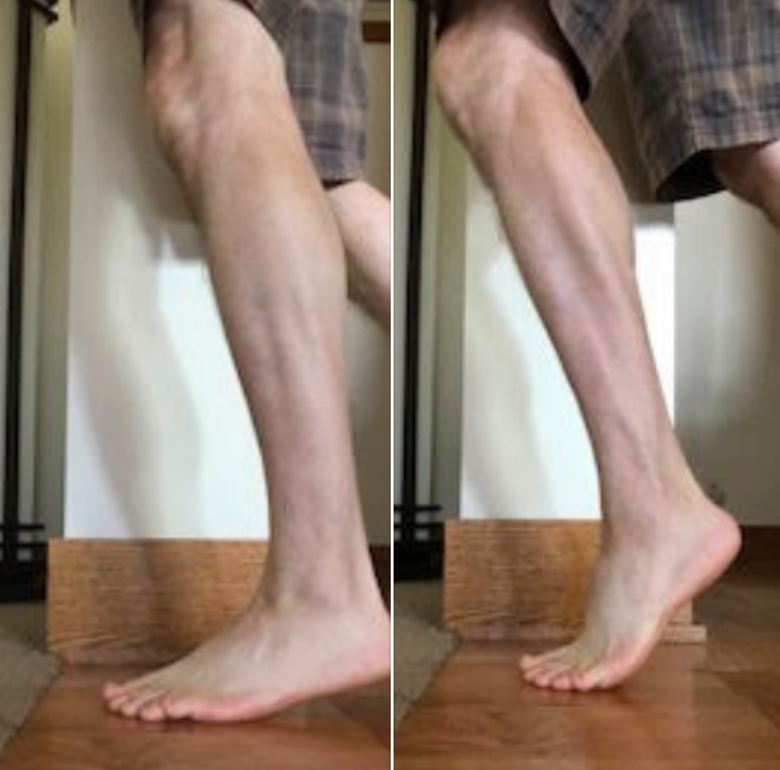
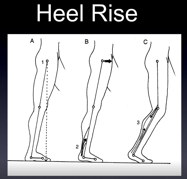
 In fact, back to the gastrocnemius another 2+ joint muscle (crosses knee, mortise and subtalar joints), we all typically think of it as a “push off” muscle.  It causes the heel to rise and accelerates push off in gait and running. But, when the foot is fixed on the ground the insertion is more stable and thus the contraction, because the origin is above the posterior joint line, can pull the femoral condyles into a posterior shear vector. It thus, like the hamstrings, needs to be adequately trained in a ACL or post-operative ACL, deficient knee to help reduce the anterior shear of normal joint loading. It is vital to note, that when ankle rocker is less than 90  degrees (less than 90 degrees of ankle dorsiflexion is available), knee hyperextension is a viable strategy to progress forward in the sagittal plane.  But in this scenarios, the posterior shear capabilites of the gastrocnemius are brought to the front of the line as a frequent strategy.  And not a good one for the menisci we should mention.

Andrews J G (1982)  On the relationship between resultant joint torques and muscular activity  Med Sci Sports Exerc  14: 361-367.

Andrews J G (1985)  A general method for determining the functional role of a muscle  J Biomech Eng  107: 348-353.

Bobbert MF, van Soest AJ (2000) Two-joint muscles offer the solution - but what was the problem? Motor Control 4: 48-52 & 97-116.

Gregor, R.J., Cavanagh, P.R., & LaFortune, M. (1985). Knee flexor moments during propulsion in cycling–a creative solution to Lombard’s Paradox. Journal of Biomechanics, 18, 307-16 .

Ingen-Schenau GJv (1989) From rotation to translation: constraints on multi-joint movement and the unique action of bi-articular muscles. Hum. Mov. Sci. 8:301-37.

Lombard, W.P., & Abbott, F.M. (1907). The mechanical effects produced by the contraction of individual muscles of the thigh of the frog. American Journal of Physiology, 20, 1-60.

Mansour J M & Pereira J M (1987)  Quantitative functional anatomy of the lower limb with application to human gait  J Biomech  20: 51-58.

Park S, Krebs DE, Mann RW (1999) Hip muscle co-contraction: evidence from concurrent in vivo pressure measurement and force estimation. Gait & Posture 10: 211-222.

Rasch, P.J., & Burke, R.K. (1978). Kinesiology and applied anatomy. (6th ed.). Philadelphia: Lea & Febiger.

Visser JJ, Hoogkamer JE, Bobbert MF & Huijing PA (1990) Length and Moment Arm of Human Leg Muscles as a Function of Knee and Hip Angles. Eur. J Appl Physiol 61: 453-460.

Zajac FE & Gordon MF (1989) Determining muscle’s force and action in multi-articular movement  Exerc Sport Sci Revs  17: 187-230.

Zajac FE, Neptune RR, Kautz SA (2003) Biomechanics and muscle coordination of human walking - Part II: Lessons from
dynamical simulations and clinical implications, Gait & Posure 17 (1): 1-17.