What does progressive weakness of the posterior compartment look like?

Look at this video carefully and what do you notice? Can you see the progressive dip in the left heel as time goes on while toe walking? This is a cardinal sign of lack of endurance in the posterior compartment, in this patient’s case tibialis posterior. Your differential, in addition to lack of type one muscle fibers, insufficient vascularity or mitochondria for whatever reason would be circulatory problems as well as conditions causing progressive motor weakness like myasthenia gravis.

Fatigue testing is very important because a lot of times the problem doesn’t come out till the person reaches say a half an hour, an hour or sometimes even many miles into the run or ride. Our job as clinicians is to try to diagnose the problem to the best of our abilities. Our job also is to “exploit their weaknesses” rather than “extol their virtues”. 

If you’re getting somebody with posterior calf pain or a foot drop, or maybe somebody who gets worse over time, consider fatigue testing.

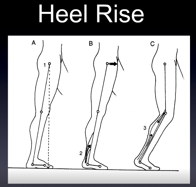
The 4 Factors of Heel Rise.

Screen Shot 2019-01-14 at 12.48.19 PM.png
Screen Shot 2019-01-14 at 12.48.08 PM.png

These SHOULD all happen to have appropriate heel rise and forward progression

1. active contraction of the posterior compartment of the calf

2. passive tension in the posterior compartment of the calf

3. knee flexion and anterior translation of the tibia ankle rocker

4. the windlass mechanism

a problem with any one of these (or more collectively) can effect heel rise, usually causing premature heel rise.

ask yourself:

  • Do you think the posterior compartment is actively contracting? not enough or too much? Remember the medial gastrocnemius adducts the heel at the end of terminal stance to assist in supination. Don't forget about the tibialis posterior as well as the flexor digitorum longs and flexor hallucinate longus.

  • Does there appear to be increased passive tension in the posterior compartment? How visible and prominent are their calf muscles?

  • Do they have forward progression of the body mass?

  • How is his windlass mechanism? Good but not good enough.

Dr Ivo Waerlop. One of The Gait Guys…

#gait, #gaitanalysis, #continuingeducation, #limp, #casestudy, #gaitparameters, #heelrise, #prematureheelrise, #windlassmechanism

The gastroc can causse ankle dorsi and plantarflexion ? Yup. What ?

The gastroc, does it cause ankle dorsiflexion and ankle plantarflexion ? Yup. What ?

You may think you know the answer, the gastrocs are ankle plantarflexors, because that is the easy one we all recognize. But I stew on things when unique cases come in and do not fit the "normal" models and it got me reviewing principles I need to always keep in mind.

Think about it, the gastroc cross the knee, so it causes knee flexion. And when the knee flexes, the proximal tibia is progressing forward in the sagittal plane. Now remember, the foot is on the ground, so the distal tibia is (relatively) fixated in relation to the upper tibia. So, as this proximal top tibial moves forward, because of gastroc contraction, the muscle is actually causing ankle dorsiflexion !

So, it is it important to know your normal gait cycle events ? Yes, Ivo and i harp on that all the time ! One has to know the normal cycles to know when abnormal gait cycles are presenting clues.
So, am I saying that the gastroc are helpers of ankle rocker and ankle dorsiflexion ? Yes, they can be. It is a timing thing. So, we have to again get out of our model of open chain events, and thinking that only the anterior compartment muscles are ankle dorsiflexors. We also have to remember that a bent knee heel raise is not the same as a straight leg (knee extension) heel raise. One can stimulate and assist in ankle dorsiflexion and the other cannot so much. So, in clients with loss of ankle dorsiflexion/ankle rocker should you be assessing the function of the gastroc at the proximal knee, for its effects of dorsiflexion at the ankle ? Yes. Go ahead and try it, bend knee and straight knee heel raises, they are different beasts. This gets more complicated, and i will go into that next week ! I have had some deeper epiphanies i wish to share.
Also, remember, single and biarticular muscles have varied and vast capabilities. Thus it is always vital to consider whole body movements where muscles have abilities to accelerate, decelerate, and control and stablize joints they span, and do not span, via dynamic coupling.
Dr. Allen

What do you know about the Ia Afferents?

This is a nice study looking at lateral gastroc activity and changing firing patterns with speed of movement. Great if you treat anyone or anything that walks...

Ia afferents

You remember them, large diameter afferent (sensory) fibers coming from muscle spindles and appraising the nervous system of vital information like length and rate of change of length of muscle fibers, so we can be coordinated. They act like volume controls for muscle sensitivity. Turn them up and the muscle becomes more sensitive to ANY input, especially stretch (so they become touchy…maybe like you get if you are hungry and tired and someone asks you to do something); turn them down and they become less or unresponsive.

Their excitability is governed by the sum total (excitatory and inhibitory) of all neurons (like interneuron’s) acting on them (their cell bodies reside in the anterior horn of the spinal cord).

If we slow things down, the rate of change of length slows as well and excitability decreases, like we see in this study (3-6% slower). We also notice that the length of contraction increases; hmmm, why doesn’t it decrease?

Remember these folks are on a treadmill. The treadmill is constantly moving, opposite the direction of travel. With the foot on the ground, this provides a constant rate of change of length of the gastroc/soleus (ie, it is putting it through a slow stretch); so , once the muscle is activated, it contracts for a longer period of time because of the treadmill putting a slow stretch on the gastroc (and soleus).

This article also talks about people with upper motor neuron lesions. An important set of inhibitory neurons come from higher centers of the brain, in the motor cortex. These tend to attenuate the signals affecting the Ia afferents, and keep us stable. When we have an upper motor neuron lesion (like a brain lesion or stroke), we lose this “attenuation” and the stretch reflexes (and muscle tone) becomes much more active (actually hyperactive), making the muscle more sensitive to stretch. This loss of attenuation, along with differing firing patterns of the gastroc are important to remember in gait rehab.

The soleus and medial gastroc begin firing in the first 10% of the gait cycle (at the beginning of loading response) and fire continuously until pre swing (peaking just after midstance). The lateral head begins firing at midstance; both heads (along with soleus) decelerate the forward momentum of the tibia, flex the knee at midstance, and the medial head assists in adducting the calcaneus to assist in supination.

Making sure these muscles fire appropriately is important and needling is just one way of helping them to function better. Don’t overlook the tricep surae on your next patient that has a “hitch in their giddyup”.


Effects of treadmill walking speed on lateral gastrocnemius muscle firing.

by Edward A Clancy, Kevin D Cairns, Patrick O Riley, Melvin Meister, D Casey Kerrigan

American journal of physical medicine rehabilitation Association of Academic Physiatrists (2004) Volume: 83, Issue: 7, Pages: 507-51 PubMed: 15213474

Abstract

OBJECTIVE: To study the electromyographic profile-including ON, OFF, and peak timing locations-of the lateral gastrocnemius muscle over a wide range of walking speeds (0.5-2.1 m/sec) in healthy young adults. DESIGN: We studied gastrocnemius muscle-firing patterns using an electromyographic surface electrode in 15 healthy subjects ambulating on a treadmill at their normal walking speed and at three paced walking speeds (0.5, 1.8, and 2.1 m/sec). Initial heel contact was determined from a force-sensitive switch secured to the skin over the calcaneous. RESULTS: For all speeds, the gastrocnemius firing pattern was characterized by a main peak, occurring 40-45% into the gait cycle, that increased in amplitude with walking speed. Speeds of > or =1.3 m/sec produced a common electromyographic timing profile, when the profile is expressed relative to the stride duration. However, at 0.5 m/sec (a speed typical of individuals with upper-motor neuron lesions), the onset of gastrocnemius firing was significantly delayed by 3-6% of the gait cycle and was prolonged by 8-11% of the gait cycle. CONCLUSION: Many patients with upper motor neuron lesions (e.g., stroke and traumatic brain injury) walk at speeds much slower than those commonly described in the literature for normal gait. At the slow walking speed of 0.5 m/sec, we have measured noticeable changes in the electromyographic timing profile of the gastrocnemius muscle. Given the importance of appropriate plantar flexor firing patterns to maximize walking efficiency, understanding the speed-related changes in gastrocnemius firing patterns may be essential to gait restoration.

Pain on the outside of one leg, inside of the other. 

Whenever you see this pattern of discomfort, compensation is almost always at play and it is your job to sort it out. 

This patient presents with with right sided discomfort lateral aspect of the right fibula and in the left calf medially. Pain does not interfere with sleep.  He is a side sleeper 6 to 8 hours. His shoulders can become numb; left shoulder bothers him more than right.

PAST HISTORY: L shoulder surgery, rotator cuff with residual adhesive capsulitis. 

GAIT AND CLINICAL EVALUATION: see video. reveals an increased foot progression angle on the right side. Diminished arm swing from the right side. A definite body lean to the right upon weight bearing at midstance on that side.

He has external tibial torsion bi-lat., right greater than left with a right short leg which appears to be at least partially femoral. Bi-lat. femoral retrotorsion is present. Internal rotation approx. 4 to 6 degrees on each side. He has an uncompensated forefoot varus on the right hand side, partially compensated on the left. In standing, he pronates more on the left side through the midfoot. Ankle dorsiflexion is 5 degrees on each side. 

trigger points in the peroneus longus, gastroc (medial) and soles. 

Weak long toe extensors and short toe flexors; weak toe abductors. 

pathomechanics in the talk crural articulation b/l, superior tip/fib articulation on the right, SI joints b/l

WHAT WE THINK:  

1.    This patient has a leg length discrepancy right sided which is affecting his walking mechanics. He supinates this extremity as can be seen on video, especially at terminal stance/pre swing (ie toe off),  in an attempt to lengthen it; as a result, he has peroneal tendonitis on the right (peroneus is a plantar flexor supinator and dorsiflexor/supinator; see post here). The left medial gastroc is tender most likely due to trying to attenuate the midfoot pronation on the left (as it fires in an attempt to invert the calcaneus and create more supination). see here for gastroc info

2.    Left shoulder:  Frozen shoulder/injury may be playing into this as well as it is altering arm swing.

WHAT WE DID INITIALLY (key in mind, there is ALWAYS MORE we can do):    

  •  build intrinsic strength in his foot in attempt to work on getting the first ray down to the ground; EHB, the lift/spread/reach exercises to perform.
  • address the leg length discrepancy with a 3 mm sole lift
  • address pathomechanics with mobilization and manipulation. 
  • improve proprioception: one leg balancing work
  • needled the peroneus longus brevis as well as medial gastroc and soles. 
  • follow up in 1 week to 10 days.

Pretty straight forward, eh? Look for this pattern in your clients and patients

So what do we see here?


a limp on the left?
a short leg on the right?
a weak gluteus medius on the left?
a shortened step length on the right?
increased arm swing on the left?

watch the push off (terminal stance/pre swing) on the right and then the left. Note how the left is weaker?
now watch the heel strike. Notice how it is shorter when the right strikes the ground than the left?
did you note the pelvic shift to the left on L stance phase? How about the subtle increased knee flexion on the left?

This gentleman has an atrophied gastroc/soleus on the left from an injury. He compensates by increasing thigh flexion on the left to clear the leg. Because he has lost gastroc/soleus strength on the left (the lateral gastrocis an important inverter of the heel after midstance and important component of rearfoot supination), the rearfoot everts more. allowing more midfoot pronation. This collapse of the midfoot brings his weight more medially, so he shifts his pelvis laterally (to the left) to keep his center of gravity over the foot.

Fix?

  • Make client aware of what is going on.
  • make sure gastroc/soleus complex strength and function is maximized through muscle work, acupuncture, muscle activation, functional gait exercise

The Gait Guys. Bringing you the meat, without the filler.

Copyright 2012: The Gait Guys/The Homunculus Group. All rights reserved. Don’t rip off our stuff!

Stretching out Plantar Fasciitis

Neuromechanics Weekly: Look to the hammy’s???

“These findings show that while we always consider the tightness of the gastrocnemius/soleus complex and the subsequent restricted ankle motion from this equinus, we also need to consider the role of the hamstrings,” said Jonathan Labovitz, DPM, lead author and associate professor at Western University of Health Sciences, Pomona, CA.

this article from Lower Extremity Review, concludes “After controlling for covariates, participants (86 of 210 feet) with hamstring tightness were 8.7 times as likely to experience plantar fasciitis (p < .0001) as participants without hamstring tightness. Patients with a BMI >35 were 2.4 times as likely as those with a BMI <35 to have plantar fasciitis.”

The question is why?

They go on to say “ If you can’t get dorsiflexion at your talo-crural joint, this often drives dorsiflexion at other joints and that is going to cause collapse of the longitudinal arch of the foot, loading the plantar fascia with increased tensile stress.”

So, loss of ankle rocker leads to increased midfoot pronation, which loads the plantar fascia. That sounds pretty logical to us. We are sure you are thinking a loss of hip extension may do the same thing. Correct. Or you may say ” The calves may be tight so the medial gastroc can invert the rearfoot to correct for too much midfoot pronation and the foot can be supinated"…and you would be correct again.

So why are the tight hammys driving the bus? Or are they?

We remember the hams are a 2 joint muscle, and with the foot in a closed chain position (ie, on the ground); they flex the thigh on the lower leg and tilt the pelvis posteriorly (ie reduce the lordosis). They are FLEXORS which are active from late swing phase, just prior to heelstrike (initial contact) and a little nudge just prior to toe off (preswing) to help extend the thigh. 

The tricep surae are FLEXORS and are supposed to be active from loading response till almost pre swing, with a burst of activity at heel lift (terminal stance). 

So they take turns, and are not firing (normally) at the same time (or maybe have a small overlap). Going from heel strike to heel strike, the hammys fire 1st.

So IF the two are related, it could be a neurological sequencing issue. How often does that happen? The literature says (and there aren’t many studies) that you can change the order of recruitment of motor units ( the nerve and the muscle fibers it innervates), but not (usually) individual muscles. So probably not.

OK, how about plan B?

The hams and tricep surae are all flexors, correct? What is the innervation to the hamstrings and tricep surae? Hmm….Hamstrings, mostly tibial branch of the sciatic nerve, short head of biceps femoris is the common peroneal: L5-S2. How about the tricep surae? Tibial nerve, mostly S1-S2. I think I see a trend here. Common neurological overlap of FLEXOR muscles.

So are the hams driving the bus? Probably not, but neither are the gastroc/ soleus. The FLEXORS are driving the bus, and excitation of that common neuronal pool is probably causing the tightness

Ivo and Shawn….Uber footgeeks of the web. Dicing and slicing through the literature so you don’t have to.

This is a nice study looking at lateral gastroc activity and changing firing patterns with speed of movement. It also melds nicely with yesterdays Neuromechanics post&hellip;
Those darn Ia afferents….
You remember them, large diameter afferent (sens…

This is a nice study looking at lateral gastroc activity and changing firing patterns with speed of movement. It also melds nicely with yesterdays Neuromechanics post…

Those darn Ia afferents….

You remember them, large diameter afferent (sensory) fibers coming from muscle spindles and appraising the nervous system of vital information like length and rate of change of length of muscle fibers, so we can be coordinated. They act like volume controls for muscle sensitivity. Turn them up and the muscle becomes more sensitive to ANY input, especially stretch (so they become touchy…maybe like you get if you are hungry and tired and someone asks you to do something); turn them down and they become less or unresponsive.

Their excitability is governed by the sum total (excitatory and inhibitory) of all neurons (like interneuron’s) acting on them (their cell bodies reside in the anterior horn of the spinal cord).

If we slow things down, the rate of change of length slows as well and excitability decreases, like we see in this study (3-6% slower). We also notice that the length of contraction increases; hmmm, why doesn’t it decrease?

Remember these folks are on a treadmill. The treadmill is constantly moving, opposite the direction of travel. With the foot on the ground, this provides a constant rate of change of length of the gastroc/soleus (ie, it is putting it through a slow stretch); so , once the muscle is activated, it contracts for a longer period of time because of the treadmill putting a slow stretch on the gastroc (and soleus).

This article also talks about people with upper motor neuron lesions. An important set of inhibitory neurons come from higher centers of the brain, in the motor cortex. These tend to attenuate the signals affecting the Ia afferents, and keep us stable. When we have an upper motor neuron lesion (like a brain lesion or stroke), we lose this “attenuation” and the stretch reflexes (and muscle tone) becomes much more active (actually hyperactive), making the muscle more sensitive to stretch. This loss of attenuation, along with differing firing patterns of the gastroc are important to remember in gait rehab.

The soleus and medial gastroc begin firing in the first 10% of the gait cycle (at the beginning of loading response) and fire continuously until pre swing (peaking just after midstance). The lateral head begins firing at midstance; both leads (along with soleus) decelerate the forward momentum of the tibia, flex the knee at midstance, and the medial head assists in adducting the calcaneus to assist in supination.

We remain, inexplicably….The Gait Guys


Effects of treadmill walking speed on lateral gastrocnemius muscle firing.

by Edward A Clancy, Kevin D Cairns, Patrick O Riley, Melvin Meister, D Casey Kerrigan

American journal of physical medicine rehabilitation Association of Academic Physiatrists (2004) Volume: 83, Issue: 7, Pages: 507-51 PubMed: 15213474

Abstract

OBJECTIVE: To study the electromyographic profile-including ON, OFF, and peak timing locations-of the lateral gastrocnemius muscle over a wide range of walking speeds (0.5-2.1 m/sec) in healthy young adults. DESIGN: We studied gastrocnemius muscle-firing patterns using an electromyographic surface electrode in 15 healthy subjects ambulating on a treadmill at their normal walking speed and at three paced walking speeds (0.5, 1.8, and 2.1 m/sec). Initial heel contact was determined from a force-sensitive switch secured to the skin over the calcaneous. RESULTS: For all speeds, the gastrocnemius firing pattern was characterized by a main peak, occurring 40-45% into the gait cycle, that increased in amplitude with walking speed. Speeds of > or =1.3 m/sec produced a common electromyographic timing profile, when the profile is expressed relative to the stride duration. However, at 0.5 m/sec (a speed typical of individuals with upper-motor neuron lesions), the onset of gastrocnemius firing was significantly delayed by 3-6% of the gait cycle and was prolonged by 8-11% of the gait cycle. CONCLUSION: Many patients with upper motor neuron lesions (e.g., stroke and traumatic brain injury) walk at speeds much slower than those commonly described in the literature for normal gait. At the slow walking speed of 0.5 m/sec, we have measured noticeable changes in the electromyographic timing profile of the gastrocnemius muscle. Given the importance of appropriate plantar flexor firing patterns to maximize walking efficiency, understanding the speed-related changes in gastrocnemius firing patterns may be essential to gait restoration.