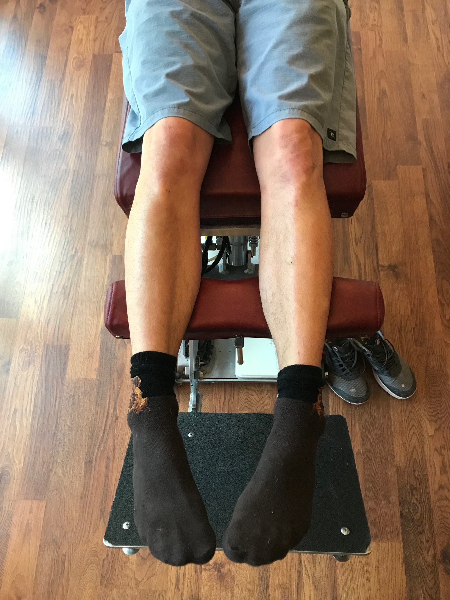
How you load and off-load your forefoot bipod matters.

Screen Shot 2020-11-14 at 8.45.37 AM.png

If you are a sprinter, how you load the forefoot bipod might be a variable for speed or injury. Tendons can change their cross sectional area, if you load them, but they don't change, if you don't.
Of course this article is not exclusive for sprinters, it pertains to any running sport, even endurance.
Maximum isometric force had increased by 49% and tendon CSA by 17% !
Tendons can change their cross sectional area, if you load them.
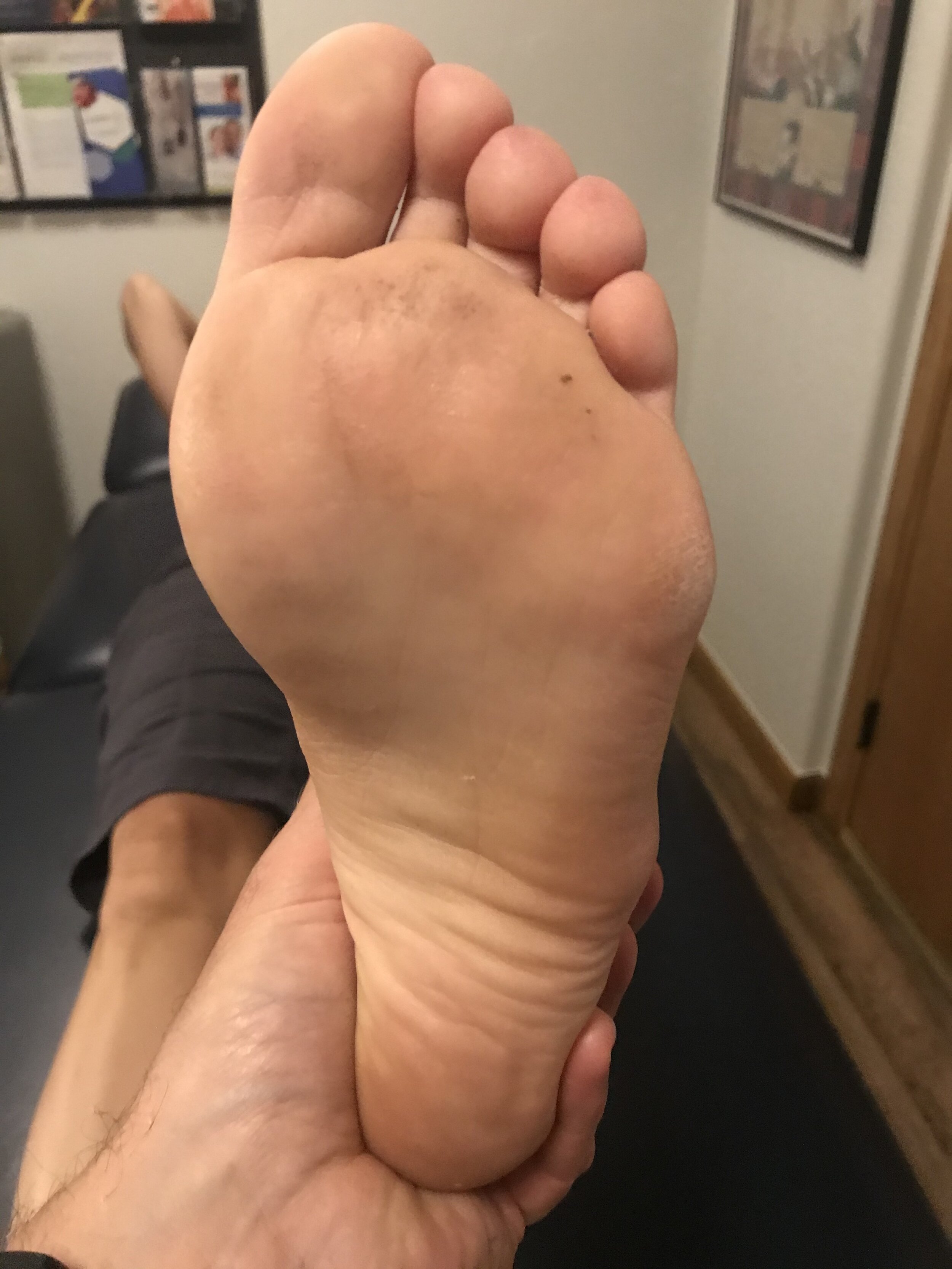
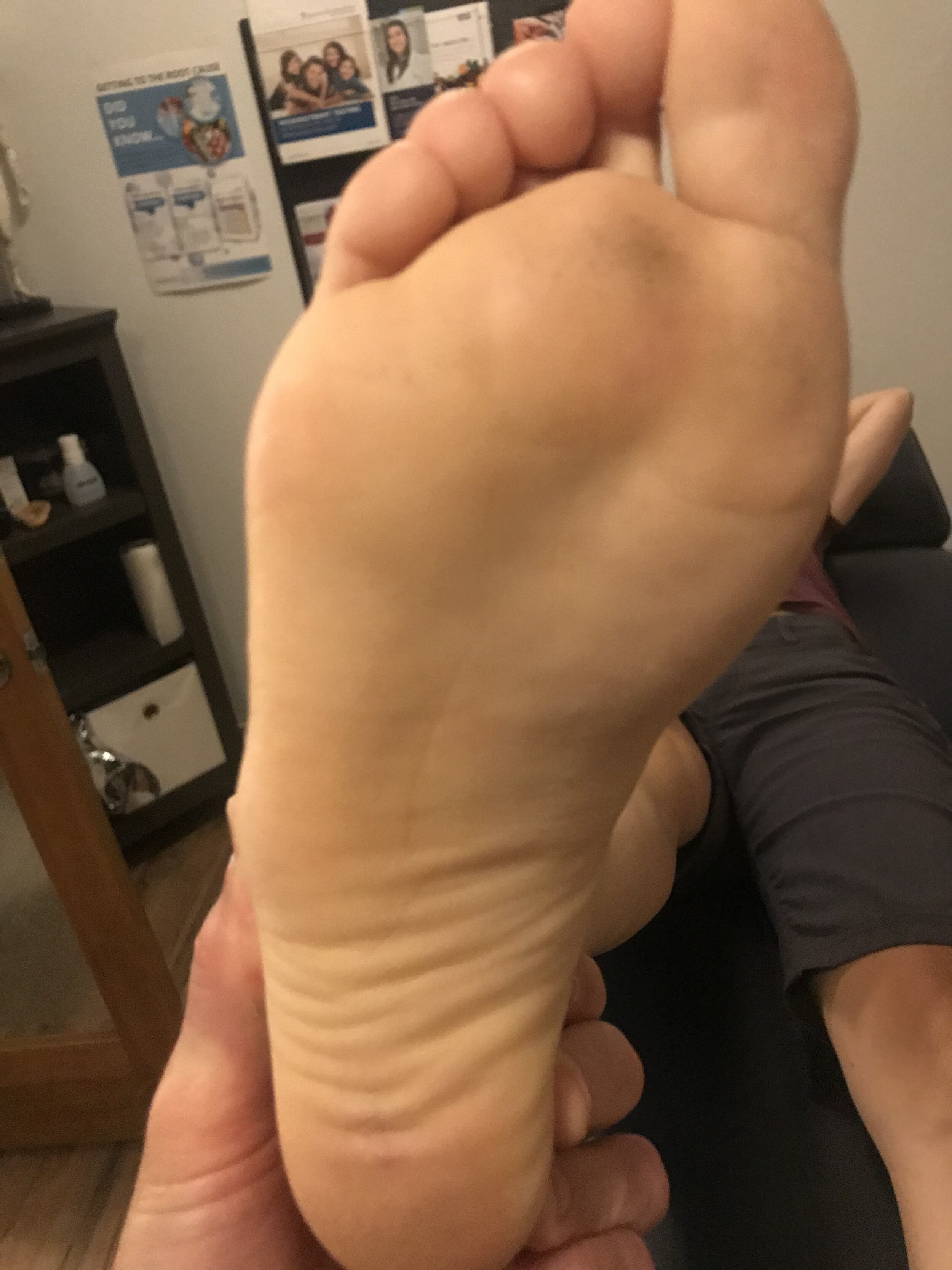
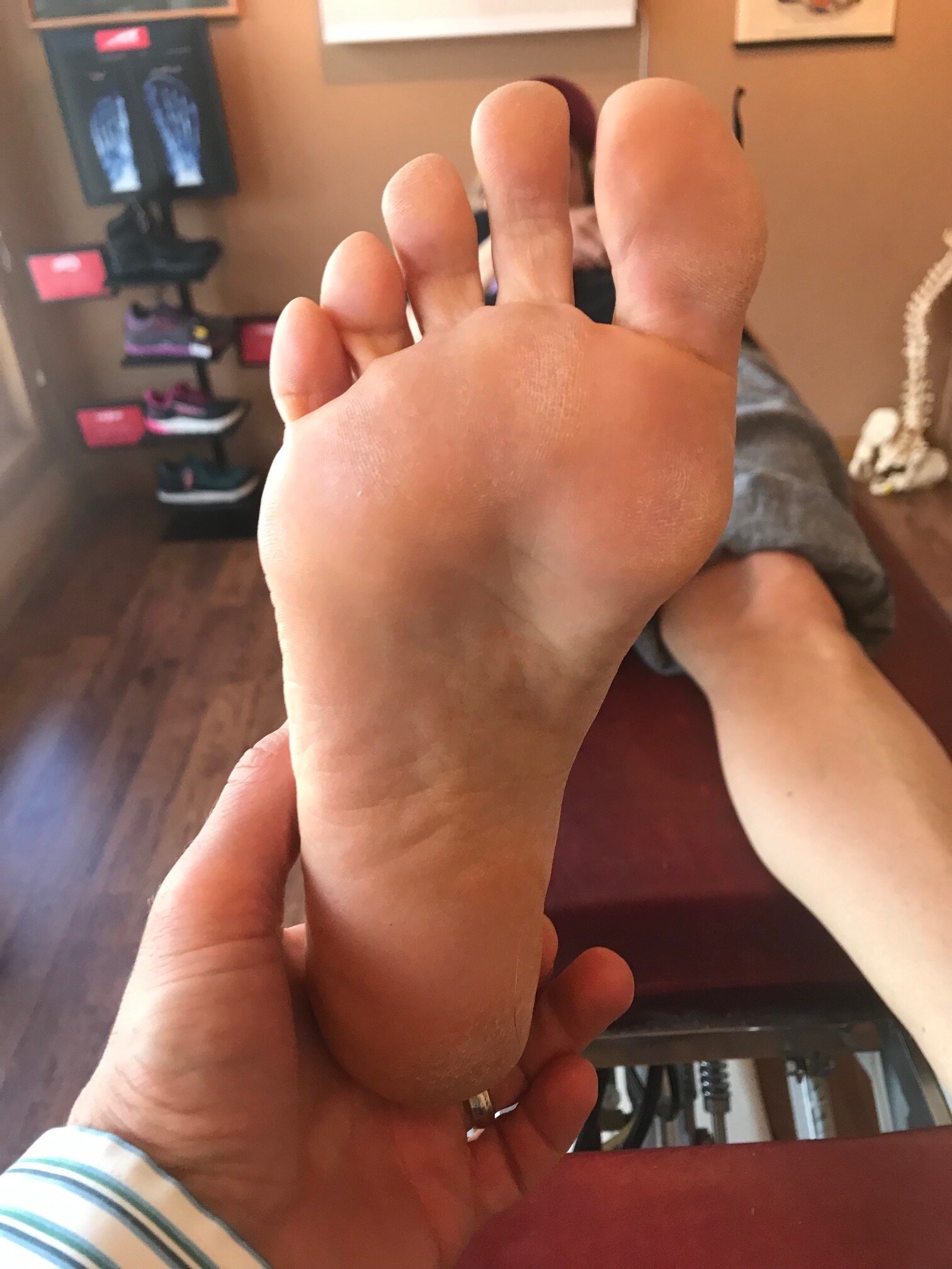
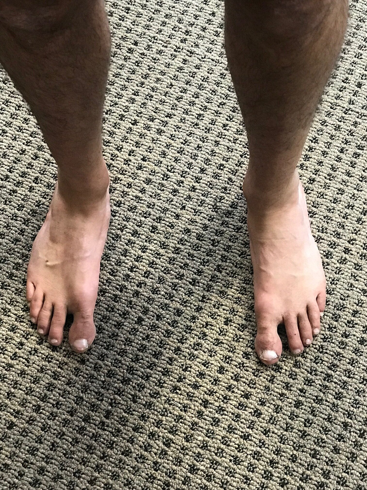
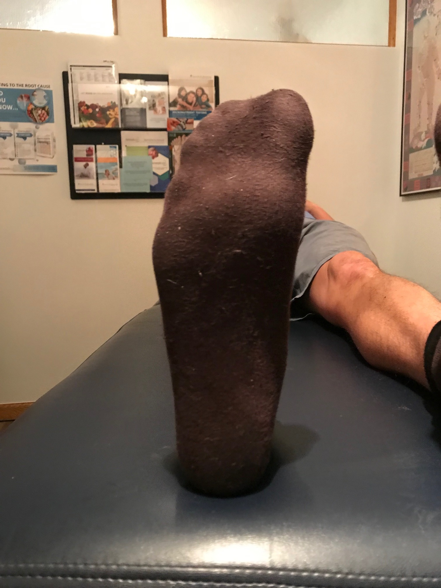
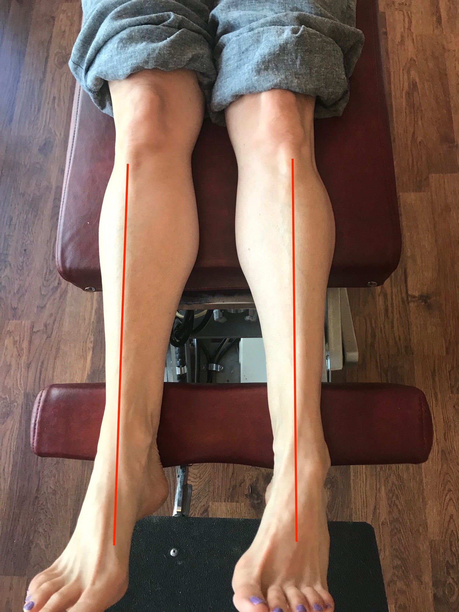
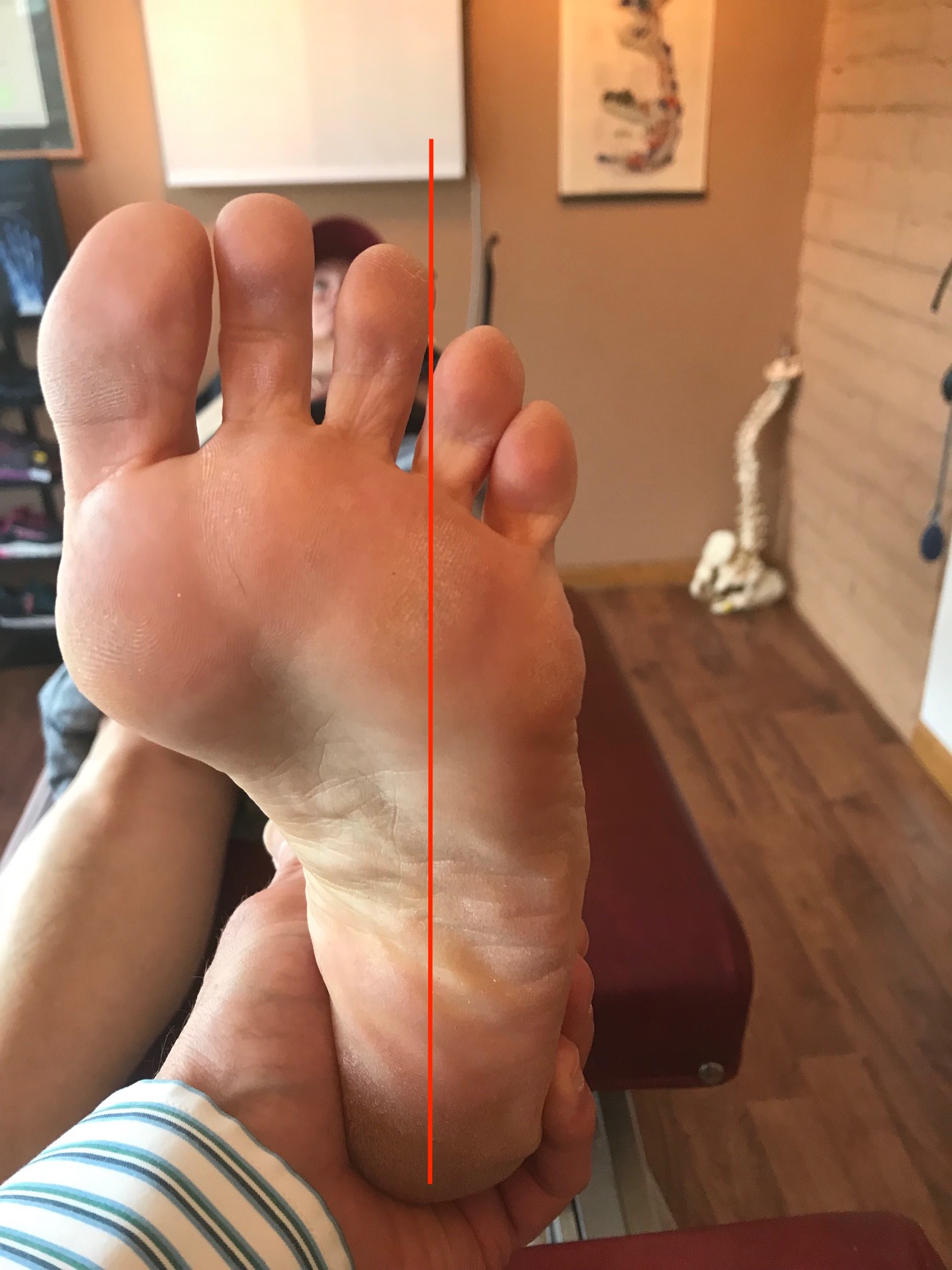
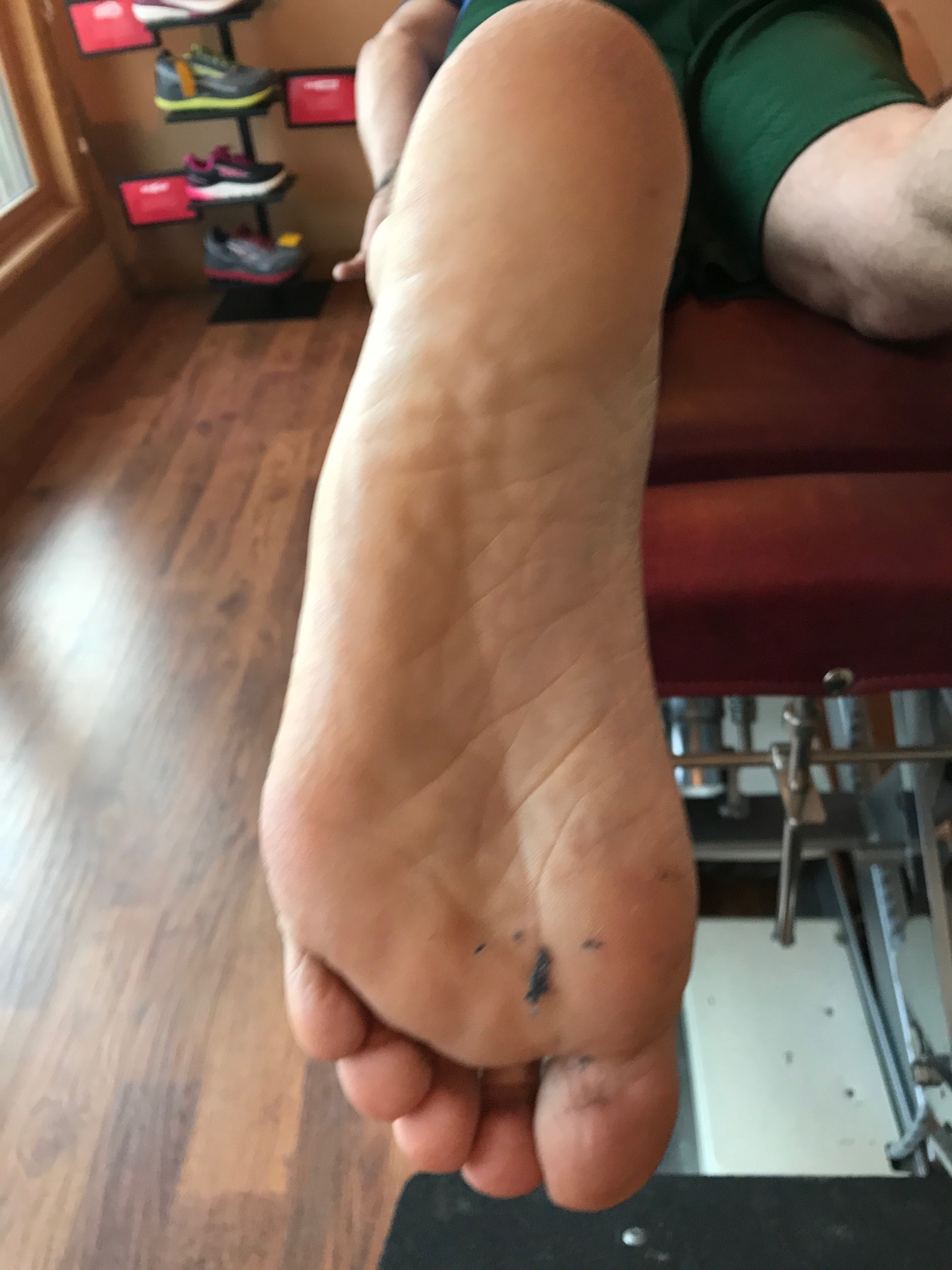
Here I show lateral forefoot loading in a heel raise, and a medial forefoot loading in heel raise. This has to be part of the discovery process outlined below. Forefoot types will play into the loading choice, and unequal strength of the medial or lateral calf compartment will also play into the loading choice made. Where do you need to put your strength ? And is the forefoot competent to take that loading challenge ? Meaning, do they have a forefoot valgus? A forefoot supinatus ? These things matter. If you are a sprinter, how you load the forefoot bipod might be a variable of foot type, asymmetrical posterior compartment strength, or foot strike pattern in the frontal plane (search our blog for cross over gait and glute medius targeting strategies for step width) ,or a combination of several or all of the above. These things matter, and why and where you put your strength matters, if you are even aware of where and how you are putting the loads, and why of course. Of course, then there are people like the recent Outside online article that says how you foot strike doesn’t matter, but it does matter. But of course, if you do not know the things we have just mentioned, it is easy to write such an article.
Isometrics are useful, they have their place. In a recent podcast we discussed the place and time to use isometrics, isotonics, eccentrics and concentrics.
One of the goals in a tendinopathy is to restore the tendon stiffness. Isometrics are a safe way to load the muscle tendon complex without engaging a movement that might have to go through a painful arc of movement. With isometrics here is neurologic overspill into the painful arc without having to actually go there.
The key seems to be load. More load seems to get most people further along. Remember, the tendon is often problematic because it is inflammed and cannot provide a stiffness across its expanse. Heavy isometric loading seems to be a huge key for most cases. But, we have to say it here, not everyone fits this mold. Some tendons, in some people, will respond better to eccentrics, and strangely enough, some cases like stretching (perhaps because this is a subset of an eccentric it seems or because there is a range of motion issue in the joint that is a subset of the problem). Now the literature suggests that stretching is foolish, but each case is unique all in its own way, and finding what works for a client is their medicine, regardless of what the literature and research says.
Finding the right load for a given tendon and a right frequency of loading and duraction of loading is also case by case specific. Part of finding the right loading position is a discovery process as well, as noted in the photos above. Finding the fascicles you want to load, and the ones you do not want to load (painful) can be a challenging discovery process for you and your client. Finding the right slice of the pie to load, and the ones not to load takes experimentation. When it is the achilles complex, finding the safe However, if one is looking for a rough template to build from, brief, often, heavy painfree loads is a good template recipe to start with.
Here, in this Geremia et al article, "ultrasound was used to determine Achilles tendon cross-sectional area (CSA), length and elongation as a function of plantar flexion torque during voluntary plantar flexion."
They discovered that, "At the end of the training program, maximum isometric force had increased by 49% and tendon CSA by 17%, but tendon length, maximal tendon elongation and maximal strain were unchanged. Hence, tendon stiffness had increased by 82%, and so had Young’s modulus, by 86%.
Effects of high loading by eccentric triceps surae training on Achilles tendon properties in humans. Jeam Marcel Geremia, Bruno Manfredini Baroni, Maarten Frank Bobbert, Rodrigo Rico Bini, Fabio Juner Lanferdini, Marco Aurélio Vaz
European Journal of Applied Physiology
August 2018, Volume 118, Issue 8, pp 1725–1736

Can you say Forefoot Adductus?

-Take a look at these tootsies. Draw an imaginary lines for the center of the hill: this should normally pass up through the foot either through the second metatarsal or between the second and third. Can you see how the foot is somewhat banana shaped?

IMG_8466.jpg

– This is also called metatarsus adductus. The deformity is at the apex of the tarsal/metatarsal joint, also known as Lisfranc joint. The fifth metatarsal base is often prominent in the foot is convex in shape with a higher arch.

-This is usually caused by intrauterine positioning and if caught early will usually spontaneously resolve. Since this gal is over 34, that’s probably not her reality. It is interesting to note that along with this congenital deformity, hip dysplasia and internal tibial torsion (which she has) are extremely common.

IMG_8468.jpg

IMG_8467.jpg

– Gait abnormalities seen with this include a decreased progression angle, and a propulsive gait often secondary to poor intrinsic function of the foot musculature

#forefoot #forefootadductus #adductus #foot #footproblem #gait #clinicalexam #thegaitguys 

The consequences of an inverted forefoot

A forefoot that is inverted with respect to the rearfoot. Whether it is a forefoot varus, forefoot supinatus or an everted rearfoot ( because the forefoot is still inverted with respect to the rearfoot), what are the biomechanical sequelae?

If we accept the premise that the foot is basically a tripod between the calcaneus, base of the first and base of the fifth metatarsal‘s, we know that all of these parts needs to be on the ground at certain points in the gait cycle. Forces should travel from the calcaneus, up the lateral aspect of the foot, across the metatarsal heads to the first metatarsal head and hopefully out through the hallux.

The foot should hit the ground in slight inversion of the entire foot at initial contact and pronate through the middle of mid stance and then supinate through the remainder of the gait cycle. There’s an intricate balance of biomechanical events that must occur, especially in the latter half of the gait cycle when the rear foot is inverting where the forefoot is everting, so that we can have high gear push off through the distal first ray.

If the forefoot remains inverted then somehow the head of the first metatarsal needs to be brought down to the ground. If there’s not adequate range of motion in the foot, particularly the first ray, then you may pronate through the midfoot, rearfoot or in cases where this is insufficient, bring them immediately over the foot to get it down. This of course shifts center of gravity to midline and the body above must compensate in someway.

Take a look at this video footage and what do you see? She strikes on the outside of her foot but does not have adequate motion in her forefoot and therefore “crashes“ down on the forefoot, forcing a valgus moment into the ankle and the need to shift immediately by the pelvis attempts to dampen it. Notice how this is worse on the right side with more medial knee shift, pelvic shift as well as a lateral bending of the body to the right. Notice also how the upper body twists more to the left than to the right.

So what’s the fix? Well the answer is, “what’s bothering the patient?” We don’t necessarily fix what we see; we correlate what we see with what the patient’s symptoms are because that’s usually why they show up in your office. Yes, we do get people from time to time that come in strictly for “performance enhancement“ but this is pretty rare.

This woman has very little motion and plantar flexion of the first Ray complex so our primary goal was to get her to descend the first ray. We accomplished this by the following:

1. Manipulation in plantar and dorsiflexion of the first ray complex
2. Soft tissue work in the first intermetatarsal interval
3. Exercises of muscles to assist in descending the first ray including the following: extensor hallucis brevis, peroneus longus, flexor digitorum brevis
4. Pelvic stability work to improve the skill, endurance and strength of the gluteus medius complex as well as abdominal endurance work.

Your rehab program should change as the patient has more functional gains, tailoring it to the patient’s deficiencies.


Dr Ivo Waerlop, one of The Gait Guys

#invertedforefoot #forefootsupinatus #forefootvarus #pronation #forefoot #gaitanalysis

The “ banana foot”

IMG_7018.jpg

So, you see at foot that looks like this and what do you think? What are some of the biomechanical characteristics of people with the foot that when, you bisect the calcaneus, the line passing forward passes lateral to the second metatarsal or a line between the second and third?

This condition can be congenital, in conditions like forefoot adductus or compensatory.

The first thing that springs to mind when we see deformities like this is “things usually occur in threes“. So we would expect to see other anatomical and/or genetic abnormalities. An adducted forefoot, like you see here, often occurs as the result of lack of internal rotation of the hip on that side so therefore will often be present with conditions like internal tibial torsion and femoral retrotorsion, which we often, but not always, see together. Because of the increased gait and foot progression angle in these individuals, the forefoot compensates and adducts to bring the center of gravity more to midline.

Feet like this are often, but not always, cavus and rigid. If it remains in relative supination (plantarflexion, abduction and inversion) it is an excellent level but poor shock absorber.

Forefoot adduction can also be a compensation pattern if an individual is unable to get the head of their first ray completely down to the ground. It could be a true forefoot varus or more commonly, a forefoot supinatus; either results in an inability to get the first ray down. This often causes the foot to adduct in compensation, and, due to the tarsal articulations, often raises the base of the first metatarsal increasing the inclination angle of the first ray. This frequently leads to limited dorsiflexion of the first metatarsophalangeal articulation.

So what is a clinician to do?

Ensure that the mechanics of the foot are clean through manipulation and mobilization

Make sure there are appropriate flexors/extensor ratios of skill, endurance, and strength of the foot musculature both intrinsically and extrinsically. This means making sure that the long flexors and extensors are in some degree of balance.

Work on balance and coordination of the lower extremity. This can be impeded if they’re unable to get ahead of the first right down to the ground. Exercises for the peroneus longus, extensor hallucis brevis and short flexors of the foot will often help with this.

“Banana foot”. Coming to your clinic, or a clinic near you. Maybe today…

Dr. Ivo Waerlop, one of The Gait Guys.

#forefootadductus #bananafoot #supination #thegaitguys

Foot Types? Do they really matter?

IMG_6218 copy.jpg

The answer is " yes, often times".

Did you miss our 3rd Wednesdays presentation last week on foot types and obligate biomechanics (and pathomechanic) that ensue? Here is the video feed that you can watch and get ce credits for:

https://www.chirocredit.com/course/Chiropractic_Doctor/Biomechanics_214

#foottypes #biomechanics #thegaitguys

Asymmetries can make all the difference…

Take a good look at these pictures of this gentleman’s feet. Can you see any differences from side to side?

If you look closely, you’ll notice that his right foot is in and abducted position (4 foot adductus) and relatively normal. Asymmetries can make all the difference…

IMG_7024.jpg
IMG_7023.jpg


Take a good look at these pictures of this gentleman‘s feet. Can you see any differences from side to side?

If you look closely, you will notice that his right foot is in an adducted position (forefoot adductus) and the left one relatively normal. If you bisect the heel, it should pass through the second or between the second and third metatarsal in his clearly falls laterally.

So what you say?

Well, putting a foot in relative supination with respect to the other causes certain biomechanical sequela. This forefoot adductus often leads to a forefoot supinatus, so he’s unable to get the head of his first ray down to the ground. Think that might make a difference in his gait cycle?

Think about all the extra internal rotation that will have to occur in that lower extremity during a normal gait cycle. Now combine that with something like external tibial torsion or a leg length discrepancy and things can really stack up and make a big difference.

Lastly, think about the asymmetrical mechanoreceptor input from the joint mechanoreceptors and muscle spindles traveling up the neuraxis. Do you think over time that that may cause some cortical remodeling and ultimately change the way he activates muscles?

Look for asymmetries, they really do matter

Dr Ivo Waerlop, one of The Gait Guys

#asymmetriesmatter #gaitanalysis #thegaitguys #forefootadductus

Determining foot types...In a nutshell

Screen Shot 2019-10-15 at 8.31.27 PM.png

We talked yesterday about how foot types (i.e., the forefoot to rear foot relationship) can often produce predictable pathomechanics. Here's How to do it. Pretty basic, eh? Its the characteristics, along with the other anatomical goodies they may have that helps to clinch the diagnosis and dictate treatment.

To find out about how to apply your newfound knowledge, join us tomorrow night on our 3rd Wednesdays tele seminar: Biomechanics 314 on online.com

5 PST, 6MST, 7CST, 8EST

Foot types: do they really matter?

forefoot varus: note how the forefoot is inverted with respect to the rear foot

forefoot varus: note how the forefoot is inverted with respect to the rear foot

Foot type. You know what we are talking about. The relation in anatomically and in space of the rear foot to the forefoot. We believe that this anatomical relationship holds key clinical insights to predictable biomechanics in that particular foot type.

Simply put, the rear foot can be either inverted, everted or neutral; Same with the forefoot. If the rear foot is inverted we call that a rearfoot varus. If the foot is inverted we call that a forefoot varus. If the rear foot is everted we call that a rear foot valgus and if the forefoot is inverted we call that a forefoot valgus.

Now think about the simple motions of pronation and supination. Pronation is dorsiflexion, eversion and abduction; supination is plantar flexion, inversion and adduction. If it remains in eversion, we say that it is in vslgus and that means they will be qualities of pronation occurring in that foot while it is on the ground. If the foot is inverted, it will have qualities of supination.

We think of pronation as making the foot into a mobile adapter and supination is making the foot into a rigid lever.

During a typical gait cycle the foot is moving from supination at initial contact/loading response to full pronation at mid stance and then into supination from mid stance to terminal stance/pre-swing. I know that if the foot remains and pronation past mid stance that it is a poor lever and if it remains in supination prior to mid stance it will be a poor shock absorbers. Foot type plays into this displaying or amplifying the characteristics of that particular foot type during the gait cycle: if this occurs at a time other than when it supposed to occur, then we can see predictable biomechanics such as too much pronation resulting in increased rear foot eversion, midfoot collapse, abduction of the forefoot and internal rotation of the knee with most often medial knee fall. Now, consider these mechanics along with any torsions or versions in the lower extremity that the patient may have.

This Wednesday night we will be discussing foot types and their biomechanics. Join us on onlinece .com for Biomechanics 314 6:00 MST

Dr Ivo Waerlop, one of The Gait Guys

Botox for plantar fasciitis? Sounds like a bad idea to us....

image source: https://commons.wikimedia.org/wiki/File:Plantar_aponeurosis_-_axial_view.png

image source: https://commons.wikimedia.org/wiki/File:Plantar_aponeurosis_-_axial_view.png

Botox..For plantar fasciitis? Really?

We found this article (1) in one of our favorite journals, Lower Extremity Review , and were a little surprised. Let us get this straight: you are going to take one of the the most poisonous biological neurotoxins known (1) and inject it into your calf and foot?

The article in LER is well written and the results (thankfully) were inconclusive regarding its usage. They do cite 3 studies (with two by the same lead author) where it has been effective (2-4). Yes, it is better than saline (5) (but not as good as extracorporeal shock wave therapy (6)), and better than placebo (7-10) but considerably more risky.

So the premise is “if the muscle is dysfunctional, then let’s just take it out of the equation”. But this really doesn’t fix the problem, it just covers up the symptom. And what about the other potential side effects since botulinum toxin acts not only at the neuromuscular junction, blocking the release of acetylcholine, but also at the autonomic ganglia, postganglionic parasympathetic nerve endings, as well as the post ganglionic sympathetics that use acetylcholine (capillaries of skin, piloerector muscles and sweat glands) (11)?.

In our experience, most cases of plantar fasciitis are secondary to lack of forefoot rocker, lack of ankle rocker, lack of hip extension or in some cases, direct trauma. Wouldn’t it make more sense to strengthen the anterior compartment to reciprocally inhibit the posterior compartment, increasing ankle dorsiflexion and hip extension? We find, oftentimes, treating only the area of chief complaint and not what is "driving the bus" can offer temporary, symptomatic relief but not long standing pathmechanics or pathoanatomy.

Just like the road to enlightenment, there are no shortcuts in treating plantar fasciitis and if you are not going to treat the cause, then be prepared to reap what you sow.

Dr Ivo Waerlop, one of The Gait Guys

#botox #plantarfascitis #badideas #gaitproblem #thegaitguys

1. https://lermagazine.com/article/botox-injection-not-just-for-celebrities-furrows-and-wrinkles

2. Elizondo-Rodriguez J, Araujo-Lopez Y, Moreno-Gonzalez JA, Cardenas-Estrada E,
Mendoza-Lemus O, Acosta-Olivo C. A comparison of botulinum toxin A and intralesional steroids for the treatment of plantar fasciitis: A randomized, double-blinded study. Foot Ankle Int.
2013;34(1):8-14.

3. Díaz-Llopis IV, Rodríquez-Ruíz CM, Mulet-Perry S, Mondéjar-Gómez FJ., Climent-Barberá JM., Cholbi-Llobel F. Randomized controlled study of the efficacy of the injection of botulinum toxin type A versus corticosteroids in chronic plantar fasciitis: results at one and six months. Clin Rehabil. 2012;26(7):594-606.

4. Díaz-Llopis IV, Gómez-Gallego D, Mondéjar-Gómez FJ, López-García A, Climent-Barberá JM, Rodríguez-Ruiz CM. (2013). Botulinum toxin type A in chronic plantar fasciitis: clinical effects one year after injection. Clin Rehabil. 2013;27(8):681-685.

5. Ahmad J, Ahmad SH, Jones K. Treatment of Plantar Fasciitis With Botulinum Toxin. Foot Ankle Int. 2017 Jan;38(1):1-7. doi: 10.1177/1071100716666364. Epub 2016 Oct 1.1.

6. Roca B, Mendoza MA, Roca M. Comparison of extracorporeal shock wave therapy with botulinum toxin type A in the treatment of plantar fasciitis. Disabil Rehabil. 2016 Oct;38(21):2114-21. doi: 10.3109/09638288.2015.1114036. Epub 2016 Mar 1

7. Babcock MS, Foster L, Pasquina P, Jabbari B. Treatment of pain attributed by plantar fasciitis with botulinum toxin A: a short-term randomized, placebo-controlled, double blinded study. Am J Phys Med Rehabil. 2005;84(9):649-654.

8. Samant PD, Kale SY, Ahmed S, Asif A, Fefar M, Singh SD. Randomized controlled study comparing clinical outcomes after injection botulinum toxin type A versus corticosteroids in chronic plantar fasciitis. Int J Res Orthop. 2018;4(4):672-675.

9. Huang YC, Wei SH, Wang HK, Lieu FK. Ultrasonographic guided botulinum toxin type A treatment for plantar fasciitis: an outcome-based investigation for treating pain and gait changes. J Rehabil Med. 2010;42(2):136-140.

10. Ahmad J, Ahmad SH, Jones K. Treatment of plantar fasciitis with botulinum toxin. Foot Ankle Int. 2017;38(1):1-7.

11. Nigam PK, Nigam A. Botulinum toxin. Indian J Dermatol. 2010;55(1):8–14. doi:10.4103/0019-5154.60343

When you see this, you should be thinking one of 3 possible etiologies...

Cardinal sign of either a forefoot supinatus/forefoot varus or collapsing midfoot

I was hiking behind this young chap over the weekend along with my son and friends. Note the amount of calcaneal eversion present on the right side that is not present on the left. Also note the increased progression angle of the right foot and subtle circumduction of the extremity.

In my experience, you would generally see this much calcaneal diversion and one of three scenarios:

1. Moderate leg length discrepancy with the increased calcaneovalgus occurring on the longer leg side. This would support the amount of circumduction were seeing on the right side.

2. When there is a forefoot supinatus present and and inadequate range of motion available in the midfoot and/or forefoot. This is most likely the case here.

3. In moderate To severe midfoot collapse. This is clearly not the case as the medial aspect of the shoe is usually “blown out”.

Next time you see an everting rearfoot, think about these three possible etiologies.

Dr Ivo Waerlop, on of The Gait Guys

#evertedrrarfoot #calcanealvalgus #shortleg #forefootsupinatus #forefootvarus #gaitanalysis #thegaitguys

The amazing power of compensation. Coming to a patient in your office… Maybe today

This gal has had a right sided knee replacement. She has an anatomical right short leg, a forefoot supinatus, an increased Q angle and a forefoot adductus. So, what’s the backstory?

When we have an anatomical short leg, we will often have a tendency to try to “lengthen“ that extremity and “shorten” the longer extremity. This is often accomplished through pelvic rotation although sometimes can be with knee flexion/extension or change in the Q angle. When the condition is long-standing, the body will often compensate in other ways, such as what we are seeing here.

IMG_6736.jpg

The fore foot can supinate in an attempt to lenthen the extremity. Note how the right extremity forefoot is in varus with respect to the rearfoot, effectively lengthening the extremity. As you can see from the picture, this is becoming a “hard“ deformity resulting in a forefoot varus.

IMG_6740.jpg

Over time, the forefoot has actually “adducted “ as you can see, again in an attempt to lengthen the extremity. Remember that supination is plantar flexion, abduction and inversion, all three which are visible here.


You will also see that the Q angle is less on the right side (se above), effectively lengthening that extremity, but not quite enough as we can see from the picture :-)


Dr Ivo Waerlop, one of The Gait Guys

#forefootadductus #shortleg #kneereplacement #tkr #forefootvarus #gait #thegaitguys

Sixth toe disease...That growth on the outside of your foot… Or on somebody’s foot is coming to see you…

IMG_6704.JPG

You know what we’re talking about. That extra growth on the lateral aspect of the foot that happens way too often and many of your clients. A Taylor’s bunion or sometimes referred to as a “bunionette”. 

What is the usual fix?

Usually in a ski boot or hiking boot, they blow out the lateral side of the shoe. This is usually not a good fix because most of these folks have internal tibial torsion and somewhat of a forefoot supinatus/varus.

IMG_6706.JPG
IMG_6707.JPG

The internal tibial torsion places the knee outside the saggital plane and an arch support without a forefoot valgus post will just push it further out, creating a conflict at the knee. The forefoot supinatus and/or varus places them on the outside of the foot as well. Remember, most of these folks are ALREADY on the outside of the foot and the foot wants to migrate laterally...so creating more space just means it migrates farther. Good thought, doesn’t work that way.

IMG_6710.JPG

So what did we do?

  • We created a valgus post for the forefoot (see picture above) tapering from lateral to medial and to help “push“ the distal aspect of the first ray down (because there was motion available that was not being used)

  • We gave him exercises to help descend the first ray like the extensor hallucis brevis exercise, toe waving as well as peroneus longus exercises

  • We gave him plenty of balance and coordination work

    Dr Ivo Waerlop, one of The Gait Guys


#6thtoe #internaltibialtorsion #forefootvarus # forefootsupinatus #gaitanalysis #thegaitguys


Holy Forefoot Flare, Batman!


Some sources say foot strike pattern does not matter. We disagree.

Look at this gal who midfoot/forefoot strikes. She also has a forefoot supinatus, a plastic condition where the forefoot is inverted with respect to the rearfoot. Take that combination and put it in a shoe with a forefoot flare and what do you get? Can you say AMPLIFICATION?

We’re not saying this is a bad shoe or even the wrong shoe. But, if she is going to run in this shoe, we will need to help her gain more ROM in her forefoot ( and some pelvic and hip stability) dodge doesn’t have to crash into eversion on each landing.

Help your patients with shoe selection. Something with less of a lateral flare in the forefoot would certainly make her life easier.

Need to know more? Consider taking our National Shoe Fit Program: link here:

Dr Ivo Waerlop, one of The Gait Guys

#badshoes #forefootflare #thegaitguys #forefootsupinatus #lateralflare #inversion

We’ve told you once and we will tell you again…

Folks with femoral retro torsion often experience lower back pain with twisting movements

This left handed hydrology engineer Presented to the office with an acute onset of lower back pain following “swinging a softball bat”. He comments that he always “hit it out of the park“ and hit “five home runs“ in the last game prior to his backs demise.

note the internal tibial torsion. drop a plumbline from the tibial tuberosity. it should pass through the 2nd met or between the 2nd and 3rd met shafts

note the internal tibial torsion. drop a plumbline from the tibial tuberosity. it should pass through the 2nd met or between the 2nd and 3rd met shafts

note the internal tibial torsion. drop a plumbline from the tibial tuberosity. it should pass through the 2nd met or between the 2nd and 3rd met shafts

note the internal tibial torsion. drop a plumbline from the tibial tuberosity. it should pass through the 2nd met or between the 2nd and 3rd met shafts

He presented antalgic with a pelvic shift to the left side, flexion of the lumbar spine with 0° extension and a complete loss of the lumbar lordosis. He could not extend his lumbar spine past 0° and was able to flex approximately 70. Lateral bending was approximately 20° on each side. Neurological exam negative. Physical exam revealed bilateral femoral retro torsion as seen above. Note above the loss of internal rotation at the hips of both legs, thus he has very limited internal rotation of the hips. Femoral retroversion means that the angle of the neck of the femur (also known as the femoral neck angle) is less than 8°, severely limiting internal rotation of the hip and often leading to CAM lesions.

Stand like you’re in a batters box and swing like you’re left handed. What do you notice? As you come through your swing your left hip externally rotates and your right hip must internally rotate. He has no internal rotation of the right hip and on a good day, the lumbar spine has about 5° of rotation with half of that occurring at the lumbosacral junction. Guess what? The facet joints are going to become compressed!

bisect the calcaneus. the line should fall though the 2nd metatarsal or between the 2nd and 3rd met shafts

bisect the calcaneus. the line should fall though the 2nd metatarsal or between the 2nd and 3rd met shafts

bisect the calcaneus. the line should fall though the 2nd metatarsal or between the 2nd and 3rd met shafts

bisect the calcaneus. the line should fall though the 2nd metatarsal or between the 2nd and 3rd met shafts

Now combine that with bilateral 4 foot adductus (see photos above). His foot is already in supination so it is a poor shock observer.

Go back to your “batters box“. Come through your swing left handed. What do you notice? The left foot goes into a greater amount of pronation in the right foot goes into a greater amount of supination. Do you think this is going to help the amount of internal rotation available to the hip?

When folks present with lower back pain due to twisting injuries, make sure to check for femoral torsions. They’re often present with internal tibial torsion, which is also present in this individual.

Remember a while ago we said “things occur in threes”. That goes for congenital abnormalities as well: in this patient: femoral retro torsion, internal tibial torsion and forefoot adductus.

What do we do? Treat locally to reduce inflammation and take steps to try to improve internal rotation of the hips bilaterally as well as having him externally rotate his right foot when he is in the batteries box to allow him to "create" more internal rotation of the right hip.

Dr Ivo Waerlop, one of The Gait Guys

#internalrotation #hipproblem #femoraltorsion #femoralversion #retroversion #retrotorsion #thegaitguys

Neuroma! Triple Threat....

Can you guess why this patient is developing a neuroma on the left foot, between the 3rd and 4th metatarsals?

IMG_6220.jpg
IMG_6218.jpg
IMG_6219.jpg

This gal presented to the office with pain in the left foot, in the area she points to as being between the 3rd and 4th metatarsals. It has been coming on over time and has become much worse this spring with hiking long distances, especially in narrower shoes. It is relieved by rest and made worse with activity.

Note the following:

  • She has an anatomical short leg on the left (tibial)

  • internal tibial torsion on the left

  • left forefoot adductus (see the post link below if you need a refresher)

Lets think about this.

The anatomical short leg on the left is causing this foot to remain in relative supination compared the right and causes her to bear weight laterally on the foot.

The internal tibial torsion has a similar effect, decreasing the progression angle and again causing her to bear weight laterally on the foot, compressing the metatarsals together.

We have discussed forefoot adductus before here on the blog. Again, because of the metararsal varus angle, it alters the forces traveling through the foot, pushing the metatarsals together and irritating the nerve root sheath, causing hypertrophy of the epineurium and the beginnings of a neuroma.

In this patients case, these things are additive, causing what I like to a call the “triple threat”.

So, what do we do?

  • give her shoes/sandals with a wider toe box

  • work on foot mobility, especially in descending the 1st ray on the left

  • work on foot intrinsic strength, particularly the long extensors

  • treat the area of inflammation with acupuncture

Dr Ivo Waerlop, one of The Gait Guys

#forefootadductus #metatarsusadductus #neuroma #gaitanalysis #thegaitguys #internaltibialtorsion

Things seem to come in 3's...

Things tend to occur in threes. This includes congenital abnormalities. Take a look this gentleman who came in to see us with lower back pain.

Highlights with pictures below:

  • bilateral femoral retrotorsion

  • bilateral internal tibial torsion

  • forefoot (metatarsus) adductus

So why LBP? Our theory is the lack of internal rotation of the lower extremities forces that motion to occur somewhere; the next mobile area just north is the lumbar spine, where there is limited rotation available, usually about 5 degrees.

Dr Ivo Waerlop, one of The Gait Guys.

#tibialtorsion #femoraltorsion #femoralretrotorsion #lowbackpain #thegaitguys #gaitproblem

this is his left hip in full internal rotation. note that he does go past zero.

this is his left hip in full internal rotation. note that he does go past zero.

full internal rotation of the right hip; note he does not go past zero

full internal rotation of the right hip; note he does not go past zero

note the internal tibial torsion. a line dropped from the tibial tuberosity should go through the 2nd metatarsal or between the 2nd and 3rd.

note the internal tibial torsion. a line dropped from the tibial tuberosity should go through the 2nd metatarsal or between the 2nd and 3rd.

ditto for the keft

ditto for the keft

a line bisecting the calcaneus should pass between the 2nd and 3rd metatarsal shafts. If talar tosion was present, the rearfoot would appear more adducted

a line bisecting the calcaneus should pass between the 2nd and 3rd metatarsal shafts. If talar tosion was present, the rearfoot would appear more adducted

less adductus but still present

less adductus but still present

look at that long flexor response in compensation. What can you say about the quadratus plantae? NO bueno…

look at that long flexor response in compensation. What can you say about the quadratus plantae? NO bueno…

Ditto!

Ditto!

1st MTP Pain? The Biomechanics of the Big Toe...

Remember the rockers? We have done a series on this in the past. Remember there are three: heel, ankle and forefoot. We are going to concentrate on the forefoot today.

As a reminder, forefoot rocker occurs at the 1st metatarsal phalangeal joint (big toe knuckle) as the tibia progresses over the forefoot during forward movement. You NEED 50 degrees to do this competently; you SHOULD have 65 degrees. When you don’t, you have a condition called hallux limitus. This could be from a number of reasons, from overpronation in the mid foot, to a bunion, to faulty firing patterns of the muscles which help to descend the 1st ray (the extensor hallucinations brevis, the peroneus longs and the short flexors off the toes). Pretty much, ANYTHING that causes a dorsal and posterior shift of the 1st MTP axis will cause limited forefoot rocker.

So, the question is, “Do you know where 1st 1st MTP pain may be coming from? How familiar are you with the mechanics of that joint?”

Take a few minutes to review it in this video with Dr Ivo Waerlop of The Gait Guys.

#gait, #gaitanalysis, #1stmtp, #forefootrocker, #thegaitguys,

Barefoot vs Shoes...It's about the strike pattern


Footnotes 7 - Black and Red.jpg

“The influence of strike patterns on running is more significant than shoe conditions, which was observed in plantar pressure characteristics. Heel-toe running caused a significant impact force on the heel, but cushioned shoes significantly reduced the maximum loading rate. Meanwhile, although forefoot running can prevent impact, peak plantar pressure was centered at the forefoot for a long period, inducing a potential risk of injury in the metatarsus/phalanx. Plantar pressure on the forefoot with RFS was lesser and push-off force was greater when cushioned shoes were used than when running barefoot.”


takeaways from the study?

  • forefoot strike reduces heel impact

  • rear foot strike reduces forefoot impact

  • forefoot strike increases and prolongs pressures (in shoes) on the forefoot which could potentially cause forefoot problems

  • cushioned shoes do not really change impact force but change (reduce) the rate of loading

  • in a forefoot strike, pressures are shifted more to the mid foot

want to know more? Join us this Wednesday, December 19th on online.com: Biomechanics 303


Sun XYang YWang LZhang XFu W. Do Strike Patterns or Shoe Conditions have a Predominant Influence on Foot Loading? J Hum Kinet. 2018 Oct 15;64:13-23. doi: 10.1515/hukin-2017-0205. eCollection 2018 Sep.

link to FREE FULL TEXT: https://www.ncbi.nlm.nih.gov/pmc/articles/PMC6231350/


Forefoot Varus vs Forefoot Supinatus

tumblr_nry1ywvvjb1qhko2so1_250.jpg

We talked about forefoot varus, forefoot supinatus and subsequent biomechanics in a recent onlinece.com course. Here is a great commentary on a review article we discussed as well as a great explanation about thew tru differences between at forefoot varus (rare) and the more common forefoot supinatus.

Take home message? FROM THE ARTICLE:

" In summary: both look the same, but they are totally different beasts:

    a forefoot varus is bony and a forefoot supinatus is soft tissue
    a forefoot varus is a cause of ‘overpronation’ and a forefoot supinatus is the result of ‘overpronation’
    a forefoot varus is rare and a forefoot supintus is common
    a forefoot varus cannot be corrected and a forefoot supinatus can be corrected"

http://www.runresearchjunkie.com/the-effect-of-forefoot-varus-on-the-hip-and-knee-and-the-effect-of-the-hip-and-knee-on-forefoot-supinatus/

More Foot Rocker Pathology Clues.

Is ankle rocker normal and adequate or is it limited ?  Is it limited in early midstance or late midstance ? How about at Toe off?  Is it even possible to distinguish this ? Well, we are splitting hairs now but we do think that it is possible. It is important to understand the pathologies on either end of the foot that can impact premature ankle rocker. 

Look at the photo above. You can see the clinical hint in the toe wear that this runner may have a premature heel rise. However, this is not solid evidence that every time you see this you must assume pathologic ankle rocker. The question is obviously, what is the cause.

Considerations:

1- weak anterior compartment, which is quite often paired with the evil neuroprotective tight calf-achilles posterior complex to offer the necessary sagittal protection at the ankle mortise.  This will cause premature heel rise from a posterior foot aspect.

2- rigid acquired blocked ankle rocker from something like “Footballer’s ankle”. This will also cause premature heel rise from a relatively posterior foot aspect.

3- there are multiple reasons for late midstance ankle rocker pathology. The client could completely avoid the normal pronation/supination phase of gait because of pain anywhere in the foot. For example, they could have plantar fascial pain, sesamoiditis, a weak first ray complex from hallux vaglus, they could have a painful bunion, they could be avoiding the collapse of a forefoot varus. There are many reasons but any of them can impair the timely pronation-supination phase in attempting to gain a rigid lever foot to toe off the big toe-medial column in “high gear” fashion. And when this happens the preparatory late midstance phase of gait can be delayed or rushed causing them to move into premature heel rise for any one of several reasons.  Rolling off to the outside and off of the lesser toes creates premature heel rise.  

4- And now for one anterior aspect cause of premature heel rise. This is obviously past the midstance phase but it can also cause premature heel rise. Turf toe, Hallux rigidus/limitus or even the dreaded fake out, the often mysterious Functional Hallux limitus (FnHL) can cause the heel to come up just a little early if the client cannot get to the full big toe dorsiflexion range.  

We could go on and on and include other issues such as altered Hip Extension Patterning, loss of hip extension range of motion, weak glutes, or even loss of terminal knee extension (from things like an incompleted ACL rehab, Osteoarthritis etc) but these are things for another time. Lets stay in the foot today.

All of these causes, with their premature heel rise component, will rush the foot to the forefoot and likely create Metatarsal head plantar loading and could cause forces appropriate enough to create stress responses to the bone. This abrupt forefoot loading thrust will often cause a reactive hammer toe effect.  Quite often just looking at the resting nature of a clients toes while they are lying down will show the underlying increase in neuro-protective hammering pattern (increased long toe flexor and short toe extensor activity paired with shortness of the opposing pairs which we review here in this short video link).  The astute observer will also note the EVA foam compressing of the shoe’s foot bed, and will also note the distal displacement of the MET head fat pad rendering the MET head pressures even greater osseously. 

Premature ankle rocker and heel rise can occur for many reasons. It can occur from problems with the shoe, posterior foot, anterior foot, toe off, ankle mortise, knee, hip or even arm swing pathomechanics.  

When premature heel rise and impaired ankle rocker rushes us to the front of the foot we drive the front half of the shoe into the ground as the foot plantarflexion is imparted into the shoe.  The timing of the normal biomechanical events is off and the pressures are altered.  instead of rolling over the forefoot and front half of the shoe after our body has moved past the foot these forces are occurring more so as our body mass is still over the foot. And the shoe can show us clues as to the torture it has sustained, just like in this photo case.

You must know the normal biomechanical gait events if you are going to put together the clues of each runner’s clinical mystery.  If you do not know normal how will you know abnormal when you see it ? If all you know is what you know, how will you know when you see something you don’t know ?

Shawn and Ivo, The Gait Guys … .  stomping out the world’s pathologic gait mechanics one person at a time.