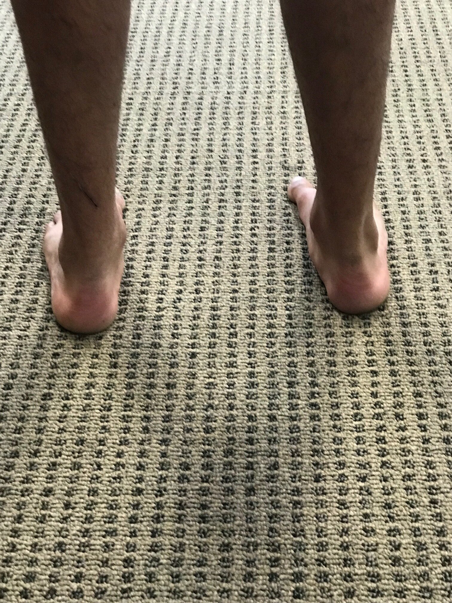
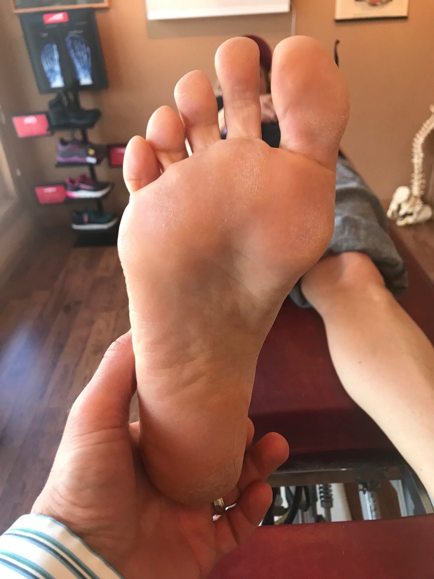
The consequences of an inverted forefoot

A forefoot that is inverted with respect to the rearfoot. Whether it is a forefoot varus, forefoot supinatus or an everted rearfoot ( because the forefoot is still inverted with respect to the rearfoot), what are the biomechanical sequelae?

If we accept the premise that the foot is basically a tripod between the calcaneus, base of the first and base of the fifth metatarsal‘s, we know that all of these parts needs to be on the ground at certain points in the gait cycle. Forces should travel from the calcaneus, up the lateral aspect of the foot, across the metatarsal heads to the first metatarsal head and hopefully out through the hallux.

The foot should hit the ground in slight inversion of the entire foot at initial contact and pronate through the middle of mid stance and then supinate through the remainder of the gait cycle. There’s an intricate balance of biomechanical events that must occur, especially in the latter half of the gait cycle when the rear foot is inverting where the forefoot is everting, so that we can have high gear push off through the distal first ray.

If the forefoot remains inverted then somehow the head of the first metatarsal needs to be brought down to the ground. If there’s not adequate range of motion in the foot, particularly the first ray, then you may pronate through the midfoot, rearfoot or in cases where this is insufficient, bring them immediately over the foot to get it down. This of course shifts center of gravity to midline and the body above must compensate in someway.

Take a look at this video footage and what do you see? She strikes on the outside of her foot but does not have adequate motion in her forefoot and therefore “crashes“ down on the forefoot, forcing a valgus moment into the ankle and the need to shift immediately by the pelvis attempts to dampen it. Notice how this is worse on the right side with more medial knee shift, pelvic shift as well as a lateral bending of the body to the right. Notice also how the upper body twists more to the left than to the right.

So what’s the fix? Well the answer is, “what’s bothering the patient?” We don’t necessarily fix what we see; we correlate what we see with what the patient’s symptoms are because that’s usually why they show up in your office. Yes, we do get people from time to time that come in strictly for “performance enhancement“ but this is pretty rare.

This woman has very little motion and plantar flexion of the first Ray complex so our primary goal was to get her to descend the first ray. We accomplished this by the following:

1. Manipulation in plantar and dorsiflexion of the first ray complex
2. Soft tissue work in the first intermetatarsal interval
3. Exercises of muscles to assist in descending the first ray including the following: extensor hallucis brevis, peroneus longus, flexor digitorum brevis
4. Pelvic stability work to improve the skill, endurance and strength of the gluteus medius complex as well as abdominal endurance work.

Your rehab program should change as the patient has more functional gains, tailoring it to the patient’s deficiencies.


Dr Ivo Waerlop, one of The Gait Guys

#invertedforefoot #forefootsupinatus #forefootvarus #pronation #forefoot #gaitanalysis

Unilateral calcaneal valgus: what can it mean?

right calcaneal valgus

right calcaneal valgus

Take a good look at this picture and what do you see? Do you see the calcaneal valgus on the right side. What runs through your mind?

Possibilities for causing this condition, as well as the clinical implications are numerous.

The short list should include:

  • A shorter leg on the contralateral side: often times we will pronate more on the longer leg side to compensate for a short leg on the opposite

  • Increased rear foot and/or fore foot pronation on the valgus side. Laxity of the spring ligament or incompetency of the musculature which helps to maintain your arch (tibialis posterior, foot intrinsics, tibialis anterior to name a few) often causes more collapse on the effected side

  • A lack of available rearfoot eversion on the contralateral side. It may be that the increase calcaneovalgus is normal and the opposite side is more rigid.

  • If you were seeing this in the middle of the gait cycle it could be that that is their strategy to get around a loss of hip extension or ankle rocker

  • External tibial torsion on that side. Go ahead, stand up and spin your right foot into external rotation and keep your left foot with a normal progression angle. Can you see how your arch collapses to a greater degree on the side with the external torsion? Remember that pronation is dorsiflexion, eversion and abduction.

  • Internal tibial torsion on the contralateral side. Internal tibial torsion puts the foot into supination which makes it into more of a rigid lever rather than mobile adapter.

    And the list goes on…

    Next time you see a unilateral deformity like this, hopefully some of these things run through your mind and will help you to pinpoint where the problem actually is.

    Dr Ivo Waerlop, one of The Gait Guys

    #calcanealeversion #rearfootvalgus
    #lowerextremitydeformities

Foot Types? Do they really matter?

IMG_6218 copy.jpg

The answer is " yes, often times".

Did you miss our 3rd Wednesdays presentation last week on foot types and obligate biomechanics (and pathomechanic) that ensue? Here is the video feed that you can watch and get ce credits for:

https://www.chirocredit.com/course/Chiropractic_Doctor/Biomechanics_214

#foottypes #biomechanics #thegaitguys

Determining foot types...In a nutshell

Screen Shot 2019-10-15 at 8.31.27 PM.png

We talked yesterday about how foot types (i.e., the forefoot to rear foot relationship) can often produce predictable pathomechanics. Here's How to do it. Pretty basic, eh? Its the characteristics, along with the other anatomical goodies they may have that helps to clinch the diagnosis and dictate treatment.

To find out about how to apply your newfound knowledge, join us tomorrow night on our 3rd Wednesdays tele seminar: Biomechanics 314 on online.com

5 PST, 6MST, 7CST, 8EST

Foot types: do they really matter?

forefoot varus: note how the forefoot is inverted with respect to the rear foot

forefoot varus: note how the forefoot is inverted with respect to the rear foot

Foot type. You know what we are talking about. The relation in anatomically and in space of the rear foot to the forefoot. We believe that this anatomical relationship holds key clinical insights to predictable biomechanics in that particular foot type.

Simply put, the rear foot can be either inverted, everted or neutral; Same with the forefoot. If the rear foot is inverted we call that a rearfoot varus. If the foot is inverted we call that a forefoot varus. If the rear foot is everted we call that a rear foot valgus and if the forefoot is inverted we call that a forefoot valgus.

Now think about the simple motions of pronation and supination. Pronation is dorsiflexion, eversion and abduction; supination is plantar flexion, inversion and adduction. If it remains in eversion, we say that it is in vslgus and that means they will be qualities of pronation occurring in that foot while it is on the ground. If the foot is inverted, it will have qualities of supination.

We think of pronation as making the foot into a mobile adapter and supination is making the foot into a rigid lever.

During a typical gait cycle the foot is moving from supination at initial contact/loading response to full pronation at mid stance and then into supination from mid stance to terminal stance/pre-swing. I know that if the foot remains and pronation past mid stance that it is a poor lever and if it remains in supination prior to mid stance it will be a poor shock absorbers. Foot type plays into this displaying or amplifying the characteristics of that particular foot type during the gait cycle: if this occurs at a time other than when it supposed to occur, then we can see predictable biomechanics such as too much pronation resulting in increased rear foot eversion, midfoot collapse, abduction of the forefoot and internal rotation of the knee with most often medial knee fall. Now, consider these mechanics along with any torsions or versions in the lower extremity that the patient may have.

This Wednesday night we will be discussing foot types and their biomechanics. Join us on onlinece .com for Biomechanics 314 6:00 MST

Dr Ivo Waerlop, one of The Gait Guys

Yep, these shoes stink for this gal...

IMG_6882.jpg

Look at the left shoe and compare it to the right. See how the upper is canted on the outsole? This “varus cant” can create lots of problems or could actually be beneficial, believe it or not, depending upon the pathology.

In this particular persons story, it was NOT a good thing. They have an anatomical short leg on the left-hand side. If you remember from following us here in the past, generally speaking, the shorter leg tide tends to be more supinated and the forefoot tends to be in more varus. This means more of a “reach” with that foot during the contact phase of gait, Whether that’s running or walking. This generally means that the forefoot will pronate more on the long leg side.

This shoe “defect“ may actually be benefit for someone who has too much rear or mid foot pronation as it would “delay” pronation by starting to rearfoot in an inverted position at heel strike.

The Fix?

You could grind the sole into varus an equal amount to equal the varus cant. In our opinion, not a good idea.

You could return the shoe (that’s what this person is doing) and get another one

In addition, you could…

Give the person a 3 mm sole lift to correct for the leg length discrepancy

Make sure they have adequate range of motion in the first ray on the short leg side to be be able to plantar flex the 1st ray and reach the ground

Make sure they have adequate control of the core musculature as well as foot intrinsic musculature during stance phase.

Dr Ivo Waerlop, one of The Gait Guys

#badshoes #theshoeistheproblem #forefootvarus #leglengthdifference
#gaitproblem

Barefoot vs Shoes...It's about the strike pattern


Footnotes 7 - Black and Red.jpg

“The influence of strike patterns on running is more significant than shoe conditions, which was observed in plantar pressure characteristics. Heel-toe running caused a significant impact force on the heel, but cushioned shoes significantly reduced the maximum loading rate. Meanwhile, although forefoot running can prevent impact, peak plantar pressure was centered at the forefoot for a long period, inducing a potential risk of injury in the metatarsus/phalanx. Plantar pressure on the forefoot with RFS was lesser and push-off force was greater when cushioned shoes were used than when running barefoot.”


takeaways from the study?

  • forefoot strike reduces heel impact

  • rear foot strike reduces forefoot impact

  • forefoot strike increases and prolongs pressures (in shoes) on the forefoot which could potentially cause forefoot problems

  • cushioned shoes do not really change impact force but change (reduce) the rate of loading

  • in a forefoot strike, pressures are shifted more to the mid foot

want to know more? Join us this Wednesday, December 19th on online.com: Biomechanics 303


Sun XYang YWang LZhang XFu W. Do Strike Patterns or Shoe Conditions have a Predominant Influence on Foot Loading? J Hum Kinet. 2018 Oct 15;64:13-23. doi: 10.1515/hukin-2017-0205. eCollection 2018 Sep.

link to FREE FULL TEXT: https://www.ncbi.nlm.nih.gov/pmc/articles/PMC6231350/


Varus anyone?

Varus anyone?

Does patello femoral pain have anything to do with rearfoot varus? Perhaps, according to this study:

" A small but significant increase in rearfoot varus was found in the patellofemoral pain group compared with the control group (8.9 vs. 6.8 degrees; p = .0002). These results suggest that increased rearfoot varus may be a contributing factor in patellofemoral pain and should be assessed when evaluating the events at the subtalar joint and the lower extremity. In addition, it has been demonstrated that consistent rearfoot measurements can be obtained by an individual clinician."


Powers CM, Maffucci R, Hampton S. Rearfoot posture in subjects with patellofemoral pain. J Orthop Sports Phys Ther. 1995 Oct;22(4):155-60.

Do you know SQUAT? Have you seen SQUAT? Have patients/clients that LIKE to squat? Seen a foot that looks like this? Can you say REARFOOT VALGUS?

 "Significant changes in lower limb kinematics may be observed during bilateral squatting when rearfoot alignment is altered. Shoe pitch alone may significantly reduce peak pronation during squatting in this population, but additional reductions were not observed in the subtalar neutral position. Further research investigating the effects of footwear and the subtalar neutral position in populations with lower limb pathology is required."
 
So, what does this study tell us?

when rearfoot aliment changes, so do the kinematics (duh)
the surface (tilted into varus or inversion) or shoes (which are medially posted) can make or break the man (or women) when it comes to "peak" pronation (we knew that already; confirmation is always nice)
inverting the rearfoot can change ankle dorsiflexion (read "ankle rocker"); inverting the rearfoot seems to reduce it
inverting the rearfoot can change knee flexion; inverting the rearfoot seems to increase knee flexion
inverting the rearfoot can change hip abduction (and thus knee valgus); reducing it

Learn about the gait kinematics and clinical findings associated with this foot type, along with video clip examples and always entertaining discussion with us tomorrow night on onlinece.com: Biomechanics 308: Focus on the Rear Foot.  5PST, 6MST, 7 CST, 8EST


Power V, Clifford AM. The Effects of Rearfoot Position on Lower Limb Kinematics during Bilateral Squatting in Asymptomatic Individuals with a Pronated Foot Type. J Hum Kinet. 2012 Mar;31:5-15. doi: 10.2478/v10078-012-0001-0. Epub 2012 Apr 3.

#rearfootvalgus #squat #foottype

2012 Mar;31:5-15. doi: 10.2478/v10078-012-0001-0. Epub 2012 Apr 3.

The Effects of Rearfoot Position on Lower Limb Kinematics during Bilateral Squatting in Asymptomatic Individuals with a Pronated Foot Type.

Power V1, Clifford AM.

Author information

Abstract

Clinicians frequently assess movement performance during a bilateral squat to observe the biomechanical effects of foot orthotic prescription. However, the effects of rearfoot position on bilateral squat kinematics have not been established objectively to date. This study aims to investigate these effects in a population of healthy adults with a pronated foot type. Ten healthy participants with a pronated foot type bilaterally (defined as a navicular drop >9mm) performed three squats in each of three conditions: barefoot, standing on 10mm shoe pitch platforms and standing on the platforms with foam wedges supporting the rearfoot in subtalar neutral. Kinematic data was recorded using a 3D motion analysis system. Between-conditions changes in peak joint angles attained were analysed. Peak ankle dorsiflexion (p=0.0005) and hip abduction (p=0.024) were significantly reduced, while peak knee varus (p=0.028) and flexion (p=0.0005) were significantly increased during squatting in the subtalar neutral position compared to barefoot. Peak subtalar pronation decreased by 5.33° (SD 4.52°) when squatting on the platforms compared to barefoot (p=0.006), but no additional significant effects were noted in subtalar neutral. Significant changes in lower limb kinematics may be observed during bilateral squatting when rearfoot alignment is altered. Shoe pitch alone may significantly reduce peak pronation during squatting in this population, but additional reductions were not observed in the subtalar neutral position. Further research investigating the effects of footwear and the subtalar neutral position in populations with lower limb pathology is required.

tumblr_n7yjg9vOg61qhko2so1_r1_1280.jpg
tumblr_n7yjg9vOg61qhko2so2_1280.jpg
tumblr_n7yjg9vOg61qhko2so3_r1_500.gif

Flat Dogs

Take a look at these pedographs. Wow!

  • No rear foot heel teardrop.
  • No midfoot arch on left foot and minimal on right.
  • An elongated 2nd metatarsal bilaterally and forces NOT getting to the base of the 1st metatarsal and stalling on the 2nd: classic sign of an uncompensated forefoot varus.
  • increased printing of the lateral foot on the right

Knowing what you know about pronation (need a review? click here) Do you think this foot is a good lever? Do you think they will be able to push off well?

What can we do?

  • foot exercises to build the intrinsic and extrinsic muscles of the foot (click here, here, here, and here for a few to get you started)
  • perhaps an orthotic to assist in decreasing the pronation while they are strengthening their foot
  • motion control shoe? Especially in the beginning as they are strengthening their feet and they fatigue rather easily

The prints do not lie. They tell the true story of how the forces are being transmitted through the foot. For more pedograph cases, click here.

The Gait Guys. Teaching you more about the feet and gait. Spreading gait literacy throughout the net! Do your part by forwarding this post to someone who needs to read it.

The debate continues. More support for mid and forefoot strikers.…
“Forefoot and midfoot strikers had significantly shorter ground contact times than heel strikers. Forefoot and midfoot strikers had significantly faster average race spe…

The debate continues. More support for mid and forefoot strikers.

“Forefoot and midfoot strikers had significantly shorter ground contact times than heel strikers. Forefoot and midfoot strikers had significantly faster average race speed than heel strikers.”

We are not saying “better”, but according to this study “faster”!

What is the ideal?  We wish we knew…Biomechanics seem to point to less impact is better, but what is actually best for the individual is probably due to genetics, training, practice, running surface and that individuals neuromuscular competence and ability to compensate.

The Gait Guys. bringing you the facts, even if you or we don’t like them…


J Sports Sci. 2012;30(12):1275-83. doi: 10.1080/02640414.2012.707326. Epub 2012 Aug 2.

Foot strike patterns and ground contact times during high-calibre middle-distance races.

Source

Department of Sport and Exercise Sciences, School of Life Sciences, Northumbria University, Newcastle-upon-Tyne NE1 8ST, UK. phil.hayes@northumbria.ac.uk

Abstract

The aims of this study were to examine ground contact characteristics, their relationship with race performance, and the time course of any changes in ground contact time during competitive 800 m and 1500 m races. Twenty-two seeded, single-sex middle-distance races totaling 181 runners were filmed at a competitive athletics meeting. Races were filmed at 100 Hz. Ground contact time was recorded one step for each athlete, on each lap of their race. Forefoot and midfoot strikers had significantly shorter ground contact times than heel strikers. Forefoot and midfoot strikers had significantly faster average race speed than heel strikers. There were strong large correlations between ground contact time and average race speed for the women’s events and men’s 1500 m (r = -0.521 to -0.623; P < 0.05), whereas the men’s 800 m displayed only a moderate relationship (r = -0.361; P = 0.002). For each event, ground contact time for the first lap was significantly shorter than for the last lap, which might reflect runners becoming fatigued.

PMID:22857152[PubMed - indexed for MEDLINE]

http://www.ncbi.nlm.nih.gov/pubmed/22857152

Support for a midfoot strike?
&ldquo;Running with a midfoot strike pattern resulted in a significant increase in gastrocnemius lateralis pre-activation (208 ± 97.4 %, P &lt; 0.05) and in a significant decrease in tibialis anterior EMG activity (56.2…

Support for a midfoot strike?

Running with a midfoot strike pattern resulted in a significant increase in gastrocnemius lateralis pre-activation (208 ± 97.4 %, P < 0.05) and in a significant decrease in tibialis anterior EMG activity (56.2 ± 15.5 %, P < 0.05) averaged over the entire stride cycle. The acute attenuation of foot-ground impact seems to be mostly related to the use of a midfoot strike pattern and to a higher pre-activation of the gastrocnemius lateralis. ”

Do these results surprise you? They didn’t really surprise us.

The lateral head of the gastroc is a midstance to preswing stabilizer and works synergistic with the medial head, with the medial head firing earlier. Sutherland talks about these muscles not being propulsive in nature, but rather maintainers of forward progression, step length and gait symmetry. Thinking this through in a closed chain (foot up) fashion, this would counter the inversion moment created by the medial gastroc for supination in the second half of contact phase. If the foot is already partially supinated (as we believe it would be in a midfoot strike), it would have to pre activate.

A decrease in tibialis anterior activity? Sure. If the foot is striking more parallel to the ground, the anterior compartment (including the tibialis anterior, extensor hallucis longus, and extensor digitorum longus) would not have to eccentrically contract to decelerate the lowering of the foot to the ground.

Better? Maybe, maybe not. We are seeing more and more literature about foot strike (if you missed our last few posts, click here, here, here and here), We still maintain that you need a competent lower kinetic chain, including the foot and an intact nervous system to drive the boat.

We remain, handsome, bald and nerdy…Ivo and Shawn


Eur J Appl Physiol. 2012 Aug 9. [Epub ahead of print]

Impact reduction during running: efficiency of simple acute interventions in recreational runners.

Source

University of Lyon, 42023, Saint-Etienne, France.

Abstract

Running-related stress fractures have been associated with the overall impact intensity, which has recently been described through the loading rate (LR). Our purpose was to evaluate the effects of four acute interventions with specific focus on LR: wearing racing shoes (RACE), increasing step frequency by 10 % (FREQ), adopting a midfoot strike pattern (MIDFOOT) and combining these three interventions (COMBI). Nine rearfoot-strike subjects performed five 5-min trials during which running kinetics, kinematics and spring-mass behavior were measured for ten consecutive steps on an instrumented treadmill. Electromyographic activity of gastrocnemius lateralis, tibialis anterior, biceps femoris and vastus lateralis muscles was quantified over different phases of the stride cycle. LR was significantly and similarly reduced in MIDFOOT (37.4 ± 7.20 BW s(-1), -56.9 ± 50.0 %) and COMBI (36.8 ± 7.15 BW s(-1), -55.6 ± 29.2 %) conditions compared to NORM (56.3 ± 11.5 BW s(-1), both P < 0.001). RACE (51.1 ± 9.81 BW s(-1)) and FREQ (52.7 ± 11.0 BW s(-1)) conditions had no significant effects on LR. Running with a midfoot strike pattern resulted in a significant increase in gastrocnemius lateralis pre-activation (208 ± 97.4 %, P < 0.05) and in a significant decrease in tibialis anterior EMG activity (56.2 ± 15.5 %, P < 0.05) averaged over the entire stride cycle. The acute attenuation of foot-ground impact seems to be mostly related to the use of a midfoot strike pattern and to a higher pre-activation of the gastrocnemius lateralis. Further studies are needed to test these results in prolonged running exercises and in the long term.

PMID:22875194 [PubMed - as supplied by publisher]


All material copyright 2013 The Gait Guys/The Homunculus Group, yada, yada, yada…