Muscle Spindles and Proprioception

image source: https://en.wikipedia.org/wiki/File:Fusimotor_action.jpg

image source: https://en.wikipedia.org/wiki/File:Fusimotor_action.jpg

And what have we been saying for the last 6 years? 

Connected to the nervous system by large diameter afferent (sensory) fibers, they are classically thought of as appraising the nervous system of vital information like length and rate of change of length of muscle fibers, so we can be coordinated. They act like volume controls for muscle sensitivity. Turn them up and the muscle becomes more sensitive to ANY input, especially stretch (so they become touchy…maybe like you get if you are hungry and tired and someone asks you to do something); turn them down and they become less or unresponsive.

Their excitability is governed by the sum total (excitatory and inhibitory) of all neurons (like interneuron’s) acting on them (their cell bodies reside in the anterior horn of the spinal cord).

Along with with Golgi tendon organs and joint mechanoreceptors, they also act as proprioceptive sentinels, telling us where our body parts are in space. We have been teaching this for years. Here is a paper that exemplifies that, identifying several proteins responsible for neurotransduction including the Piezo2 channel as a candidate for the principal mechanotransduction channel. Many neuromuscular diseases are accompanied by impaired  muscle spindle function, causing a decline of motor performance and coordination. This is yet another key finding in the kinesthetic system and its workings. 

Remember to include proprioceptive exercises and drills (on flat planar surfaces, like we talked about here) in your muscle rehab programs


Kröger S Proprioception 2.0: novel functions for muscle spindles. Curr Opin Neurol. 2018 Oct;31(5):592-598. 

Woo SH, Lukacs V, de Nooij JC, Zaytseva D, Criddle CR, Francisco A, Jessell TM, Wilkinson KA, Patapoutian A. Piezo2 is the principal mechanotransduction channel for proprioception.Nat Neurosci. 2015 Dec; 18(12):1756-62. Epub 2015 Nov 9.

Fusimotor control of proprioceptive feedback during locomotion and balancing: can simple lessons be learned for artificial control of gait?

Hulliger M. Fusimotor control of proprioceptive feedback during locomotion and balancing: can simple lessons be learned for artificial control of gait? Prog Brain Res. 1993; 97:173-80.

tumblr_ne2sg64OZ11qhko2so1_400.gif
tumblr_ne2sg64OZ11qhko2so3_500.jpg
tumblr_ne2sg64OZ11qhko2so2_1280.jpg

More thoughts on stretching

   We get a lot of interest in our posts on stretching. Seems like this is a pretty hot subject and there is a lot of debate as to whether it is injury preventative or not. Are you trying to physically lengthen the muscle or are you trying to merely bring it to its physiological limit?  There’s a big difference in what you need to do to accomplish each of these goals. Lets take a look at each, but 1st we need to understand a little about muscles and muscle physiology.

 Muscles are composed of small individual units called sarcomeres. Inside of these “sarcomeres” there are interdigitating fibers of actin and myosin (proteins) which interact with one another like a ratchet when a muscle contracts.  Sarcomeres can be of various lengths, depending on the muscle, and are linked and together from one end of the muscle to the other. When a muscle contracts concentrically (the muscle shortening while contracting) the ends of the sarcomere (called Z lines or Z discs) are drawn together, shortening the muscle fiber over all (see the picture above).
 
 Signals are sent from the brain (actually the precentral gyrus of the cerebral cortex areas 4, 4s and 6) down the corticospinal tract to the spinal cord to synapse on motor neurons there.  These motor neurons (alpha motor neurons) then travel through peripheral nerves to the muscles to cause them to contract (see picture above).

   The resting length of the muscle is dependent upon two factors:
The physical length of the muscle
2. The “tone” of the muscle in question.

The physical length of the muscle is determined by the length of the sarcomeres and the number of them in the muscle.   The “tone” of the muscle determined by an interplay of neurological factors and the feedback loops between the sensory (afferent) receptors in the muscle (Ia afferents, muscle spindles, Golgi tendon organs etc.), relays in the cerebellum and basal ganglia as well as input from the cerebral cortex.

 If you’re trying to “physically lengthen” a muscle, then you will need to actually add sarcomeres to the muscle. Research shows that in order to do this with static stretching it must be done 20 to 30 minutes per day per muscle.

 If you were trying to “bring a muscle to its physiological limit” there are many stretching methods to accomplish this.  Pick your favorite whether it be a static stretch, contract/ relax, post isometric relaxation etc. and you’ll probably be able to find a paper to support your position.

  Remember with both not to ignore neurological reflexes (see above). Muscle spindle loops are designed to provide feedback to the central nervous system about muscle length and tension. Generally speaking, slow stretch activates the Ia afferent loop which causes causes physiological contraction of the muscle (this is one of the reasons you do not want to do slow, steady stretch on a muscle in spasm). This “contraction” can be fatigued overtime, causing the muscle to be lengthened to it’s physiological limit.  Do this for an extended period of time (20-30 mins per day) and you will physically add sarcomeres to the muscle.

 Next time you are stretching, or you were having a client/patient stretch, think about what it is that you’re actually trying to accomplish  because there is a difference.

We are and remain The Gait Guys.  Bald, good-looking, and above-average intelligence. Spreading gait literacy with each post we publish.

thanks to scienceblogs.com for the corticospinal tract image

On the topic of endurance training.....

On the topic of endurance training (which we discussed on this weeks PODcast, forthcoming in the next day or so; we have both been extraordinarily busy in our clinics); if you are a well trained athlete (ie endurance junkie), how might this effect your running gait?

So, you run 103 miles with an elevation change of over 31,000 feet, how do you think you would fare? These folks were tested pre and 3 hours post race on a 22 foot long pressure walkway at about 7.5 miles per hour. Here’s how this group of 18 folks did:

  1. increased step frequency
  2. decreased “aerial” time
  3. no change in contact time
  4. decrease in downward displacement of the center of mass
  5. decrease in peak vertical ground reactive force
  6. increased vertical oscillation
  7. leg stiffness remained unchanged

So what does this tell us?

  • wow, that is a lot of vertical
  • holy smokes, that is really far
  • don’t know how I would do with a race like that
  • they are fatigued (1, 2, 6)
  • they are trying to attenuate impact forces (2, 3, 4, 5, 7)

The system is trying to adapt the best it can. If you were to do a standard hip screen test (like we spoke about here)  you would probably see increased horizontal drift due to proprioceptive fatigue. Remember that proprioception (our bodies ability to sense its position in space) makes the world go round. Proprioception is dependent on an intact visual system (see our post yesterday) , an intact vestibular system and muscle and joint mechanoreceptors functioning appropriately). We would add here that central nervous system fatigue (ie central processing both at the cord and in the cortex) would probably play a role as well.

The take home message? The human machine is a neuro mechanical marvel and much more complex than having the right shoe or the right running technique. Training often makes us more competent and efficient, but everything has it limits.

The Gait Guys. Making it real with each and every post.

all material copyright 2013 The Gait Guys/ The Homunculus Group

J Biomech. 2011 Apr 7;44(6):1104-7. doi: 10.1016/j.jbiomech.2011.01.028. Epub 2011 Feb 20.

Changes in running mechanics and spring-mass behavior induced by a mountain ultra-marathon race.

Source

Université de Lyon, F-42023 Saint-Etienne, France. jean.benoit.morin@univ-st-etienne.fr

Abstract

Changes in running mechanics and spring-mass behavior due to fatigue induced by a mountain ultra-marathon race (MUM, 166km, total positive and negative elevation of 9500m) were studied in 18 ultra-marathon runners. Mechanical measurements were undertaken pre- and 3h post-MUM at 12km h(-1) on a 7m long pressure walkway: contact (t©), aerial (t(a)) times, step frequency (f), and running velocity (v) were sampled and averaged over 5-8 steps. From these variables, spring-mass parameters of peak vertical ground reaction force (F(max)), vertical downward displacement of the center of mass (Δz), leg length change (ΔL), vertical (k(vert)) and leg (k(leg)) stiffness were computed. After the MUM, there was a significant increase in f (5.9±5.5%; P<0.001) associated with reduced t(a) (-18.5±17.4%; P<0.001) with no change in t©, and a significant decrease in both Δz and F(max) (-11.6±10.5 and -6.3±7.3%, respectively; P<0.001). k(vert) increased by 5.6±11.7% (P=0.053), and k(leg) remained unchanged. These results show that 3h post-MUM, subjects ran with a reduced vertical oscillation of their spring-mass system. This is consistent with (i) previous studies concerning muscular structure/function impairment in running and (ii) the hypothesis that these changes in the running pattern could be associated with lower overall impact (especially during the braking phase) supported by the locomotor system at each step, potentially leading to reduced pain during running.

Copyright © 2011 Elsevier Ltd. All rights reserved.

http://www.ncbi.nlm.nih.gov/pubmed/21342691

tumblr_mg6a1wNO4i1qhko2so1_1280.jpg
tumblr_mg6a1wNO4i1qhko2so2_400.jpg

A little neuro, anyone?

Welcome to Monday, and yes, it is a NEURO day. In fact, if you got up this morning, you too are having a NEURO day. Dr Allen thinks it’s all about the ORTHOPEDICS, but without NEURO, there would not be any orthopedics : )

A dialogue from one of our avid readers, Dr. Ryan.

Dr. Ryan: Hey Ivo,

I just read this article on Mercola’s site which is an interview with Dr. Craig Buhler who does muscle activation techniques.  Can you check this for accuracy?  This must be a mistake b/c I always thought spindle activation will facilitate the muscle to contract.  Also, I always wondered why the O/I attachment points are tender in muscles that are inhibited.  Does his description sound right to you.  If not, do you have a better explanation?

“Your muscle system and nervous system relate to each other from within tiny muscle fibers called spindle cells, which monitor stretch. If your muscle is overloaded too rapidly, the spindle cells will temporarily inhibit the muscle. The next time you contract the muscle, it will fire again. Similarly, cells within your tendons called Golgi tendon organs also measure and monitor stretch. If your tendon is stretched too rapidly or exceeds its integrity, the Golgi tendon organs will temporarily inhibit the muscle. But the next time the muscle fires, it will again fire appropriately.

"But there’s a fail-safe system," Dr. Buhler explains. "It’s where the tendon attaches into the periosteum of the bone and the little fibers there are called Sharpey’s fibers. Those fibers are loaded with little receptors that monitor tension. And if the integrity of those fibers are exceeded, they inhibit the muscle, just like a circuit breaker would inhibit an electrical circuit.

Once that happens, the muscle will still fire under passive range of motion. But if you load the muscle, it gives way. If you continue to load the muscle, your body creates pain at the attachment points to protect you. What the central nervous system does at that point is compute an adaptive strategy by throwing stress into the muscle next to it. Other tissues begin to take on more of the load for the muscle that’s been injured.”

Here is a link to the entire article if you want to check it out:

http://fitness.mercola.com/sites/fitness/archive/2013/01/04/advanced-muscle-integration-technique.aspx?e_cid=20130104_DNL_art_1"

Dr Ivo: Thanks Dr. Ryan.

Spindles monitor length and GTO’s monitor tension. My understanding is spindles, when activated, stimulate the alpha motor neuron(at the cord) and cause contraction of that muscle or motor unit. GTO’s, when activated, cause inhibition of the muscle they are associated with. I am not aware of them being inhibitory, only GTO’s. They are believed to be GABAnergic synapses. The impulse (at least in cats) can be smaller or inhibited if the muscle is held in contraction for an extended period of time (see attached)

Perhaps he is talking about spindle dysfunction, where the intrafusal portion of the spindle (which is innervated by a gamma motor neuron) is either excited or inhibited. The gamma’s are more of a slave to the interneuronal pool (in the cord), which would be the sum total of all excitatory and inhibitory input to that area (ie the central integrated state). This not only reflects local receptor input but also cortical information descending (from areas 4s and 6 in the precentral gyrus) AND descending information from the caudal reticular formation.

Based on what you sent, I do not agree with the 1st 2 sentences. I was not aware about increased receptor density of Sharpeys fibers. I did a quick search and found this: http://www.ncbi.nlm.nih.gov/pmc/articles/PMC2100202/  , which eludes to it and here: http://www.ncbi.nlm.nih.gov/pmc/articles/PMC3098959/. I will have to dive in more when I have time.

Not sure why O/I attachments are tender in inhibited muscles. I find them tender in most folks. Maybe because inhibited muscles ave altered receptor function and that preloads the nociceptive afferent pathway or at least that neuronal pool? Are they closer to threshold for some reason? Not sure. LMK what you find.

Thanks for getting me jazzed about sharpeys fibers!

for those of you who need to know YES, there will be a forthcoming Sharpeys fibers article

Dr. Ryan: That’s what I thought.  Thanks for looking into it and I will check out those links.  You have jazzed me plenty of times over the years.  Glad I could jazz you up for a change.  Have a great weekend.


Yes, Dr Ivo is definitely an uber neuro geek, especially when he spends time on the weekend talking about spindles!


all material copyright 2013 The Homunculus Group/ The Gait Guys. All rights reserved. Please as before you lift our stuff.

Its a great day to be a neuro geek
So if the receptors on the bottom of the foot aren&rsquo;t involved aren&rsquo;t involved in 2 joint muscles staying coordinated (like the hamstring and rectus femoris in this study), how do we determine the approp…

Its a great day to be a neuro geek

So if the receptors on the bottom of the foot aren’t involved aren’t involved in 2 joint muscles staying coordinated (like the hamstring and rectus femoris in this study), how do we determine the appropriate muscle length and ratios? How about our built in muscle length receptors? Lets hear it for muscle spindles! Hooray for Ia and type II afferents!

Sifting through the science so you don’t have to. We are The Gait Guys…

Exp Brain Res. 1998 Jun;120(4):479-86.

Coordination of two-joint rectus femoris and hamstrings during the swing phase of human walking and running.

Prilutsky BI, Gregor RJ, Ryan MM.

Source

Department of Health and Performance Sciences, Center for Human Movement Studies, The Georgia Institute of Technology, Atlanta 30332-0110, USA.

Abstract

It has been hypothesized previously that because a strong correlation was found between the difference in electromyographic activity (EMG) of rectus femoris (RF) and hamstrings (HA; EMG(RF)-EMG(HA)) and the difference in the resultant moments at the knee and hip (Mk-Mh) during exertion of external forces on the ground by the leg, input from skin receptors of the foot may play an important role in the control of the distribution of the resultant moments between the knee and hip by modulating activation of the two-joint RF and HA. In the present study, we examined the coordination of RF and HA during the swing phase of walking and running at different speeds, where activity of foot mechanoreceptors is not modulated by an external force. Four subjects walked at speeds of 1.8 m/s and 2.7 m/s and ran at speeds of 2.7 m/s and 3.6 m/s on a motor-driven treadmill. Surface EMG of RF, semimembranosus (SM), and long head of biceps femoris (BF) and coordinates of the four leg joints were recorded. An inverse dynamics analysis was used to calculate the resultant moments at the ankle, knee, and hip during the swing phase. EMG signals were rectified and low-pass filtered to obtain linear envelopes and then shifted in time to account for electromechanical delay between EMG and joint moments. During walking and running at all studied speeds, mean EMG envelope values of RF were statistically (P<0.05) higher in the first half of the swing (or at hip flexion/knee extension combinations of joint moments) than in the second half (or at hip extension/knee flexion combinations of joint moments). Mean EMG values of BF and SM were higher (P<0.05) in the second half of the swing than in the first half. EMG and joint moment peaks were substantially higher (P<0.05) in the swing phase of walking at 2.7 m/s than during the swing phase of running at the same speed. Correlation coefficients calculated between the differences (EMG(RF)-EMG(HA)) and (Mk-Mh), taken every 1% of the swing phase, were higher than 0.90 for all speeds of walking and running. Since the close relationship between EMG and joint moments was obtained in the absence of an external force applied to the foot, it was suggested that the observed coordination of RF and HA can be regulated without a stance-specific modulation of cutaneous afferent input from the foot. The functional role of the observed coordination of RF and HA was suggested to reduce muscle fatigue.

tumblr_lq7m4hUHRb1qhko2so1_400.jpg
tumblr_lq7m4hUHRb1qhko2so2_500.jpg

The Truth About Treadmills: A Neurological Perspective

Gender differences in walking and running on level and inclined surfaces. 

Chumanov ES, Wall-Scheffler C, Heiderscheit BC. Clin Biomech (Bristol, Avon). 2008 Dec;23(10):1260-8. Epub 2008 Sep 6.


What the Gait Guys have to say about this article:

This article highlights some of the differences in gait between males and females on treadmills. Though treadmills don’t necessarily represent real life, they are an approximation. While reading this article, please keep the following in mind:

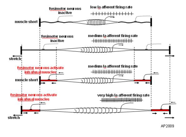
1. the treadmill pulls the hip into extension and places a pull on the anterior hip musculature, especially the hip flexors including the rectus femoris, iliopsoas and iliacus. This causes a slow stretch of the muscle, activating the muscle spindles (Ia afferents) and causing a mm contraction (ie the stretch reflex). This acts to inhibit the posterior compartment of hip extensors (especially the glute max) through reciprocal inhibition, making it difficult to fire them.

 2. Because the deck is moving, the knee is brought into extension, with stretch of the hamstrings, the quads become reciprocally inhibited (same mechanism above).

 3. The moving deck also has a tendency to put the ankle in dorsiflexion, initiating a stretch reflex in the tricep surae (gastroc/soleus) facilitating toe off through here and pushing you through the gait cycle, rather than pulling you through (with your hip extensors).

 4. the moving deck forces you to flex the thigh forward for the next footstrike (ie footstance), firing the RF, IP and Iliacus, and reciprocally inhibit the g max

If your core isn’t engaged, the pull of the rectus femoris and iliopsoas/iliacus pulls the ilia and pelvis into extension (ie increases the lordosis) and you reciprocally inhibit the erectors and increase reliance on the multifidus and rotatores, which have short lever arms and are supposed to be more proprioceptive in function. Can you say back pain?


In summary, treadmills are not the scourge of humanity, but do have some pitfalls for training, and equal amounts of “backwards” running should be employed (with great caution, mind you)

With that being said, lets look at the results: increased hip internal rotation and adduction, as well as more glute activity for the ladies. Not surprising considering women generally have a larger Q angle (17 +/- 3 degrees for females, 14 +/-3 degrees for males) and greater amounts of hip anteversion (average 14 degrees in females vs 8 in males). The larger Q angle places more stress at the medial knee (compression of the medial femoral condyle and usually increased pronation as the center of gravity over the foot is moved medially) and thus more control needed to slow pronation (from the glutes to control/augment internal rotation). Greater hip anteversion means the angle of the femoral head is greater than 12 degrees to the shaft of the femur. This moves the lower extremity into a more internally rotated position, approximating the origin and insertion of the adductors, making them easier to access. With an increased Q angle and easier access, greater demands are placed on adductors in single leg stance (which is considerably greater in running), This increased adductory moment places more demand on the gluteus medius (and contralateral QL) as well, to stabilize the pelvis and this correlates with speed and incline, also found in the study.

The take home message? Don’t throw away your treadmill! The treadmill can be an excellent diagnostic tool! Gluteal and adductor insufficiencies will be more visible (and probably more prevalent) in females, especially those running or walking on treadmills. The hip extension and ankle dorsiflexion moment created by a treadmill works against some of the stabilizing mechanisms (glute inhibition, ankle dorsiflexor inhibition) and help to highlight some of the subtle gait abnormailities you may miss otherwise.


Abstract from Article

BACKGROUND: Gender differences in kinematics during running have been speculated to be a contributing factor to the lower extremity injury rate disparity between men and women. Specifically, increased non-sagittal motion of the pelvis and hip has been implicated; however it is not known if this difference exists under a variety of locomotion conditions. The purpose of this study was to characterize gender differences in gait kinematics and muscle activities as a function of speed and surface incline and to determine if lower extremity anthropometrics contribute to these differences.

METHODS: Whole body kinematics of 34 healthy volunteers were recorded along with electromyography of muscles on the right lower limb while each subject walked at 1.2, 1.5, and 1.8m/s and ran at 1.8, 2.7, and 3.6m/s with surface inclinations of 0%, 10%, and 15% grade. Joint angles and muscle activities were compared between genders across each speed-incline condition. Pelvis and lower extremity segment lengths were also measured and compared.

FINDINGS: Females displayed greater peak hip internal rotation and adduction, as well as gluteus maximus activity for all conditions. Significant interactions (speed-gender, incline-gender) were present for the gluteus medius and vastus lateralis. Hip adduction during walking was moderately correlated to the ratio of bi-trochanteric width to leg length.

INTERPRETATION: Our findings indicate females display greater non-sagittal motion. Future studies are needed to better define the relationship of these differences to injury risk.

PMID: 18774631 [PubMed - indexed for MEDLINE]

Yup, we’re gait nerds….Don’t laugh….You are too if you are reading this…..

The Gait Guys: finding other uses for treadmills, other than for hanging the laundry…..