Keep your eyes up and your toes up...,And it doesn’t hurt to use your abs

IMG_5822.jpg
IMG_5820.jpg
IMG_5818.jpg
IMG_5817.jpg


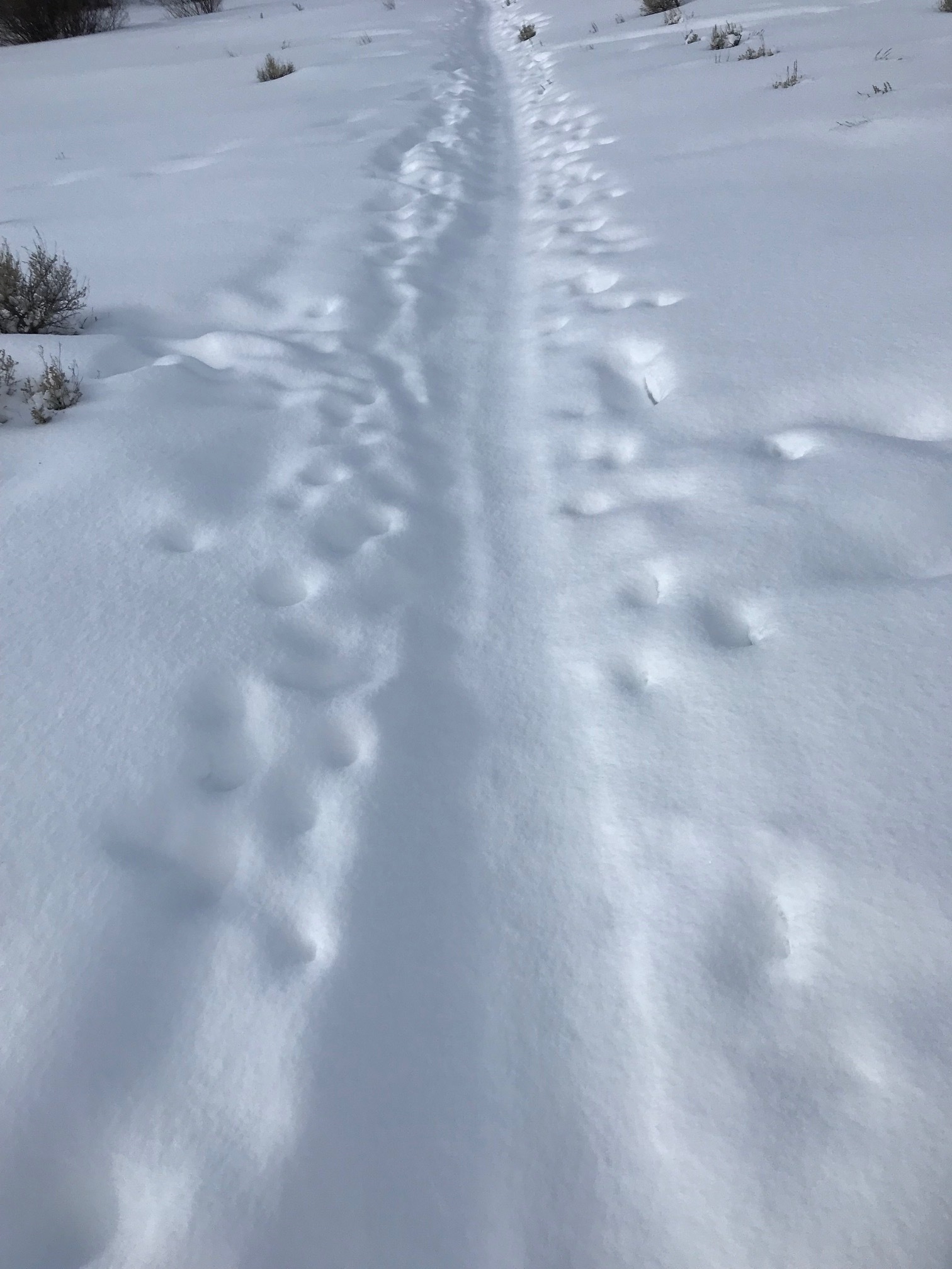
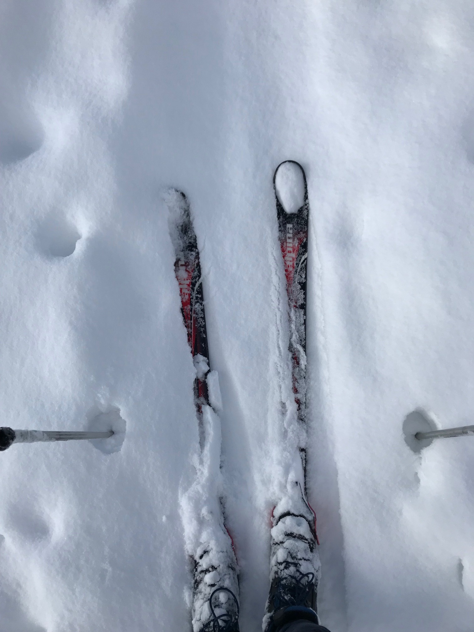
While out cross country skiing after a few inches of fresh fallen snow it dawned on me, especially when going uphill on my cross-country skis, lifting your toes up definitely pushes the head of the first metatarsal down and helps you to gain more purchase with the scales on the bottom of the skis. It also helps to press the center portion of the camber of the ski downward so that you can get better traction. Thinking about this further, lifting your toes up also helps you to engage your glutes to a greater degree.

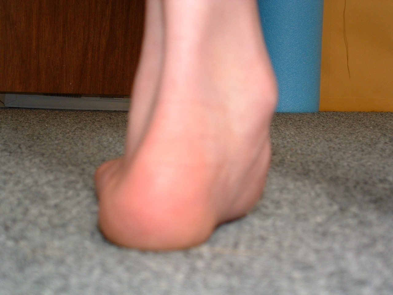
Try this: stand comfortably with your knees slightly flexed. Lift up your toes leaving the balls of your feet on the ground. Do you feel the first metatarsal head going down and making better contact with the ground? Can you feel your foot tripod between the head of the first metatarsal, head of the fifth metatarsal and the calcaneus? Now let your toes go down. Squeeze your glute max muscles. You should still be able to fart so don’t squeeze the sphincter. You can palpate these muscles to see if you’re actually getting to them. You can do this by placing your hands on top of your hips with your fingers calling around forward like when your mom used to put her hands on her hips and yell at you. Now relax with your toes up again leaving the balls of your feet on the ground. Now engage your glutes. See how much easier it is?

Now stand with your feet flat on the ground and put your hands on your abs, specifically your external obliques. Now raise your right leg. Do you feel your external oblique engage? Now, lift your toes up leaving the balls of your feet on the ground. Now lift your leg. Do you feel how much more your abs engage?

Little tricks of the trade. That’s why you listen here and why your patients/clients come to see you. Now go out and do it!

Dr. Ivo, one of The Gait Guys

#gaitanalysis, #crosscountryskiing, #skiing, hallux, #engage, #abs

tumblr_nka9cydN3k1qhko2so3_400.jpg
tumblr_nka9cydN3k1qhko2so1_400.jpg
tumblr_nka9cydN3k1qhko2so4_500.jpg
tumblr_nka9cydN3k1qhko2so2_1280.jpg

The partial truth about the Foot Tripod. The HEXApod.

The gait guys have talked about the foot tripod for a very long time. But the truth of the matter is that it is really a HEXApod. HEXA means 6. And when the foot is properly orientated and engaged on the ground, the 5 metatarsal heads and the heel should all be engaged with the ground, truly making it an asymmetrical hexapod. In an ideal scenario, the foot would be most stable if it looked like the strange symmetrical hexapod above with the contact points equally distributed around a center point. But that is not the human foot and this version of a hexapod is far simpler and likely inferior to the foot hexapod when human locomotion is to be attempted. The human foot is engineering marvel when it works properly.  

Perhaps the best example of what I mean by the foot being a HEXApod is in the pressure diagram above. In that first picture, on the right of that picture, we see multiple pressure points under the metatarsal heads of the right foot.  Minus the missing 1st metatarsal head pressure point (taken over by increased flexor hallucis longus activity represented by increased pressure at the big toe),  this pretty much confirms that the foot is not a tripod, rather a hexapod. The theory of the tripod, the 1st and 5th metatarsal heads and the heel, is only crudely accurate and honest. In this picture case, this person has increased lateral foot weight bearing (possibly why the 1st MET head pressure is absent) and possibly represented by pressure under the base of the 5 metatarsal. This is not normal for most people and if this person could get the 1st MET head down, they might even have a HEPTApod foot structure because of the 5th metatarsal base presentation (which sometimes represents peroneal muscle weakness). 

Where did we lead you astray after all these years of “tripod” talk ? We have always discussed the foot tripod. We have always discussed the imperative need to keep the limb’s plumb line forces within the area represented by the tripod.  If your forces fall more laterally within the tripod, as in this first discussed picture, one is at increased risk of inversion events and the myriad of compensations within the entire body that will occur to prevent that inversion. So again, why the tripod?  Well, it is easier to understand and it serves our clients well when it comes to finding active foot arch restoration as seen in this video of ours here.  But, the truth of the matter is that all of the metatarsal heads should be on the ground. The 2nd METatarsal is longer, the 3rd a little shorter, and the 4th and 5th even a little short than those. With the 1st MET shorter, these 5 form a kind of parabolic arc if you will. Each metatarsal head still should contact the ground and then each of those metatarsals should be further supported/anchored by their digits (toes) distally.  So the foot is actually more truly a HEXAPOD. Take the old TRIPOD theory we have always spoken about and extend a curved line beyond the forefoot bipod points (1st and 5th metatarsals) to incorporate contact points on the 2, 3 and 4th metatarsal heads. These metatarsals help to form the TRANSVERSE arch of the foot. It is this transverse arch that has given us the easily explainable foot TRIPOD because if a line is drawn between just the shorter 1st and 5th metatarsals, we do not see contact of the 2-4 metatarsal heads when we only look for pressure between these two bipod landmarks, but the obvious truth is that the 2-4 metatarsals are just longer and extend to the ground further out beyond this theoretical line drawn between the 1st and 5th MET heads.   

So, the foot is a HEXAPOD. Make no mistake about it. It is more stable than a tripod because there are more contact points inside the traditionally discussed foot tripod model. And frankly, the tripod theory is just a lie and just too fundamentally simple, unless you are an American 3 toed woodpecker.

Dr. Shawn Allen,     www.doctorallen.co

one of the gait guys

tumblr_mo22vpR5zv1qhko2so1_1280.jpg
tumblr_mo22vpR5zv1qhko2so2_1280.jpg
tumblr_mo22vpR5zv1qhko2so3_1280.jpg

So, what’s in a test? The standing tripod test

Many of you probably recognize this as the standing tripod test (see here for video of standing tripod exercise). You have the individual stand on both foot tripods (center of calcaneus, head of 1st metatarsal and head of 5th metatarsal). Then have the person lift one leg and remain on the other tripod. This individual was featured in last thurdays post.

watch for:
collapse of arch
body lean
hip sway
falling to either side
spontaneous combustion (OK, this is a RARE complication).

What do we see here?

top picture, L leg

  • collapse of arch
  • forefoot eversion
  • valgus angulation of knee
  • pelvic shift to L
  • arm moves to compensate on right

middle picture, R leg

  • mild collapse of arch
  • pronounced pelvic shift to left
  • body lean to R
  • compensatory arm movement on L

Bottom picture

  • note the pronounced appearance of the head of the 1st met on the L foot
  • bilateral hallux abducto valgus most likely means bilateral uncompensated forefoot varus
  • more hammering (flexion) of digits on the R foot
  • note the prominence of the tail or tubercle of the 5th metatarsal on the L foot

Some questions for you:

Q: why does he have a pelvic shift to the left in both r and L leg standing?

A: look at the feet. He is able t make a better tripod on the L foot, probably because of the prominence of the head of the 1st metatarsal. also note the valgus angulation of the knee, which helps to shift the center of mass to the midline. this is most likely a long term compensation

Q: Why does he have more body lean to the R during r leg standing?

A: see previous question AND he probably has weaker hip abductor muscles on the right

Q: did you notice that the hand and forearm were more supinated in the top (L standing) picture than the middle (r standing) picture (where he is more pronated)? What gives?

A: Wow, this is some subtle stuff, eh? Look to the brain. remember coordinate arm swing? (if not, look here and here) Supination accesses more of the extensors of the arm and pronation more of the flexors. When we have less extensor activity (remember flexor dominance? if not, click here) you have a tendency to use your flexors more to compensate (you use what you have available to you). It appears that he has a much tougher time standing on his r leg (judging from the increased compensation)

Q: Wow,  nice floors! Are they hardwood?

A: No, laminate

The Gait Guys. Helping you help others each and every post. Keep your eyes and your mind open : )

tumblr_mny9smEGnB1qhko2so1_1280.jpg
tumblr_mny9smEGnB1qhko2so2_1280.jpg
tumblr_mny9smEGnB1qhko2so3_1280.jpg
How good is your tripod? Looks can be deceiving

You have heard us here on the blog talking about the foot tripod. For those of you who may not remember; click here and here for a refresher.
In the right foot (far left image) pedograph, you notice increased ink under the three points of the tripod (pass your mouse or click on the image to enlarge): The center of the calcaneus, the head of the 1st metatarsal and the head of the 5th metatarsal. Looks pretty good, correct ? The left one (center image) shows more weight on the lateral aspect of the foot.

Note now the picture of the feet that go with this tripod (far right). Pretty scary, huh ? Their left foot actually looks like a better tripod, especially the medial tripod.  So, what does that tell you? It tells you that from the pedograph print (remember the person is walking across the pedograph), they are able to compensate better on the right than on the left.  Remember what we always say, what you see is not what is wrong or what is actually truthfully going on.
So, what do you do?
consider exercises to increase the foot tripod (tripod standing, the Extensor hallucis brevis exercise,  lift spread reach ) and try and make the weight distribution equal from side to side.

The Gait Guys. Making sure you are firing on all your cylinders (or walking on all 3 points of the tripod). 

Want to know more? Consider taking the 3 part National Shoe Fit Program. Email us at thegaitguys@gmail.com for more details. 

Podcast #31: Walking Straight, Mastalgia & Shoes


podcast link:

http://thegaitguys.libsyn.com/podcast-31-walking-straight-matalgia-queen

iTunes link:

https://itunes.apple.com/us/podcast/the-gait-guys-podcast/id559864138

Gait Guys online /download store:

http://store.payloadz.com/results/results.aspx?m=80204

other web based Gait Guys lectures:

www.onlinece.com   type in Dr. Waerlop or Dr. Allen  Biomechanics

Today’s show notes:

1. Neuroscience Piece:

http://www.cell.com/current-biology/abstract/S0960-9822(09)01479-1

http://www.npr.org/blogs/krulwich/2011/06/01/131050832/a-mystery-why-can-t-we-walk-straight

Today we have a neuroscience piece on “turning”, in a matter of speaking. So why, when blindfolded, can’t we walk straight?

These “Turning” field studies appear in Chris McManus’ book, Right Hand, Left Hand, The Origins of Asymmetry in Brains, Bodies, Atoms and Cultures (Phoenix, 2002). 

NPR Story Produced by Jessica Goldstein, Maggie Starbard.

2. neuroscience 2 at the end of the show.
The myth of the 8 hour sleep
3. Blog reader asks:
Any shoe recommendations for an uncompensated forefoot varus?

4. and another from the Blog:
Hi The Gait Guys, what can I do to regain medial tripod? I have a forefoot varus and when I am standing it compensates and my rearfoot everts and gets valgus. I have been having some pain lately and it is annoying me a lot. Please help. Thank you.

5. FACEBOOK readers asks:

Bringing the Foot Back To Life: Restoring the Extensor Hallucis Brevis Muscle.

http://youtu.be/1iZg_e4veWk
6. PUBMED

Foot loading patterns can be changed by deliberately walking with in-toeing or out-toeing gait modifications.

Gait Posture. 2013 Apr 25. pii: S0966-6362(13)00190-2.

7. The Gait Guys are always talking about ankle rocker, dorsiflexion strength and the importance of the anterior compartment of the lower leg. Here is another study to add fuel to our fire.

Ankle dorsiflexor strength relates to the ability to restore balance during a backward support surface translation

Gait & Posture

———-
8. Shoes:

NB new Minimus 10V2

The Minimus 10 is back - and better than ever. The MR10v2 is the latest version of the previous Minimus Road 10,


9.
http://www.runnersworld.com/health/study-one-third-female-marathoners-report-breast-pain

Study: One-Third of Female Marathoners Report Breast Pain

10. Painkiller meds taken before marathons

http://www.labspaces.net/127827/Painkillers_taken_before_marathons_linked_to_potentially_serious_side_effects

from the British Medical Journal

11. The myth of the 8 hour sleep

http://www.bbc.co.uk/news/magazine-16964783
By Stephanie Hegarty BBC World Service

One simple hip screen that gives you lots of information.

This is the one leg standing test. We use it as a hip function (abduction) screen(as well as an exercise), to see if a person’s gluteus medius is working in a functional situation (as opposed to manual muscle testing).

As you may remember (don’t remember? Click here), the gluteus medius fires throughout stance phase (ie; when the foot is on the ground). It keeps the pelvis level while the foot is on the ground and works in conjunction with the opposite quadratus lumborum muscle (if you have not read up on this, please see our groundbreaking work on the problematic cross over gait, found here, here and here).

The test is simple; try it on yourself while watching yourself in a mirror. Stand on one leg on your foot tripod (the heel, base of big toe and base of little toe). Raise the opposite foot off the ground by flexing the thigh. Observe.

You should see the pelvis remaining level with no shift of the torso or hips. 

Watch for:

  • ·      Pelvic drift to the side you are standing on
  • ·      Pelvis drop on the side opposite you are standing on
  • ·      Body lean to the side you are standing on
  • ·      Excessive hiking of the opposite, non weight bearing hip
  • ·      Any combination of the above


Seeing any (or all) of these means the gluteus medius is probably having some trouble.  The reason we say probably is that a person with a hip problem (like arthritis) or an anatomically short leg may do some of these things in compensation.

The question you are hopefully asking is why do they drift, lean, hike, etc? Not everything you see is muscle weakness per se.

  • ·      Maybe they have a balance issue
  • ·      Maybe they have a disc injury
  • ·      Maybe they have injury to the nerve going to the gluteus medius
  • ·      Maybe they have a knee/ankle/foot issue
  • ·      And the list goes on…

So, if it were a muscle weakness, how could you fix it? Determine the cause. Begin at the bottom with foot exercises: tripod standing, lift/spead/reach with the toes etc. Then have them repeat the exercise IN A MIRROR, maintaining a level pelvis. Yes, it is that simple. Now see if they can translate that to their gait cycle. If so, great. If not, start again and repeat till they can.

The Gait Guys. Making it real, each and every day.

all material copyright 2013 The Gait Guys/ The Homunculus Group. All rights reserved. Please ask before using!

tumblr_mee0ffpHp51qhko2so1_1280.jpg
tumblr_mee0ffpHp51qhko2so2_r1_1280.jpg
tumblr_mee0ffpHp51qhko2so3_1280.jpg
tumblr_mee0ffpHp51qhko2so4_1280.jpg

“I’ll plead the 1st … ."   More foot geek stuff from The Gait Guys.

The 1st Ray that is!

The "1st ray” consists of the 1st metatarsal and the medial cunieform, essentially the long bones associated with the big toe. It is a functional unit we often refer to when discussing foot biomechanics.

You have heard us speak of the 1st ray needing to descend to form the medial tripod of the foot (tripod review: head of 1st metatarsal, head of 5th metetarsal, center of calcaneus). This action depends to some degree on the competency of the peroneus longus, which attaches from the upper lateral fibula and the associates interosseous membrane; curves around the lateral malleolus, crosses under the foot and attaches to the base of the 1st metatarsal and medial cunieform. The tibialis posterior is supportive to this action. This action is opposed (or modulated, for every Yin there is a Yang; it’s all about balance) is the tibialis anterior, which attaches to the top of the base of the 1st metatarsal and 1st cunieform.

As a result, 1st rays can be elevated or depressed. (here is a latin term to impress your friends with: Metatarsus Primus Elevatus, or elevation/dorsiflexion of the 1st ray/metatarsal). Clinically, we see more that are elevated, resulting in a faulty (collapsing) medial tripod of the foot. The important thing is isn’t necessarily its position, but rather its flexibility. The inflexible ones (isn’t it always?) are the problem children, because they result in altered (notice I didn’t say bad) biomechanics. The further we move from ideal, the closer we seem to move to some compensation pattern. The flexible ones are still a problem but we can control and dampen their rate of flexible collapse.

Generally speaking, a plantar flexed 1st ray that is rigid, has a tendency to throw your center of gravity (an often your knee) to the outside of the foot tripod (think of a rigid cavus foot) and a dorsiflexed to the inside of the foot tripod. Sure, there are LOTS of other factors, but we are talking in generalities here.

Look carefully at the images above and note the position of the 1st metatarsal heads. In the top set, the 1st is depressed (or plantarflexed). In the bottom set they are elevated (or dorsiflexed). Cool, eh? 

NOTE: please refrain from using the term “dropped metatarsal”. Nothing gets dropped, it is correctly stated as plantarflexed (rigid or flexible).

Be on the look out for these on your clinical exam.

Ivo and Shawn. Bringing you one step closer to foot geekdom each day!

copyright 2012 The Homunculus Group/The Gait Guys. All rights reserved. If you rip off our stuff, you will be plagued with the curse of Toelio…..

The Almighty Foot Tripod

You have heard us talk time and time again about the importance of the foot tripod. To review, it consists of the center of the calcaneus, the base of the 1st metatarsal and the base of the 5th metatarsal.  To see some of our other posts on the foot tripod, including other exercises, click here

Join Dr Ivo in this brief and informative video demonstrating an exercise that most people with an inadequate foot tripod will benefit from.

Remember Skill, Endurance and Strength. There are many nuances to this simple exercise, don’t take it lightly!

The Gait Guys: Hammering it out, daily, to give you the goods!

Is Your Foot Tripod Stable Enough to Walk or Run without Injury or Problem ?

The all to common case of the Wobbling Tripod.

Note the music we have chosen today. We tried to match the rate of the dancing tibialis anterior tendon to the tempo of the song, just for fun of course. Well, actually, for neurological reasons as well, as with a steady tempo or beat, your nervous system can learn better. Why do you think we teach kids songs to learn (or you can’t get the theme from the “Jetsons” out of your head).

This is a great video. This client has an obvious problem stabilizing the foot tripod during single leg stance as seen here.  There is also evidence of long term tripod problems by the degree of redness and size (although difficult to see on this plane of view) of the medial metatarsophalangeal (MTP) joint (the MPJ or big knuckle joint) just proximal to the big toe.  This is the area of the METatarsal head, the medial aspect of the foot tripod.

As this client moves slowly from stance into a mild single leg squat knee bend the challenges to the foot’s stability, the tripod, become obvious.  Stability is under duress. There is much frontal plane “Checking” or shifting and the tibial and body mass is rocking back and forth on a microscopic level as evidenced by the dancing tibialis tendon at the ankle level.  The medial foot tripod is loading and unloading multiple times a second. 

Is it any shock to you that this person has chronic foot problems which are exacerbated by running ?  Every time this foot hits the ground the foot is trying to find stability. The medial tripod fails and the big knuckle joint (the 1st MPJ or big toe joint) is enlarging from inflammation, uncontrolled loading through the joint, and early cartilage wear and decay, not to mention the knee falling medially to the foot line as well.  Hallux limitus (turf toe) is subclinical at this time, but it is on the menu for a later date. A dorsal crown of osteophytes (the turf toe ridge on the top of the foot) is developing steadily, soon to block out the range necessary for adequate toe off in this client.  And that means a limitation in  hip extension sometime down the road (and premature heel rise……. did you read Wednesday’s blog post on that topic ?).

*addendum:

Take the time to develop the skill. We ask our clients to work on standing with the toes up to find a clean tripod and do some shallow squats working on holding the tripod quietly. Be sure your glutes are in charge, spin of the limb is in part controlled at the core-hip level so that can a primary location to hunt as well. Eventually work into toes pressed flat but be sure the tripod is still valid, esp the medial tripod. Don’t be what Dr. Allen refers to as a “knuckle popper”. No toe curling/hammering either. Keep that glute on. Move the swing leg forward during a lunge, and then behind you during a squat (mimicking early and late midstance phases of gait/running). This will help your brain realize when it needs this stability and it will also act to press you off balance and will make the foot check and challenge. Do this until you feel the foot fatigue on the bottom. Then Stop. Repeat later. If the medial tripod collapses, the knee will drop inwards and excess pronation is inevitable. We modified this with our prescription of the “100 ups”…..combine the two !

Shawn and Ivo … .  comfortably numb.

Once you have been to the Dark Side of the Moon  (and hopefully you didn’t have any Brain Damage) you will know it well and know what to expect when you return again.  Meaning, when you have seen these issues over and over again, hopefully in your daily work if not regularly here at The Gait Guys, you will quickly know what things to assess and look for in your athletes.  And you might just turn into a Pink Floyd fan at the same time, or at least crave some Figgy Pudding (but you have to eat yer’ meat! How can you have any pudding if you don’t eat  yer’ meat?).

tumblr_m9sytjXhaA1qhko2so1_540.png
tumblr_m9sytjXhaA1qhko2so2_540.png

The Toe Waving Exercise: Part 3  The Lumbricals

Welcome to Friday, Folks. Part 3 of this series, just in time for the weekend.

In part 1 of this series, we looked at the flexor digitorum longus muscle, and in part 2 the  extensor digitorum longus. In part 3, we will look at some often overlooked muscles, the plantar lumbricals.

Watch the video from the 1st post again. Note the flexion at the metatarsal phalangeal joint.

The lumbricals of the foot attach proximally to the sides of adjacent  tendons of the flexor digitorum longus (with the exception of the 1st, which only attaches to the medial side) and attach distally to the medial aspect of the head of the proximal phalynx; they then continue on to the extensor hoods in toes 2-5. Their open chain function (ie. the foot is off the ground) is described as flexion of the metatarsal phalangeal joint and extension of the proximal and distal interphalangeal joints. They also compress the metatarsal-phalangeal and inerpahlangeal joints, providing stability to the foot. There is also a small adductory moment to counteract abductory shear, due to the tendon passing medial to the metatarsal-phalangeal joints.

The lumbricals are most active from midstance to preswing. That means they act predominantly in closed chain (or, when the foot is on the ground). Thinking along these lines, the lumbricals (along with the other intrinsic muscles of the foot) play a role in maintaining the medial longitudinal arch of the foot and stabilization of the forefoot during stance phase and rearfoot during preswing.

Thinking now, as we are sure you are, in a closed chain fashion, from a distal to proximal orientation, they actually flex the metatarsal on the phalynx, assist in dorsiflexion of the ankle, and help to keep the toes from clawing from over recruitment of the flexor digitorum longus (which we say happen in last weeks post here).

Now think about the changes which can occur with in the gait cycle due to dysfinction of this muscle. A shortened step length, diminished ankle rocker, increased forefoot rocker and premature heel rise. Now we will need increased extension at the metatarsophalangeal joints (particularly the 1st), shifting the tendon of the lumbricals upward and behind the transverse metatarsal joint axis, causing even more extension now at this joint. Chronically over time, this causes displacement of the fat pads anteriorly from under the metatarsal heads.How many patients have you seen with metatarsal head pain?

What about the changes up the kinetic chain and the musculoskeletal implications of muscle inhibition, overfacilitation and joint dysfunction, often with neurological sequalae. Because of the lack of ankle dorsiflexion, you have less hip extension, so you borrow some from the lumbar spine, with increased compressive forces there and an increase in the lordosis, which causes an increase in the thoracic kyphosis and cervical lordosis. We need to get this leg up and forward to continue our progression ahead, so now we fire our hip flexors instead of the obliques. This further fuels inhibition of the glutes, compounding the loss of hip extension. How about a little increased shoulder flexion on the contralateral side to assist getting that leg forward? Don’t forget that we have altered the thoracic kyphosis and thus changed scapulo humeral mechanics. Neck/ shoulder pain from bad feet? Maybe.

Look to the base; it is where many problems begin. Consider function in the context of where it occurs. Have and promote an adequate foot tripod. The Toe Wave is one step in the right direction.

The Gait Guys….Foot Geeks….Gait Geeks…..Shoe Nerds……Yup. If you are reading this, then you are one of us now. Help us to spread the word….


all material copyright 2012 The Homunculus Group/The Gait Guys. All rights reserved. Don’t rip off our stuff! If you ask us nicely we will probably let you use it.

The Toe Waving Exercise, Part 2

In part one of this series, we talked about the importance of the short flexors of the toes (FDB or flexor digitorum brevis) in forming and maintaining the foot tripod. In this installment, we discuss another important muscle used in this exercise,  the extensor digitorum longus (EDL).

We have shown you time and time again, dominance of the long flexors in gait, which cause biomechanical imbalances. We remember that through reciprocal inhibition, the log flexors will reciprocally inhibit the long extensors, so increased activity in the former, means decreased activity and activation in the latter.

The balanced activity of the long flexors and long extensors helps to create harmony during gait. Working the long extensors with this exercise (along with others, like tripod standing, toes up walking and the shuffle gait exercises) helps one to achieve this balance.

The Gait Guys; promoting foot and gait competency every day here, on Youtube, Facebook, Twitter, and in our offices and yours.


all material copyright 2012 The Homunculus Group/The Gait Guys. All rights reserved. If you rip off our stuff, you will never have an adequate foot tripod and will have gait problems for the remainder of your days.

tumblr_m9t1byPpdc1qhko2so1_400.jpg
tumblr_m9t1byPpdc1qhko2so2_1280.gif

The Mighty Extensor Digitorum Longus  (EDL): a pedograph case.

We have long been promoting appropriate function of the long extensors of the toes (predominantly the EDL, but also the Extensor hallucis longus) here, in our practices, our lectures, on Youtube, in our book……You get the idea. Lets tale a closer look at this muscle (picture left above)

The EDL has a proximal attachment in the lower leg up at the condyle of the tibia, proximal fibula, the interosseus membrane, and the connective tissues between the muscles. It travels down the leg, under the extensor retinaculum and attach to the base of the distal phalanges of toes 2-4. These muscles act from initial contact (to help eccentrically lower the foot to the ground), loading response (to continue to lower the foot slow or attenuate pronation), midstance and terminal stance (to provide compression of the metatarsal phalangeal and interphalangeal joints, and to offset the long flexors (which are often overactive, due to flexor dominance)).

What does it look like when the long extensors don’t work so well? Have a look at the pedograph on the right. what do we see? First we notice the lack of printing under the head of the 1st metatarsal and increased printing of the second metatrsal head. Looks like this individual has an uncompensated forefoot varus (cannot get the head of the 1st metatarsal to the ground, and thus a weak medial tripod). Next we see increased printing of the distal phalanges of digits 2-4. Looks like the long flexors are dominant, which means the long extensors are inhibited. What about the lack of printing of the 5th toe? I thought the flexors were overactive? They are, but due to the forfoot varus, the foot is tipped to the inside and the 5th barely contacts the ground!

How do you fix this?

Help make a better foot tripod using the toe wave, tripod standing and extensor hallucis brevis exercises. Make sure the articulations are mobile with joint mobilization, manipulation and massage. Make sure the knee and hip are functioning appropriately. Give the client lots of homework and put them in footwear that will allow the foot to function better (a less rigid, less ramp delta shoe). As a last resort, if they cannot make an adequate tripod (because of lack of motivation or anatomical constraints), use a foot leveling orthotic.

Ivo and Shawn. Two guys, making a difference, every day.


all material copyright 2012 The Homunculus Group/The Gait Guys. All rights reserved. If you rip off our stuff, you will be plagued with foot fungus and bunions for all your days.

The Toe Waving Exercise: Part 1

Welcome to Friday, Folks. A little exercise here for you today that we use all the time.

There are at least 3 muscles important in forming and maintaining the foot tripod. The short flexors of the lesser digits (Flexor Digitorum Brevis or FDB) are one of the important component sfor creating and maintaining the foot tripod (the tripod between the head of the 1st metatarsal, head of 5th metatarsal and center of calcaneus).

It arises by a narrow tendon from the medial process of the calcaneal tuberosity the plantar aponeurosis, and from the connective tissue between it and the adjacent muscles. As it passes forward, and divides into four tendons, one for each of the four lesser toes which divide into 2 slips ( to allow the long flexor tendons to pass through), unite and divides a second time, inserting into the sides of the second phalanx.

Because the axes of the tendons passe anterior to the metatarsal phaalngeal joint (MTP), they also provide an upward (or dorsal) movement of the MTP joint complex, moving it posterior (or dorsal) with respect to the 1st metatarsal heal (thus functionally moving the 1st met head “down”). This is a boon for people with a forefoot varus, as it can help create more mobility of the 1st ray, as well as help descend the head of 1st ray to form the medial tripod (and assist the peroneus longus in anchoring the base of the big toe). It also helps the lumbricals to promote flexion of the toes at the MTP, rather than the distal interphalangeal joint.

In this brief video, Dr Ivo explains the exercise to a patient (Thank you N, for allowing us to use this footage).

The Toe Wave: try it. Use it with your patients. Spread the tripod. We know you want to….

Ivo and Shawn


all material copyright 2012: The Homunculus Group/The Gait Guys

Part 2 of the EHB: Bringing the Extensor Hallucis Brevis of the Foot Back to Life.

Today we show you a proprietary exercise we developed here at The Gait Guys. It was developed out of necessity for those clients who are too EHL dominant (long big toe extensor muscle) and big toe short flexor dominant (FHB). These two muscles are what we call a foot functional pair.  Big toes like these will be dysfunctional and will not be able to gain sufficient purchase on the ground to produce stability and power without impacting the joint (1st metatarsophalangeal joint).  Imbalances like these lead to altered joint loading responses and can be a possible predictor for premature damage to the joint over time. These imbalances are also what lead to injuries to the big toe, the arch and other areas of the foot. After all, when the big is weak or dysfunctional gait will be compensated.  When imbalance at this joint occurs because of EHB weakness the medial tripod anchor (the head of the 1st metatarsal) is compromised possibly transmitting stress into the foot, arch and medial stabilizers such as the tibialis posterior for example.

This exercise is to be weaned back to less and less yellow band resistance until the EHB can be engaged on its own. Then the gait retraining must begin. Simply reactivating and strengthening the skill and muscle is not enough. The pattern must be then taken to the floor and learned how to be used in the gait cycle.

Do we need to mention the critical function this muscle plays in extension of the 1st MPJ, of its importance in hallux rigidus/limitus, in bunions, hallux valgus, toe off function, arch height and function ? We hope not.

It is a process restoring gait. All too often the “Devil is in the Details”.
If you liked this video, visit our daily blog: www.thegaitguys.tumblr.com
or our website: www.thegaitguys.com
See our other free videos here on youtube on our “The Gait Guys Channel”.
Or our other videos here: http://store.payloadz.com/results/results.asp?m=80204

Shawn and Ivo
The Gait Guys

all material copyright 2012 The Gait Guys/ The Homunculus Group: all rights reserved.

Doing Squats, Lunges as well as Walking and Running using the Big Toe Ineffectively.

This is an important video.
Here in the initial frames you should see that this fella is using his big toe muscles incorrectly.  There is a long flexor and short flexor of the big toe, just like there is a long and short extensor muscle.
You should clearly see that the big toe sort of curls upwards in the early frames before he is coached to correct in the later frames. In these early frames his medial tripod stabilizing strategy is to use the short toe flexor (FHB - flexor halucis brevis) and more long toe extensor (EHL- extensor hallucis longus). This is what is giving the upward curl presentation. The problem with this strategy is that it is ineffective and uneconomical. It does not help to engage the medial tripod of the foot (ie. keep the big toe knuckle, the metatarsal head, down and purchased well on the ground) nor does it effectively assist the arch posturing of the foot.

You can see at the 17 second mark, with our coaching, he begins to learn and teach himself about the differing uses of the long and short hallux flexors. You can see him over correct from too much short flexor (FHB) into too much long flexor (FHL) where he claws the toe into the ground. You can then see in subsequent frames that he begins to play with the relationship to find a balance between the two. Then, you see that he loses the purchase of the medial tripod at 21 seconds where you see our hand enter the picture and queue the metatarsal head/knuckle down. When done correctly a double arch will form, one in the longitudinal arch of the foot and a second one just under the big toe. This big toe arch should be subtle but visible. If the client collapses this “toe arch” as we call it, they are driving the toe down with abundant short flexor (FHB). This can be easily seen on a pedograph mapping or foot scan represented by too much ink or pressure mapping at the proximal toe and little to no pressure distally through the pad of the big toe. These folks will struggle with adequate anchoring and purchase of the medial tripod (the 1st metatarsal head) and will challenge the longitudinal arch of the foot and thus the tibialis posterior as well as other structures. They can pronate too much and challenge the ankle mortise dorsiflexion range.  Rear foot eversion can become abundant as well. 

Balance of the long and short flexors of the big toe in concert with the long and short extensors. Too much short flexor usually couples with too much long toe extensor (hence the upward curl of the toe as we saw in the early video frames). Too much long flexor couples with too much short extensor, forming a claw-hammer toe presentation. There is a science to this. Balance must be achieved.  Just running barefoot or in minimalism does not guarantee a stronger foot or better form. It may in fact get you a more strength in a bad pattern (as you saw in the first few seconds of the video) which leads to injury  and it may get you stronger into many bad running and walking forms, both at the foot and higher up into your body.

There is more to this game than shoes and random exercises. This is a specific science, if you care to look beyond the basics that allow alot of injuries.  This is how detailed our game is with our athletes and patients, because it is the way the game should be played.

The devil is in the details
Shawn and Ivo………Uber gait geeks.

tumblr_m491hiJaBd1qhko2so1_1280.jpg
tumblr_m491hiJaBd1qhko2so2_1280.jpg
tumblr_m491hiJaBd1qhko2so3_1280.jpg
tumblr_m491hiJaBd1qhko2so6_r1_1280.jpg
Saturday quickie:

Hmmm…. Rearfoot Valgus 
(Make sure to hover mouse over each image to examine more closely)

When the rearfoot is everted with respect to the fore foot. (wondering what this means? maybe you need to view our upcoming video course on foot types!)

Cardinal signs and pathomechanics
  • Everted (heel is collapsed inward as in the pics above)
  • midfoot/arch collapse: insufficient foot tripod
  • due to midfoot collapse, the foot is in excessive pronation and is poor lever for toe off and propulsion
  • Excessive internal rotation of the limb during gait cycle
 
 Should you give up on fixing this? NO!
Should you put them in orthotics? Maybe
Should you sterilize them so they can’t reproduce? Definitely not!
Is there help for this? Of course! 
 
What would you do?  Think about that this weekend and tune in next week for some  treatment ideas. 
 
See you on the blog next week
 
Have a great Saturday.
 
Ivo and Shawn
Follow up post on yesterdays FOOT TRIPOD VIDEO
Good Day Fellow foot geeks !
Yesterday we posted a quick video  of a young  teenager who came to us for orthotic prescription.  As you  can see in the early part of the video he had a flat foot posturin…

Follow up post on yesterdays FOOT TRIPOD VIDEO

Good Day Fellow foot geeks !

Yesterday we posted a quick video of a young  teenager who came to us for orthotic prescription.  As you can see in the early part of the video he had a flat foot posturing and increased foot progression angle (feet pointing too much east and west). 

The increased foot progression posturing can be a problem, and accentuate pronation strategies,  particularly if it is outside the normative values of 5-15 degrees. This is because during midstance the limb is internally rotating.  If the foot progression angle is increased as the limb internal rotation occurs while the body mass is progressing over the foot in midstance, the positioning of the medial tripod of the foot is far off the forward/sagittal progression line (the direction of walk). When the tibia and femur internally rotate on such a foot posturing the degree of pronation is accelerated and accentuated. In another way of explaining it, the subtalar joint is almost falling medially outside of the tripod boundaries and thus cannot be controlled by the tripod. It would be like placing a camera directly on the letter “c” in the triangle diagram above, where the points of the triangle represent the positions of the camera tripod legs. The camera is at risk of tipping over because the mass of the cameral is not within the solid boundaries of the triangle.  In the foot, these tripod leg points would be represented by the 1st and 5th Metatarsal heads and the heel forming a triangle.  The goal is to stabilize the tripod on level ground and place the camera  (foot) in the middle of the tripod for maximal stability.  But, when the foot progression angle is increased, the triangle and foot position take on the triangle appearance above, risking pronation excesses.

The problem is that many folks do not know they have developed this problem posturing until symptoms occur.  This young lad was brought into our offices by an aware mom who had heard of similar successes we have had with other children and adults. 

It took all of 10 minutes to retrain his awareness of the foot tripod and posturing of the feet underneath the body (where he noticed that he could not pronate as much as seen at the end of the video clip).  HE did awesome as you can see.  For the first time in his life he saw an arch and knew how to correct his foot posturing. He became aware of the use and need for good toe extension to raise the arch (a phenomenon known as The Windlass Mechanism of Hicks).  The last stage would be to help  him retrain these strategies in gait and various movements. 

We will see if we can find that video somewhere.

Bottom line, …….did this kid need an orthotic……. NO !  It would have kept absent the strength development of the muscles needed to make the correction you see in the video.  This kid now has a fighting chance to develop normally.

Hope this helps to explain what was going on in yesterdays video.

We are………foot nerds…….

Shawn and Ivo