Muscle Spindles and Proprioception

image source: https://en.wikipedia.org/wiki/File:Fusimotor_action.jpg

image source: https://en.wikipedia.org/wiki/File:Fusimotor_action.jpg

And what have we been saying for the last 6 years? 

Connected to the nervous system by large diameter afferent (sensory) fibers, they are classically thought of as appraising the nervous system of vital information like length and rate of change of length of muscle fibers, so we can be coordinated. They act like volume controls for muscle sensitivity. Turn them up and the muscle becomes more sensitive to ANY input, especially stretch (so they become touchy…maybe like you get if you are hungry and tired and someone asks you to do something); turn them down and they become less or unresponsive.

Their excitability is governed by the sum total (excitatory and inhibitory) of all neurons (like interneuron’s) acting on them (their cell bodies reside in the anterior horn of the spinal cord).

Along with with Golgi tendon organs and joint mechanoreceptors, they also act as proprioceptive sentinels, telling us where our body parts are in space. We have been teaching this for years. Here is a paper that exemplifies that, identifying several proteins responsible for neurotransduction including the Piezo2 channel as a candidate for the principal mechanotransduction channel. Many neuromuscular diseases are accompanied by impaired  muscle spindle function, causing a decline of motor performance and coordination. This is yet another key finding in the kinesthetic system and its workings. 

Remember to include proprioceptive exercises and drills (on flat planar surfaces, like we talked about here) in your muscle rehab programs


Kröger S Proprioception 2.0: novel functions for muscle spindles. Curr Opin Neurol. 2018 Oct;31(5):592-598. 

Woo SH, Lukacs V, de Nooij JC, Zaytseva D, Criddle CR, Francisco A, Jessell TM, Wilkinson KA, Patapoutian A. Piezo2 is the principal mechanotransduction channel for proprioception.Nat Neurosci. 2015 Dec; 18(12):1756-62. Epub 2015 Nov 9.

Fusimotor control of proprioceptive feedback during locomotion and balancing: can simple lessons be learned for artificial control of gait?

Hulliger M. Fusimotor control of proprioceptive feedback during locomotion and balancing: can simple lessons be learned for artificial control of gait? Prog Brain Res. 1993; 97:173-80.

Better gait AFTER rhizotomies?

Nothing surprised me more than reading this paper and finding out that folks that have had rhizotomies, which removes the afferent input from the dorsal horn and sensory information from the reflex loops in the cord, actually had better gait. Of course these children had severe spastic diplegia, which means they have lost descending inhibition from higher center's and most likely had increased flexor tone in the lower extremities. 

image credit: http://realtyconnect.me/spinal-cord-cross-section-tracts/background-information-musculoskeletal-key-within-spinal-cord-cross-section-tracts/

image credit: http://realtyconnect.me/spinal-cord-cross-section-tracts/background-information-musculoskeletal-key-within-spinal-cord-cross-section-tracts/

Remember that the fibers entering the dorsal horn not only go to the dorsal columns but also to the spinocerebellar pathways. When someone has spasticity, the feedback loops are skewed and flexor drive coming from the rostral reticular formation generally is increased are often kept in check by the cerebellar and vestibular feedback loops. Perhaps the interruption of this feedback loop and lack of information from type IA and II afferents of the muscle spindles as well as Ib afferents from the globe tendon organs modulated the tone sufficiently to improve gait. This study did a selective dorsal rhizotomy which means only a portion of it was ablated. 

The somatotopic organization  of the dorsal horn of the spinal cord (i.e.: certain areas of the dorsal horn correspond to certain body parts) is well documented in humans; It would make sense that the dorsal root itself (i.e.: the afferent fibers in the nerve going into the dorsal horn) would be as well, as they are that way in murines (2) and felines (3). 

So, how does this apply to gait? People with strokes, cortical lesions, diseases like cerebral palsy and even possibly increased flexor tone, may benefit from altered input into the dorsal horn. It would have been really cool to see if they increased extensor activity in this individuals, if they would be benefited further. 


Abstract

OBJECTIVE: To identify factors associated with long-term improvement in gait in children after selective dorsal rhizotomy (SDR).

DESIGN: Retrospective cohort study SETTING: University medical center PARTICIPANTS: 36 children (age 4-13y) with spastic diplegia (gross motor classification system level I (n=14), II (n=15) and III (n=7) were included retrospectively from the database of our hospital. Children underwent selective dorsal rhizotomy (SDR) between January 1999 and May 2011. Patients were included if they received clinical gait analysis before and five years post-SDR, age >4 years at time of SDR and if brain MRI-scan was available.

INTERVENTION: Selective dorsal rhizotomy MAIN OUTCOME MEASURES: Overall gait quality was assessed with Edinburgh visual gait score (EVGS), before and five years after SDR. In addition, knee and ankle angles at initial contact and midstance were evaluated. To identify predictors for gait improvement, several factors were evaluated including: functional mobility level (GMFCS), presence of white matter abnormalities on brain-MRI, and selective motor control during gait (synergy analysis).

RESULTS: Overall gait quality improved after SDR, with a large variation between patients. Multiple linear regression analysis revealed that worse score on EVGS and better GMFCS were independently related to gait improvement. Gait improved more in children with GMFCS I & II compared to III. No differences were observed between children with or without white matter abnormalities on brain MRI. Selective motor control during gait was predictive for improvement of knee angle at initial contact and midstance, but not for EVGS.

CONCLUSION: Functional mobility level and baseline gait quality are both important factors to predict gait outcomes after SDR. If candidates are well selected, SDR can be a successful intervention to improve gait both in children with brain MRI abnormalities as well as other causes of spastic diplegia.


1. Oudenhoven LM, van der Krogt MM, Romei M, van Schie PEM, van de Pol LA, van Ouwerkerk WJR, Harlaar Prof J, Buizer AI. Factors associated with long-term improvement of gait after selective dorsal rhizotomy. Arch Phys Med Rehabil. 2018 Jul 4. pii: S0003-9993(18)30442-8. doi: 10.1016/j.apmr.2018.06.016. [Epub ahead of print]

2. Wessels WJ1, Marani E. A rostrocaudal somatotopic organization in the brachial dorsal root ganglia of neonatal rats. Clin Neurol Neurosurg. 1993;95 Suppl:S3-11.

3. Koerber HRBrown PB. Somatotopic organization of hindlimb cutaneous nerve projections to cat dorsal horn. J Neurophysiol. 1982 Aug;48(2):481-9.

What do you know about the Ia Afferents?

This is a nice study looking at lateral gastroc activity and changing firing patterns with speed of movement. Great if you treat anyone or anything that walks...

Ia afferents

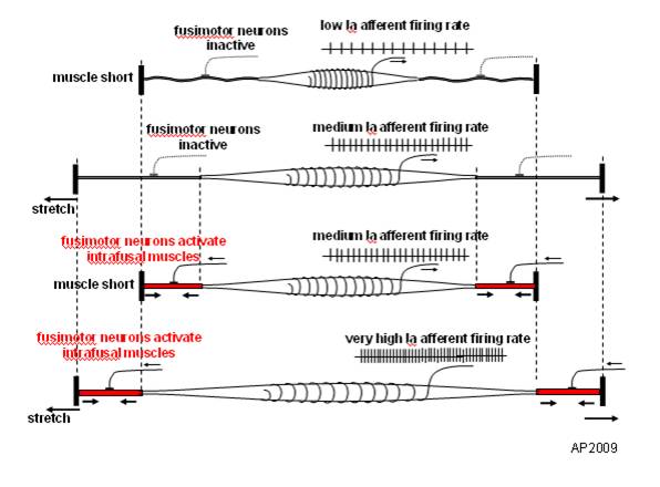
You remember them, large diameter afferent (sensory) fibers coming from muscle spindles and appraising the nervous system of vital information like length and rate of change of length of muscle fibers, so we can be coordinated. They act like volume controls for muscle sensitivity. Turn them up and the muscle becomes more sensitive to ANY input, especially stretch (so they become touchy…maybe like you get if you are hungry and tired and someone asks you to do something); turn them down and they become less or unresponsive.

Their excitability is governed by the sum total (excitatory and inhibitory) of all neurons (like interneuron’s) acting on them (their cell bodies reside in the anterior horn of the spinal cord).

If we slow things down, the rate of change of length slows as well and excitability decreases, like we see in this study (3-6% slower). We also notice that the length of contraction increases; hmmm, why doesn’t it decrease?

Remember these folks are on a treadmill. The treadmill is constantly moving, opposite the direction of travel. With the foot on the ground, this provides a constant rate of change of length of the gastroc/soleus (ie, it is putting it through a slow stretch); so , once the muscle is activated, it contracts for a longer period of time because of the treadmill putting a slow stretch on the gastroc (and soleus).

This article also talks about people with upper motor neuron lesions. An important set of inhibitory neurons come from higher centers of the brain, in the motor cortex. These tend to attenuate the signals affecting the Ia afferents, and keep us stable. When we have an upper motor neuron lesion (like a brain lesion or stroke), we lose this “attenuation” and the stretch reflexes (and muscle tone) becomes much more active (actually hyperactive), making the muscle more sensitive to stretch. This loss of attenuation, along with differing firing patterns of the gastroc are important to remember in gait rehab.

The soleus and medial gastroc begin firing in the first 10% of the gait cycle (at the beginning of loading response) and fire continuously until pre swing (peaking just after midstance). The lateral head begins firing at midstance; both heads (along with soleus) decelerate the forward momentum of the tibia, flex the knee at midstance, and the medial head assists in adducting the calcaneus to assist in supination.

Making sure these muscles fire appropriately is important and needling is just one way of helping them to function better. Don’t overlook the tricep surae on your next patient that has a “hitch in their giddyup”.


Effects of treadmill walking speed on lateral gastrocnemius muscle firing.

by Edward A Clancy, Kevin D Cairns, Patrick O Riley, Melvin Meister, D Casey Kerrigan

American journal of physical medicine rehabilitation Association of Academic Physiatrists (2004) Volume: 83, Issue: 7, Pages: 507-51 PubMed: 15213474

Abstract

OBJECTIVE: To study the electromyographic profile-including ON, OFF, and peak timing locations-of the lateral gastrocnemius muscle over a wide range of walking speeds (0.5-2.1 m/sec) in healthy young adults. DESIGN: We studied gastrocnemius muscle-firing patterns using an electromyographic surface electrode in 15 healthy subjects ambulating on a treadmill at their normal walking speed and at three paced walking speeds (0.5, 1.8, and 2.1 m/sec). Initial heel contact was determined from a force-sensitive switch secured to the skin over the calcaneous. RESULTS: For all speeds, the gastrocnemius firing pattern was characterized by a main peak, occurring 40-45% into the gait cycle, that increased in amplitude with walking speed. Speeds of > or =1.3 m/sec produced a common electromyographic timing profile, when the profile is expressed relative to the stride duration. However, at 0.5 m/sec (a speed typical of individuals with upper-motor neuron lesions), the onset of gastrocnemius firing was significantly delayed by 3-6% of the gait cycle and was prolonged by 8-11% of the gait cycle. CONCLUSION: Many patients with upper motor neuron lesions (e.g., stroke and traumatic brain injury) walk at speeds much slower than those commonly described in the literature for normal gait. At the slow walking speed of 0.5 m/sec, we have measured noticeable changes in the electromyographic timing profile of the gastrocnemius muscle. Given the importance of appropriate plantar flexor firing patterns to maximize walking efficiency, understanding the speed-related changes in gastrocnemius firing patterns may be essential to gait restoration.

Rewind double feature! Part 2

(for part 1, click here)

In conjunction with the latest PODcast talking about efferent copy, we thought it appropriate to talk about the cerebellum here. In this capsule we talk about the efferent pathways

Enjoy! and have a nice weekend (not that we are telling you what to do…)

Ivo and Shawn

Just when you thought it was safe to watch a Neuromechanics Weekly episode, Dr Ivo throws a curveball. Check out the interesting clinical asides about myelopathy (pressure on the spinal cord causing ataxic gait) and the importance of which modality to check 1st, when doing an exam.

Keep these things in mind the next time you are evaluating someone’s gait.

In this Neuromechanics weekly, Dr Waerlop Introduces the cerebellum and talks about its importance clinically, since it contains more than ½ of the neurons in the brain! It’s anatomy and inputs from the periphery are discussed. The take home message is the cerebellum is the key to understanding and directing movement, since it receives feedback from most ascending and descending pathways.

tumblr_lqrsr8mark1qhko2so1_250.gif
tumblr_lqrsr8mark1qhko2so2_250.gif

And now, some light reading for a Saturday….

Review of knee proprioception and the relation to extremity function after an anterior cruciate ligament rupture.

J Orthop Sports Phys Ther. 2001 Oct;31(10):567-7

http://www.ncbi.nlm.nih.gov/pubmed/11665744

What the Gait Guys say about this article:

Aren’t you glad you have mechanoreceptors?

As we have discussed in other posts, proprioception is subserved by cutaneous receptors in the skin (pacinian corpuscles, Ruffini endings, etc.), joint mechanoreceptors (types I,II,III and IV) and muscle spindles (nuclear bag and nuclear chain fibers) . It is both conscious and unconscious and travels in two  main pathways in the nervous system.

Conscious proprioception (awareness of where a joint or body part is in space or action) arises from the peripheral mechanoreceptors in the skin and joints and travels in the dorsal column system (an ascending spinal cord information highway) to ultimately end in the thalamus of the brain, where the information is relayed to the cerebral cortex.

Unconscious proprioception arises from joint mechanoreceptors and muscle spindles and travels in the spino-cerebellar pathways to end in the midline vermis and flocculonodular lobes of the cerebellum.

Conscious proprioceptive information is relayed to other areas of the cortex and the cerebellum. Unconscious proprioceptive information is relayed from the cerebellum to the red nucleus to the thalamus and back to the cortex, to get integrated with the conscious proprioceptive information. This information is then sent down the spinal cord to effect a response in the periphery. As you can see, there is a constant feed back loop between the proprioceptors, the cerebellum and the cerebral cortex. This is what allow us to be balanced and coordinated in our movements and actions.

The ACL is blessed with type I, II and IV mechanoreceptors (Knee Surgery, Sports Traumatology, Arthroscopy Volume 9, Number 6)   We remember that type I mechanoreceptors exist in the periphery of a joint capsule (or in this case, the periphery of the ACL) and are largely tonic in function (ie: they fire all the time) and type II are located deeper in the joint (or deeper in the ACL) and are largely phasic (ie they fire with movement). Type IV mechanoreceptors are largely pain receptors and anyone who has injured his knee can tell you all about them.

The article does a great job reviewing the importance of proprioception and how it relates to knee function and concludes A higher physiological sensitivity to detecting a passive joint motion closer to full extension has been found both experimentally and clinically, which may protect the joint due to the close proximity to the limit of joint motion. Proprioception has been found to have a relation to subjective knee function, and patients with symptomatic ACL deficiency seem to have larger deficits than asymptomatic individuals.”  Bottom line, never quit on the rehab and training of an ACL deficient knee until the absolute best outcome has unequivocally been achieved with certainty that no further improvement can be achieved…… absolute certainty.  Too many stop shy of certainty, and your brain will know it.  And it will show it in small gait, running and athletic skills.

Yup, this is some heavy stuff, but hey…you’re reading it, right?  If we didn’t explain it in detail you might not believe that WE are The Gait Guys ……. more than just foot and shoe guys. After all, there is a brain attached to the other end calling the shots.

Sorting it out so you don’t have to…We remain…The Gait Guys

tumblr_lqq4mwhPBY1qhko2so1_500.jpg
tumblr_lqq4mwhPBY1qhko2so2_500.gif

Ah yes, the Ia and type II afferents.

One of our favorites! Acting as a sentinel from the muscle spindle, concentrated in the antigravity and extensor musculature, Ia and type II afferents live in the belly of the muscle and send information regarding length and rate of change of length to the CNS via the spino cerebellar and inferior olivary pathways. In more simpler terms, think of muscle spindles as small computer chips embedded in the muscle and using la and type II afferents the team act as volume controls helping to set the tone of the muscle and it responsiveness to stretch. If they are active, they make a muscle more sensitive to stretch.

So what does that mean? Muscle spindles turn up the volume or sensitivity of the muscles response to stretch. Remember when we stretch a muscle, it’s response is to contract. Think about when a doctor tests your reflexes. What makes them more or less reactive? You guessed it, the muscle spindle; which is a reflection of what is going on in the higher centers of the brain. The muscle spindles level of excitation is based on the sum total of all information acting on the gamma motor neuron (ie the neuron going to the muscle spindle) in the spinal cord. That includes all the afferent (ie. sensory) information coming in (things like pain can make it more or less active) as well as information descending from higher centers (like the brain, brainstem and cerebellum) which will again influence it at the spinal cord level.

So we found this cool study that looks at spindles and supports their actions:

_____________________________________________________________________

http://www.ncbi.nlm.nih.gov/pubmed/19451207

J Physiol. 2009 Jul 1;587(Pt 13):3375-82. Epub 2009 May 18.

Mechanical and neural stretch responses of the human soleus muscle at different walking speeds.

Cronin NJ, Ishikawa M, Grey MJ, af Klint R, Komi PV, Avela J, Sinkjaer T, Voigt M.

At increased speeds of walking, the muscles themselves (particularly the soleus in this study) become stiffer due to changes in spindle responsiveness. The decline in amplitude and velocity of stretch of the soleus muscle fasicles with increasing walking speeds was NOT accompanied by a change in muscle spindle amplitude, as was hypothesized.

Clinically, this means that the spindles were STILL RESPONSIVE to stretch, even though the characteristics of the muscle changed with greater speeds of action. This may be one of the reasons you may injure yourself when moving or running quickly; the muscle becomes stiffer and the spindle action remains constant (the volume is UP).

Thankfully, we have another system that can intervene (sometimes) when the system is overloaded, and take the stress of the muscle. This is due to the golgi tendon organ; but that is a post for another day…

Geeking out and exploring the subtleties of the neurology as it relates to the system, we remain…The Gait Guys