Early motor experiences.

The early locomotor experience , a free play spontaneous study
Once again, we learn from our mistakes, or we should at least.
This natural locomotion study suggests that better walkers spontaneously walk more and fall less.

"Twelve- to 19-month-olds averaged 2,368 steps and 17 falls per hour. Novice walkers traveled farther faster than expert crawlers, but had comparable fall rates, which suggests that increased efficiency without increased cost motivates expert crawlers to transition to walking. After walking onset, natural locomotion improved dramatically: Infants took more steps, traveled farther distances, and fell less. Walking was distributed in short bouts with variable paths--frequently too short or irregular to qualify as periodic gait. Nonetheless, measures of periodic gait and of natural locomotion were correlated, which indicates that better walkers spontaneously walk more and fall less. Immense amounts of time-distributed, variable practice constitute the natural practice regimen for learning to walk."

Psychol Sci. 2012;23(11):1387-94. doi: 10.1177/0956797612446346. Epub 2012 Oct 19.
How do you learn to walk? Thousands of steps and dozens of falls per day.
Adolph KE1, Cole WG, Komati M, Garciaguirre JS, Badaly D, Lingeman JM, Chan GL, Sotsky RB.

Proprioceptive Clues in Children’s Gait.

This goes along with Mondays post. We can learn a lot about gait from watching our children walk. An immature nervous system is very similar to one which is compensating meaning cheating around a more proper and desirable movement pattern; we often resort to a more primitive state when challenges beyond our ability are presented. This is very common when we lose some aspect of proprioception, particularly from some peripheral joint or muscle, which in turn, leads to a loss of cerebellar input (and thus cerebellar function). Remember, the cerebellum is a temporal pattern generating center so a loss of cerebellar sensory input leads to poor pattern generation output. Watch this clip several times and then try and note each of the following:

  • wide based gait; this is because proprioception is still developing (joint and muscle mechanoreceptors and of course, the spino cerebellar pathways and motor cortex)
  • increased progression angle of the feet: this again is to try and retain stability. External rotation allows them to access a greater portion of the glute max and the frontal plane (engaging an additional plane is always more stable).
  • shortened step length; this keeps the center of gravity close to the body and makes corrections for errors that much easier (remember our myelopathy case from last week ? LINK. This immature DEVELOPING system is very much like a mature system that is REGRESSING. This is a paramount learning point !)
  • decreased speed of movement; this allows more time to process proprioceptive clues, creating accuracy of motion


Remember that Crosby, Still, Nash and young song “Teach Your Children”? It is more like, “teach your parents”…


Proprioceptive clues are an important aspect of gait analysis, in both the young and old, especially since we tend to revert back to an earlier phase of development when we have an injury or dysfunction.


Headbonking and gait

A great article (see reference below) just came out looking at the gait changes that come along with a concussion. Basically it says that folks that have concussions have more coronal plane (i.e. side to side) sway and they walk slower. This reminded us of some of the "decomposition of gait" pieces that we have done and one post on proprioceptive clues in children gait that we did about 5 years ago. Having a concussion causes decomposition of gait, and we move toward a more primitive pattern, just like we see in kids. Here was the post:

We can learn a lot about gait from watching our children walk. An immature nervous system is very similar to one which is compensating meaning cheating around a more proper and desirable movement pattern; we often resort to a more primitive state when challenges beyond our ability are presented. This is very common when we lose some aspect of proprioception, particularly from some peripheral joint or muscle, which in turn, leads to a loss of cerebellar input (and thus cerebellar function). Remember, the cerebellum is a temporal pattern generating center so a loss of cerebellar sensory input leads to poor pattern generation output. Watch this clip several times and then try and note each of the following:

  • wide based gait; this is because proprioception is still developing (joint and muscle mechanoreceptors and of course, the spino cerebellar pathways and motor cortex)
  • increased progression angle of the feet: this again is to try and retain stability. External rotation allows them to access a greater portion of the glute max and the frontal plane (engaging an additional plane is always more stable).
  • shortened step length; this keeps the center of gravity close to the body and makes corrections for errors that much easier (remember our myelopathy case from last week ? LINK.  This immature DEVELOPING system is very much like a mature system that is REGRESSING.  This is a paramount learning point !)
  • decreased speed of movement; this allows more time to process proprioceptive clues, creating accuracy of motion

Remember that Crosby, Still, Nash and young song “Teach Your Children”? It is more like, “teach your parents”…

Proprioceptive clues are an important aspect of gait analysis, in both the young and old, especially since we tend to revert back to an earlier phase of development when we have an injury or dysfunction.


Manaseer TSGross DPDennett LSchneider KWhittaker JL1. Gait Deviations Associated With Concussion: A Systematic Review.  Clin J Sport Med. 2017 Nov 21. doi: 10.1097/JSM.0000000000000537. [Epub ahead of print]

What? Gait and hearing....

No, not how does the gait SOUND, but can they hear? We have long payed attention to the mechanics of gait, muscles and proprioception, and many times, if you have been following us for any length of time, the interplay of all three. But how often have you considered someones hearing in your evaluation?

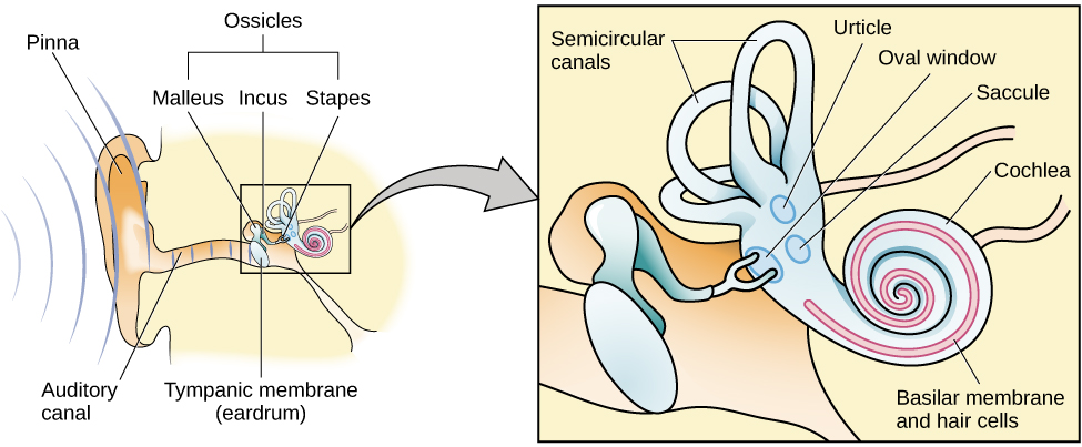
Anatomically, the hearing organs (hair cells, otolithic apparatus, cochlea) are conveniently located right next store to the vestibular system (utricle, saccule and semicircular canals). The hearing organs start with mechanical vibrations of the tympanic membrane (ear drum) which move 3 auditory bones (or ossicles) called the incus, malleus and stapes. The vibrations move the stapes moves in and out of the fluid filled cochlea and vibrate crystals laying on a platform (otolith) in which hair cells are imbedded and if the vibration sufficient, the nerves attached to the hair cells fire and you can hear your favorite baseline by Cliff Williams (or Cliff Burton, John Myung, Bootsy Collins or whomever you like to listen to).

The vestibular system, as you probably know, monitors position and velocity of movement of the head. There are three hula hoop type structures called “semicircular canals” (see picture above) that monitor rotational and tilt position and angular acceleration, as well as two other structures, the utricle and saccule, which monitor tilt and linear acceleration.

The vestibular apparatus (the canals and the utricle and saccule) feed into a part of the brain called the floccular nodular lobe of the cerebellum, which as we are sure you can imagine, have something to do with balance and coordination. This area of the cerebellum feeds back to the vestibular system (actually the vestibular nucleii); which then feed back up to the brain as well as (you guessed it) down the spinal cord and to predominantly the extensor muscles.

Can you see how the 2 systems are “attached” to one another and could conceivably be interrelated? When you move fluid in one system (endo or peri lymph) you HAVE to move fund in the other. The fluid displaces, (like air, in air filled shoes or insoles), it does not deform (like EVA in an insole)

It is well established that hearing impaired folks (especially kids) have gait (and often balance) problems (1-3) and the worse the hearing loss, the worse the gait disturbance (1). This can occur in middle aged (4), as well as older adults as well (5-7). Perhaps this is due to viscosity changes in the endo or perilymph (8), otosclerosis of the ossicles (9,10) or other age related changes in utricle and saccule function (5-7). Also, for the 1st time, we are documenting a “sensory reweighting” which occurs when auditory cues are changed (4), with a greater reliance on visual cues. Hmmm. We wonder how this plays out with listening to music while walking or running?

So, The next time someone you see has a gait or balance problem, check their hearing and adjust your treatment program accordingly : )


References:

1. Melo RS.  Gait performance of children and adolescents with sensorineural hearing loss.
Gait Posture. 2017 Jun 3;57:109-114. doi: 10.1016/j.gaitpost.2017.05.031.

2. Jafarnezhadgero AA, Majlesi M, Azadian E. Gait ground reaction force characteristics in deaf and hearing children. Gait Posture. 2017 Mar;53:236-240. doi: 10.1016/j.gaitpost.2017.02.006. Epub 2017 Feb 14.

3. Janky KL, Givens D.Vestibular, Visual Acuity, and Balance Outcomes in Children With Cochlear Implants: A Preliminary Report. Ear Hear. 2015 Nov-Dec;36(6):e364-72. doi: 10.1097/AUD.0000000000000194.

4. Maheu M, Sharp A, Landry SP, Champoux F. Sensory reweighting after loss of auditory cues in healthy adults. Gait Posture. 2017 Mar;53:151-154. doi: 10.1016/j.gaitpost.2017.01.015. Epub 2017 Jan 24.

5. Agmon M, Lavie L, Doumas M. The Association between Hearing Loss, Postural Control, and Mobility in Older Adults: A Systematic Review. J Am Acad Audiol. 2017 Jun;28(6):575-588. doi: 10.3766/jaaa.16044. Review.

6. Layman AJ1, Li C, Simonsick E, Ferrucci L, Carey JP, Agrawal Y. Association Between Saccular Function and Gait Speed: Data From the Baltimore Longitudinal Study of Aging. Otol Neurotol. 2015 Jan 7. [Epub ahead of print]

7. Otol Neurotol. 2012 Jul;33(5):832-9. doi: 10.1097/MAO.0b013e3182545061.
Decline in semicircular canal and otolith function with age.
Agrawal Y1, Zuniga MG, Davalos-Bichara M, Schubert MC, Walston JD, Hughes J, Carey JP.

8. Wu T, Marcus DC. Age-Related Changes in Cochlear Endolymphatic Potassium and Potential in CD-1 and CBA/CaJ Mice . JARO: Journal of the Association for Research in Otolaryngology. 2003;4(3):353-362. doi:10.1007/s10162-002-3026-6.

9. Grayeli AB1, Sterkers O, Toupet M. Audiovestibular function in patients with otosclerosis and balance disorders. Otol Neurotol. 2009 Dec;30(8):1085-91. doi: 10.1097/MAO.0b013e3181b0fd5d.

10. Ozmen AO1, Aksoy S, Ozmen S, Saraç S, Sennaroğlu L, Gürsel B. Balance after stapedotomy: analysis of balance with computerized dynamic posturography. Clin Otolaryngol. 2009 Jun;34(3):212-7. doi: 10.1111/j.1749-4486.2009.01915.x.


Children: Postural control of balance

From the study:

“From these indexes it was established that the postural capacity needed just to control balance with the leg muscles was not attained before 4-5 years of independent walking, i.e., at about 5-6 years of age.” -Breniere

reference link:

Exp Brain Res. 1998 Aug;121(3):255-62.Development of postural control of gravity forces in children during the first 5 years of walking.Brenière Y1, Bril B.

http://www.ncbi.nlm.nih.gov/pubmed/9746131/

tumblr_n2n3xcTGG81qhko2so1_250.jpg
tumblr_n2n3xcTGG81qhko2so2_r1_250.jpg

Things may not always be how they appear.

What can you notice about all these kids that you may not have noticed before?

Look north for a moment. What do you notice about all the kids with a head tilt? We are talking about girl in pink on viewers left, gentleman in red 2nd from left, blue shirt all the way on viewers right. Notice how the posture of the 2 on the left are very similar and the one on the right is the mirror image?

What can be said about the rest of their body posture? Can you see how the body is trying to move so that the eyes can be parallel with the horizon? This is part of a vestibulo cerebellar reflex. The system is designed to try and keep the eyes parallel with the horizon. The semicircular canals (see above), located medial to your ears, sense linear and angular acceleration. These structures feed head position information to the cerebellum which then forwards it to the vestibular nucleii, which sends messages down the vestibulo spinal tract and up the medial longitudinal fasiculus to adjust the body position and eye position accordingly. 

Can you see how when we add another parameter to the postural position (in this case, running; yes, it may be staged, but the reflex persists despite that. Neurology does not lie), that there can be a compensation that you may not have expected?

What if one of these 3 (or all three) kids had neck pain. Can you see how it may not be coming from the neck. What do you think happens with cortical (re)mapping over many years of a compensation like this? Hmmm. Makes you think, eh?

Ivo and Shawn. The Gait Guys. Taking you a little further down the rabbit hole, each and every post.

Well, how convenient. A fantastic picture for teaching from the cover of one of our favorite magazines.
For this post, lets start with the gal on the left in the pink shirt. 1st of all, she is running in flip flops. Since these require so much long …

Well, how convenient. A fantastic picture for teaching from the cover of one of our favorite magazines.

For this post, lets start with the gal on the left in the pink shirt. 1st of all, she is running in flip flops. Since these require so much long flexor activity to keep them on, not the best footwear choice, in our opinion. Check out that exaggerated left sided arm swing. This goes to propel herself forward. Why the extra effort? Check out her right (stance phase leg). What do you see? The knee points outward while the foot is planted. We are looking at either external tibial torsion or a femoral retrotorsion. Did you pick up the compensatory head tilt to the left? The vestibular system has become involved, and the trapezius and levator scapula seem to be it’s target (thus the shoulder hike and ipsilateral rotation), as well as the ipsilateral lateral benders and rotators of the cervical spine, namely the splenius cervicis and capitis (the multifidus/rotatores are contralateral rotators).

How about the subtle pelvic shift to the right? and the mild crossover gait (note the adduction of the left knee across midline).

It would be great to see a shot of her barefoot to see what changes, as increased long flexor activity has both local (impaired ankle rocker, excessive forefoot inversion, reciprocal inhibition of the anterior compartment muscles of the lower leg) as well as long distance (namely increased flexor drive to the brainstem and cerebellum) implications. We would want to see this (as well as examine her) before making any recommendations other than LOSE THE FLIP FLOPS GIRLFRIEND.

Wow, all that and we have only scratched the surface.

We remain the geeks of gait: Ivo and Shawn

tumblr_n2ac0gX4j01qhko2so1_r1_1280.jpg
tumblr_n2ac0gX4j01qhko2so2_r1_1280.jpg
tumblr_n2ac0gX4j01qhko2so3_r1_1280.jpg
tumblr_n2ac0gX4j01qhko2so4_r1_1280.jpg
tumblr_n2ac0gX4j01qhko2so5_r1_1280.jpg
tumblr_n2ac0gX4j01qhko2so6_r1_1280.jpg

Remember this kiddo?

We have been following the natural development of this little guy for some time now. For a review, please see here (1 year ago) and here (2 years ago) for our previous posts on him.

In the top 2 shots, the legs are neutral. The 3rd and 4th shots are full internal rotation of the left and right hips respectively. The last 2 shots are full external rotation of the hips.

Well, what do you think now?

We remember that this child has external tibial torsion and pes planus. As seen in the supine photo, when the knees face forward, the feet have an increased progression angle (they turn out). We are born with some degree / or little to none, tibial torsion and the in-toeing of infants is due to the angle of the talar neck (30 degrees) and femoral anteversion (the angle of the neck of the femur and the distal end is 35 degrees).  The lower limbs rotate outward at a rate of approximately 1.5 degrees per year to reach a final angle of 22 degrees….. that is of course if the normal de rotation that a child’s lower limbs go through occurs timely and completely.

He still has a pronounced valgus angle at the the knees (need a review on Q angles? click here). We remember that the Q angle is negative at birth (ie genu varum) progresses to a maximal angulation of 10-15 degrees at about 3.5 years, then settles down to 5-7 degrees by the time they have stopped growing. He is almost 4 and it ihas lessend since the last check to 15 degrees.

His internal rotation of the hips should be about 40 degrees, which it appears to be. External rotation should match; his is a little more limited than internal rotation, L > R. Remember that the femoral neck angle will be reducing at the rate of about 1.5 degrees per year from 35 degrees to about 12 in the adult (ie, they are becoming less anteverted).

At the same time, the tibia is externally rotating (normal tibial version) from 0 to about 22 degrees. He has fairly normal external tibial version on the right and still has some persistent internal tibial version on the left. Picture the hips rotating in and the lower leg rotating out. In this little fellow, his tibia is outpacing the hips. Nothing to worry about, but we do need to keep and eye on it.

What do we tell his folks?

  • He is developing normally and has improved significantly since his original presentation to the office
  • Having the child walk barefoot has been a good thing and has provided some intrinsic strength to the feet
  • He needs to continue to walk barefoot and when not, wear shoes with little torsional rigidity, to encourage additional intrinsic strength to the feet
  • He should limit “W” sitting, as this will tend to increase the genu valgus present
  • We gave him 1 leg balancing “games” and encouraged agility activities, like balance beam, hopping, skipping and jumping on each leg individually

We are the Gait Guys, promoting gait and foot literacy, each and every post.

tumblr_mumzdeHw9w1qhko2so1_1280.jpg
tumblr_mumzdeHw9w1qhko2so2_1280.jpg
tumblr_mumzdeHw9w1qhko2so3_1280.jpg
tumblr_mumzdeHw9w1qhko2so4_1280.jpg
tumblr_mumzdeHw9w1qhko2so5_1280.jpg

The power to bend bones.

What have we here? Hmmm. This little girl was brought in by her mother because of intermittent knee pain and “collapsing” of the knees while walking, for no apparent reason.

The ankle dorsi flexion (or ankle rocker; see last 2 pictures; we are fully dorsiflexing the ankles)  needs to occur somewhere, how about the knees? Or in this case, the tibia. Wow!

You are looking at a 4 year year with a condition called genu (and tibial) recurvatum. Genu recurvatum is operationally defined as knee hyperextension greater than 5 degrees. The knee is hyperextended, and in this case, the tibia is literally “bent backward”. Look at the 2 pictures of her tibia.

Generally speaking, the tibial plateau usually has a slight posterior inclination (as it does in this case; look carefully at the 1st picture) causing the knee to flex slightly when standing. Sometimes, if it is parallel with the ground and the center of gravity is forward of the knees, the knee will hyperextend (or in this case, the tibia will bend) to compensate.

In this particular case, the tibia has compensated more, rather than the knee itself. The knee joint is stable and there is no ligamentous laxity as of yet. She does not have a neurological disorder, neuromuscular disease or connective tissue disorder. She has congenitally tight calves.

As you can imagine, her step length is abbreviated and ankle rocker is impaired.

So what did we tell her Mom?

  • keep her barefoot as much as possible (incidentally, she loves to be barefoot most of the time, gee, go figure!)
  • have her walk on her heels (she’s a kid, make a game of it)
  • showed her how to do calf stretches
  • balance on 1 leg with her eyes open and closed
  • keep her out of backless shoes (like the clogs she came in with)
  • keep her out of flip flops and sandals where she would have to “scrunch” her toes to keep them on.
  • follow back in 3 months to reassess

There you have it. Next time you don’t think Wolff’s (or Davis’s) law* is real, think about this case. Want to know more? Consider taking our National Shoe Fit Program, available by clicking here.

The Gait Guys. Making you gait IQ higher with each post.

*Wolff’s law: Bone will be deposited in areas of stress and removed in areas of strain. or put another way: bone in a healthy person or animal will adapt to the loads under which it is placed

Davis’s law: soft tissue will adapt to the loads that are placed on it

Treat your children well.
We think Crosby, Stills, Nash and Young had it right…
look at this conclusion: “Shoes affect the gait of children. With shoes, children walk faster by taking longer steps with greater ankle and knee motion and …

Treat your children well.

We think Crosby, Stills, Nash and Young had it right…

look at this conclusion: “Shoes affect the gait of children. With shoes, children walk faster by taking longer steps with greater ankle and knee motion and increased tibialis anterior activity. Shoes reduce foot motion and increase the support phases of the gait cycle. During running, shoes reduce swing phase leg speed, attenuate some shock and encourage a rearfoot strike pattern."                                                                   

let’s break that down a bit:

"Shoes affect the gait of children.” Shoes effect EVERYONE’S gait, not just kids. They alter the ground reactive forces, limit some ranges of motion and thus can promote a compensation or mechanics that you may not have seen previously. Take off one of your shoes. Lift your toes up slightly so you are centered on your tripod. Stand on your “barefoot” leg with your eyes closed. See how long you can stand without faltering. Now repeat that with your shod foot. Some difference, eh? I thought shoes dampened proprioception…They do. But they also give you more support and mechanics that you didn’t have previously, so the foot doesn’t have to work as hard. 

“With shoes, children walk faster by taking longer steps with greater ankle and knee motion and increased tibialis anterior activity.”Remember we are talking about kids here. Longer steps because with a shoe we promote heel rocker and because of the added support, more stability (or at least more perceived stability). This means more confidence. Greater knee and ankle motion because of the increased stride length. Greater tibialis anterior activity because of greater dorsiflexion of the foot because of the increased weight (the shoe adds ounces and this muscle must work harder to attenuate the foot as it approached midstance) and increased heel and ankle rocker.

Shoes reduce foot motion and increase the support phases of the gait cycle.” Shoes constrain the foot and reduce available ranges of motion (yes, even non motion control shoes). Less motion (and thus proprioception) means less feedback to the brain about muscles length and tension (via muscle spindles and golgi tendon organs). The brain will need to have the foot have more contact with the ground to know where it is in space. 

“During running, shoes reduce swing phase leg speed,probably due to the increased weight so it takes more to start the process of initial (early) swing

attenuate some shock we know shoes attenuate at least initial ground reactive forces

…and encourage a rearfoot strike pattern.” most likely due to the cushioning (remember from the recent Kenyan study about barefoot heel strikers? (click here if you need a reminder) They were more likely to heel strike on softer surfaces) AND the increased stride length (which would require more ankle dorsiflexion). 

Wow. Shoes really do make the, er….kid.

The Gait Guys. Making it real and increasing your shoe and gait IQ with each post.                                                                                 

J Foot Ankle Res. 2011 Jan 18;4:3. doi: 10.1186/1757-1146-4-3.

Effect of children’s shoes on gait: a systematic review and meta-analysis.

Source

Discipline of Exercise and Sports Science, Faculty of Health Sciences, The University of Sydney, Cumberland Campus, PO Box 170, Lidcombe, 1825, NSW, Australia. cweg6974@uni.sydney.edu.au.

Abstract

BACKGROUND:

The effect of footwear on the gait of children is poorly understood. This systematic review synthesises the evidence of the biomechanical effects of shoes on children during walking and running.

METHODS:

Study inclusion criteria were: barefoot and shod conditions; healthy children aged ≤ 16 years; sample size of n > 1. Novelty footwear was excluded. Studies were located by online database-searching, hand-searching and contact with experts. Two authors selected studies and assessed study methodology using the Quality Index. Meta-analysis of continuous variables for homogeneous studies was undertaken using the inverse variance approach. Significance level was set at P < 0.05. Heterogeneity was measured by I2. Where I2 > 25%, a random-effects model analysis was used and where I2 < 25%, a fixed-effects model was used.

RESULTS:

Eleven studies were included. Sample size ranged from 4-898. Median Quality Index was 20/32 (range 11-27). Five studies randomised shoe order, six studies standardised footwear. Shod walking increased: velocity, step length, step time, base of support, double-support time, stance time, time to toe-off, sagittal tibia-rearfoot range of motion (ROM), sagittal tibia-foot ROM, ankle max-plantarflexion, Ankle ROM, foot lift to max-plantarflexion, ‘subtalar’ rotation ROM, knee sagittal ROM and tibialis anterior activity. Shod walking decreased: cadence, single-support time, ankle max-dorsiflexion, ankle at foot-lift, hallux ROM, arch length change, foot torsion, forefoot supination, forefoot width and midfoot ROM in all planes. Shod running decreased: long axis maximum tibial-acceleration, shock-wave transmission as a ratio of maximum tibial-acceleration, ankle plantarflexion at foot strike, knee angular velocity and tibial swing velocity. No variables increased during shod running.

CONCLUSIONS:

Shoes affect the gait of children. With shoes, children walk faster by taking longer steps with greater ankle and knee motion and increased tibialis anterior activity. Shoes reduce foot motion and increase the support phases of the gait cycle. During running, shoes reduce swing phase leg speed, attenuate some shock and encourage a rearfoot strike pattern. The long-term effect of these changes on growth and development are currently unknown. The impact of footwear on gait should be considered when assessing the paediatric patient and evaluating the effect of shoe or in-shoe interventions.

http://www.ncbi.nlm.nih.gov/pubmed/21244647

all material copyright 2013 The Gait Guys/ The Homunculus Group. All rights reserved. Yea, that means ask before you touch!

Speed Matters: Brief Thoughts on Gait and Running.


The journal article below sparked a few thoughts for a blog post today.

Have you ever tried to walk slower than your normal pace ? How about running slower than your normal pace (  you know, running with that person who is clearly a minute slower pace) ? Why are both so uncomfortable and labor intensive ? Why does your balance, energy and stability become challenged ? After all, slower should be easier right ?!
There are many reasons and this study hints at a few issues but the bottom line is that speed matters.  Have you ever been driving down the road and you see a big pot hole in the road that you just cannot get around because it is either too big or you do not have time to steer around it ?  What is your first reaction ?  Many will press down on the gas pedal. Why is that ? Well, logic for many is that speeding up will possibly enable you to launch across the void and reduce the impact issues of dropping down into the void.  Men will rationalize the “launch across the pothole” theory, and in some respects they are not wrong.
Running and walking slowly sort of bring out some of the same issues.  When we move slowly the body is more likely to drift into the frontal/coronal (side to side) plane.  Moving more quickly ensures that the dominant path is forward. Slowing down does not ensure that forward will occur. side to side sway enters the picture. And when side to side sway enters as an option we have to spend more time and strategies negotiating the side sway.  This is why we see all kinds of corrections with the limbs and core when we attempt to stand on one foot, but we do not see these issues when we walk or run.  When running we are mostly trying to get the next foot underneath our body so that we do not fall forward flat on our face. Locomotion is a strategy of nothing more than trying to stay upright.  When we run the predominant motion is forward. But when we slow down and reduce the advantage of speed to blur out these issues the challenges begin and other planes of movement become an option and thus planes we need to control. It is much why the elderly have more difficulty moving about, because they have to negotiate and control so many other planes of movement.
So, if you want to bring out some faulty motor patterns, move more slowly and see where your deficits lie. One of our assessments for patients and athletes is to have them walk at a 3second pace meaning each foot fall must be held for 3 seconds before the next step can be initiated. This means stance and swing must be slowed to 3 seconds.  Amazing things will show up if you just slow things down and allow weaknesses to percolate to the surface.  Speed blurs them and keeps them suppressed. It is really a form of cheating and compensation.
So, like in your car, speed matters.
Think about this next time you have to walk or run with a slower person. It may be one of the issues, but there are others and we will eventually get to them.


Gait and Speed on Child Development
J Biomech. 2008;41(8):1639-50. Epub 2008 May 7. The effect of walking speed on the gait of typically developing children. Schwartz MH, Rozumalski A, Trost JP. Abstract

Many gait studies include subjects walking well below or above typical self-selected comfortable (free) speed. For this reason, a descriptive study examining the effect of walking speed on gait was conducted. The purpose of the study was to create a single-source, readily accessible repository of comprehensive gait data for a large group of children walking at a wide variety of speeds. Three-dimensional lower extremity joint kinematics, joint kinetics, surface electromyographic (EMG), and spatio-temporal data were collected on 83 typically developing children (ages 4-17) walking at speeds ranging from very slow (>3 standard deviations below mean free speed) to very fast (>3 standard deviations above mean free speed). The resulting data show that speed has a significant influence on many measures of interest, such as kinematic parameters in the sagittal, coronal, and transverse planes. The same was true for kinetic data (ground reaction force, moment, and power), normalized EMG signals, and spatio-temporal parameters. Examples of parameters with linear and various nonlinear speed dependencies are provided. The data from this study, including an extensive electronic addendum, can be used as a reference for both basic biomechanical and clinical gait studies.

Neuromechanics weekly: proprioceptive clues in Children’s Gait.

We can learn a lot about gait from watching our children walk. An immature nervous system is very similar to one which is compensating meaning cheating around a more proper and desirable movement pattern; we often resort to a more primitive state when challenges beyond our ability are presented. This is very common when we lose some aspect of proprioception, particularly from some peripheral joint or muscle, which in turn, leads to a loss of cerebellar input (and thus cerebellar function). Remember, the cerebellum is a temporal pattern generating center so a loss of cerebellar sensory input leads to poor pattern generation output. Watch this clip several times and then try and note each of the following:

  • wide based gait; this is because proprioception is still developing (joint and muscle mechanoreceptors and of course, the spino cerebellar pathways and motor cortex)
  • increased progression angle of the feet: this again is to try and retain stability. External rotation allows them to access a greater portion of the glute max and the frontal plane (engaging an additional plane is always more stable).
  • shortened step length; this keeps the center of gravity close to the body and makes corrections for errors that much easier (remember our myelopathy case from last week ? LINK.  This immature DEVELOPING system is very much like a mature system that is REGRESSING.  This is a paramount learning point !)
  • decreased speed of movement; this allows more time to process proprioceptive clues, creating accuracy of motion

Remember that Crosby, Still, Nash and young song “Teach Your Children”? It is more like, “teach your parents”…

Proprioceptive clues are an important aspect of gait analysis, in both the young and old, especially since we tend to revert back to an earlier phase of development when we have an injury or dysfunction.

Ivo and Shawn. Still bald, still good looking, with intact cerebellums and neocortices : )

Foot maturation in children is reached by 5 years old

source article: http://www.ncbi.nlm.nih.gov/pubmed/21257173

Gait development begins early! Start good mechanics in your kids! “Based on these results, it would appear that foot maturation during gait is fully achieved at 5 years”

Foot mechanics during the first six years of Independent Walking J Biomech. 2011 Jan 21. [Epub ahead of print]

“the MP joint biomechanical parameters were similar between children (older than 2 years) and adults, hinting at a quick maturation of this joint mechanics. The ankle joint parameters and the GRFs (except for the frontal plane) showed an adult-like pattern in 5-year-old children. Some ankle joint parameters, such as the joint power and the 3D angle α(M.ω) still evolved significantly until 3.5 years. Based on these results, it would appear that foot maturation during gait is fully achieved at 5 years.”

tumblr_lxeuq7JYjB1qhko2so1_400.jpg
tumblr_lxeuq7JYjB1qhko2so2_1280.jpg
tumblr_lxeuq7JYjB1qhko2so3_500.jpg

Genu valgum in kids: What you need to know

We have all seen this. The kid with the awful “knock knees”.  It is a Latin word “which means “bent” or “knock kneed”. It appears to have 1st been used in 1884.

This condition, where the Q angle angle exceeds 15 degrees, usually presents maximally at age 3 and should resolve by age 9. It is usually physiologic in development due to obliquity of the femur, when the medial condyle is lower than the lateral. Normal development and weight bearing lead to an overgrowth of the medial condyle of the femur. This, combined with varying development of the medial and lateral epiphysies of the tibial plateau leads to the valgus development. Gradually, with increased weight bearing, the lateral femoral condyle (and thus the tibial epiphysis) bear more weight and this appears to slow, and eventually reverse the valgum.

Normal knee angulation usually progresses from 10-15 degrees varus at birth to a maximal valgus angle of 10-15 degrees  at 3-3.5 years (see picture).  The valgus usually decreases to an adult angle of 5-7 degrees.  Remember that in women, the Q angle should be less than 22 degrees with the knee in extension and in men, less than 18 degrees. It is measured by measuring the angle between the line drawn from the ASIS to the center of the patella and one from the center of the patella through the tibial tuberosty, while the leg is extended.

Further evaluation of a child is probably indicated if:

  • The angle is greater than 2 standard devaitions for their age (see chart) 
  • If their height is > 25th percentile 
  • If it is increasing in severity 
  • If it is developing asymmetrically

Management is by serial measurement of the intermalleolar distance (the distance between ankles when the child’s knee are placed together) to document gradual spontaneous resolution (hopefully). If physiologic genu valgum persists beyond 7-8 years of age, an orthopaedic referral would be indicated but certainly intervention with attempts at corrective exercises and gait therapy should be employed. Persistence in the adult can cause a myriad of gait, foot, patello femoral and hip disorders, and that is the topic on another post.

Promotion of good foot biomechanics through the use of minimally supportive shoes, encouraging walking on sand (time to take that trip to the beach!), walking on uneven surfaces (like rocks, dirt and gravel), gentle massage (to promote muscle facilitation for those muscles which test weak (origin/insertion work) and circulation), gait therapeutic exercises and acupuncture when indicated, can all be helpful.

Ivo and Shawn…  The Gait Guys…Promoting foot and gait literacy for everyone.

tumblr_luh7t7zoZU1qhko2so1_500.gif
tumblr_luh7t7zoZU1qhko2so2_500.gif

We hope you are standing up while you read this….

A newborn’s brain is only about one-quarter the size of an adult’s. It grows to about 80 percent of adult size by three years of age and 90 percent by age five (see above). This growth is largely due to changes in individual neurons and their connections, or synapses.

The truth is, most of our brain cells are formed at birth, In fact, we actually have MORE neurons BEFORE we are born. It is the formation of synapses, or connections between neurons, that actually accounts for the size change (see 1st picture above). This is largely shaped by experience and interaction with the environment.

Do you think children’s brains are less active than adults? Think again, your 3 year old’s brain is twice as active as yours! It isn’t until later in life that you actually start dialing back on some of those connections and those pathways degenerate or fade away…a process scientists call “pruning”.

How does this apply to gait? Gait depends on proprioception, or body position awareness. Your brain needs to know where your foot is, what it is standing on and so on. Proprioception, as we have discussed in other posts, is subserved by muscle and joint receptors called mechanoreceptors (muscle spindles, golgi tendon organs and type 1-4 joint mechanoreceptors to be exact). This information is fed to 2 main areas of the brain: the cerebral cortex and the cerebellum. These 2 parts of the central nervous system are interconnected on many levels.

The cerebellum is intimately associated with learning. Try this experiment. you will need a tape recorder (guess we are showing our ages, digital recorder), a timer and a moderately difficult book.

Sit down and pick a section of the book to read. start the recorder and timer and read aloud for 2 minutes. Stop reading, stop the recorder and stop the timer.

Stand up, somewhere you won’t get hurt if you fall. Stand on 1 leg (or if available, stand on a BOSU or rocker board). Open the book to a different spot. Start the timer, the recorder and start reading again for 2 minutes.

Sit back down and grab a snack. Listen to the 2 recordings and pay attention to the way you sound when you were reading, the speed, fluency and flow of words. Now think about recall. Which passage do you remember better?

The brain works best at multitasking and balance and coordination activities intimately affect learning. Having children sit in a class room and remain stationary and listen to a lecture is not the best way to learn. We always tel our students to get up and move around…

This article looks at this relationship in a slightly different way.

The Gait Guys….Sorting it out so you don’t have to.

We hope you are still standing : )

 Scand J Med Sci Sports. 2011 Oct;21(5):663-9. doi: 10.1111/j.1600-0838.2009.01027.x. Epub 2010 Mar 11

Motor coordination as predictor of physical activity in childhood.

Lopes VP, Rodrigues LP, Maia JA, Malina RM.

Source

Department of Sports Science, Research Center in Sports Sciences, Health Sciences and Human Development (CIDESD), Bragança, Portugal. vplopes@ipb.pt

Abstract

This study considers relationships among motor coordination (MC), physical fitness (PF) and physical activity (PA) in children followed longitudinally from 6 to 10 years. It is hypothesized that MC is a significant and primary predictor of PA in children. Subjects were 142 girls and 143 boys. Height, weight and skinfolds; PA (Godin-Shephard questionnaire); MC (Körperkoordination Test für Kinder); and PF (five fitness items) were measured. Hierarchical linear modeling with MC and PF as predictors of PA was used. The retained model indicated that PA at baseline differed significantly between boys (48.3 MET/week) and girls (40.0 MET/week). The interaction of MC and 1 mile run/walk had a positive influence on level of PA. The general trend for a decrease in PA level across years was attenuated or amplified depending on initial level of MC. The estimated rate of decline in PA was negligible for children with higher levels of MC at 6 years, but was augmented by 2.58 and 2.47 units each year, respectively, for children with low and average levels of initial MC. In conclusion MC is an important predictor of PA in children 6-10 years of age.

© 2009 John Wiley & Sons A/S.