Motion control Shoes + Internal Tibial Torsion = Knee Pain

Thinking about putting a motion control shoe under that foot to control pronation? You had better make sure you make friends with the knee, as it will often (depending on the compensation) be placed OUTSIDE the SAGGITAL PLANE. Like Dr Allen has said many times before , the knee is basically a hinge joint placed between 2 ball and socket joints, and it is usually the one to start grumbling...

Learn more as Dr Ivo Waerlop of The Gait Guys explains in this brief video

#gait #Gaitanalysis #gaitguys #thegaitguys #kneepain #motioncontrolshoes #internaltibialtorsion

https://vimeo.com/154496722

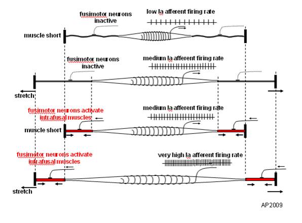
Muscle Spindles and Proprioception

image source: https://en.wikipedia.org/wiki/File:Fusimotor_action.jpg

image source: https://en.wikipedia.org/wiki/File:Fusimotor_action.jpg

And what have we been saying for the last 6 years? 

Connected to the nervous system by large diameter afferent (sensory) fibers, they are classically thought of as appraising the nervous system of vital information like length and rate of change of length of muscle fibers, so we can be coordinated. They act like volume controls for muscle sensitivity. Turn them up and the muscle becomes more sensitive to ANY input, especially stretch (so they become touchy…maybe like you get if you are hungry and tired and someone asks you to do something); turn them down and they become less or unresponsive.

Their excitability is governed by the sum total (excitatory and inhibitory) of all neurons (like interneuron’s) acting on them (their cell bodies reside in the anterior horn of the spinal cord).

Along with with Golgi tendon organs and joint mechanoreceptors, they also act as proprioceptive sentinels, telling us where our body parts are in space. We have been teaching this for years. Here is a paper that exemplifies that, identifying several proteins responsible for neurotransduction including the Piezo2 channel as a candidate for the principal mechanotransduction channel. Many neuromuscular diseases are accompanied by impaired  muscle spindle function, causing a decline of motor performance and coordination. This is yet another key finding in the kinesthetic system and its workings. 

Remember to include proprioceptive exercises and drills (on flat planar surfaces, like we talked about here) in your muscle rehab programs


Kröger S Proprioception 2.0: novel functions for muscle spindles. Curr Opin Neurol. 2018 Oct;31(5):592-598. 

Woo SH, Lukacs V, de Nooij JC, Zaytseva D, Criddle CR, Francisco A, Jessell TM, Wilkinson KA, Patapoutian A. Piezo2 is the principal mechanotransduction channel for proprioception.Nat Neurosci. 2015 Dec; 18(12):1756-62. Epub 2015 Nov 9.

Fusimotor control of proprioceptive feedback during locomotion and balancing: can simple lessons be learned for artificial control of gait?

Hulliger M. Fusimotor control of proprioceptive feedback during locomotion and balancing: can simple lessons be learned for artificial control of gait? Prog Brain Res. 1993; 97:173-80.

Are you a control freak?

While working with a post surgical ACL patient that has +2 laxity and  graft pain, I was reminded of something Dr. Allen and I were talking about while discussing this case. 

One of the primary goals post ACL is stated as improving range of motion, particularly getting to full extension. If you look at the mechanics of the anterior cruciate ligament, you'll see that placing the knee in full extension places this ligament under stretch. We often will try to increase range of motion by hyper extending the knee, or using it as a fulcrum, which can cause undue stretch to this ligament. This means the burden of oweness is on the musculature surrounding the joint to provide stability, similar to what we are seeing in my patient.

I asked him to perform a one legged stand keeping his knee over his second metatarsal and just hold it. I then had him perform a mini squat, but rather than a traditional knee forward squad I had him do a potty squat (tibia remains vertical, while flexion occurs at the knee by moving the femur and glutes backward). Note that his foot is in a tripod position and his toes are up. (see video here)  He was able to maintain good control of the knee for about the first 10° of flexion and then his motion started to degrade. Our goal will be to keep him in a range of motion where he has good neuromotor (find the first 10° of motion) and expand upon that. We remember from our principles of exercise that isotonic exercises (like a potty squat) have a physiological overflow of 15° on each side of the point of application. If I can get him to flex to 10° and be in control, I'm actually getting effects up to 25° flexion.

Simple? Yes. Important? Incredibly! If you can't control the range of motion that you have, why should you have more? Remember in your rehab procedures, keep it in a safe range.

What Are Motion Control Features, anyway?

In this brief video, Dr Ivo talks about common motion control features found in many shoes shoes. terms like “medial posting” “dual density midsoles” and “lateral flares” are discussed

The Pitfalls of Motion Control Features.

Welcome to Monday, folks. Today Dr Ivo discusses why not all shoes are created equal and why you need to understand and educate your peeps about shoes!

Internal tibial torsion is when the foot is rotated internally with respect to the tibia. When the foot is straight (like when you are walking, because the brain will not let you walk too internally rotated because you will trip and fall), the knee will rotated OUTSIDE the saggital plane (knee points out). Putting a medially posted shoe on that foot rotates the foot EVEN FURTHER laterally. Since the knee is a hinge joint, this can spell disaster for the meniscus.

need to know more? email us or send us a message about our National Shoe Fit Program.

Got Motion Control? Sometimes too much of a good thing is a bad thing!

Welcome to Monday and News You can Use, Folks.

Today we look at short video showing what someone with internal tibial torsion looks like in a medially posted (ie motion control) running shoe. Note how the amount of internal rotation of the lower leg decreases when the shoe is removed and when he runs. Be careful what shoes you recommend, as a shoe like this is likely to cause damage down the road.

You can follow along listening to Dr Ivo’s commentary. This was filmed at a recent seminar he was teaching.

tumblr_mfjhywerr21qhko2so1_1280.jpg
tumblr_mfjhywerr21qhko2so2_1280.jpg

Too much pressure for the holidays? Take a look at that midsole of yours…

In the vein of last weeks post on plantar pressures, we find that midsoles DO DECREASE plantar pressures, especially across the midfoot (30% less pressure in this study), again dependent on foot type (In this study, low vs high arched individuals). They also INCREASE plantar contact area. Contact area can be useful for helping to influence biomechanics of different foot types (often more contact area = more force attenuation)

We also saw that they increase pressures LATERALLY (see our post here).

Bottom line? You need to look at foot type and remember that “shoes are medicine”. Watch what you are prescribing and think about what you are trying to accomplish. There is no substitute for good biomechanics.

We are The Gait Guys. Bringing you the best of gait, each week.  


Shoe Types and plantar pressures
J Am Podiatr Med Assoc. 2009 Jul-Aug;99(4):330-8. Effect of running shoe type on the distribution and magnitude of plantar pressures in individuals with low- or high-arched feet. Molloy JM, Christie DS, Teyhen DS, Yeykal NS, Tragord BS, Neal MS, Nelson ES, McPoil T. Source

US Army-Baylor University Doctoral Program in Physical Therapy, Ft Sam Houston, TX 78234-6138, USA. Joseph.Molloy@amedd.army.mil

Abstract BACKGROUND:

Research addressing the effect of running shoe type on the low- or high-arched foot during gait is limited. We sought 1) to analyze mean plantar pressure and mean contact area differences between low- and high-arched feet across three test conditions, 2) to determine which regions of the foot (rearfoot, midfoot, and forefoot) contributed to potential differences in mean plantar pressure and mean contact area, and 3) to determine the association between the static arch height index and the dynamic modified arch index.

METHODS:

Plantar pressure distributions for 75 participants (40 low arched and 35 high arched) were analyzed across three conditions (nonshod, motion control running shoes, and cushioning running shoes) during treadmill walking.

RESULTS:

In the motion control and cushioning shoe conditions, mean plantar contact area increased in the midfoot (28% for low arched and 68% for high arched), whereas mean plantar pressure decreased by approximately 30% relative to the nonshod condition. There was moderate to good negative correlation between the arch height index and the modified arch index.

CONCLUSIONS:

Cushioning and motion control running shoes tend to increase midfoot mean plantar contact area while decreasing mean plantar pressure across the low- or high-arched foot.


all material copyright 2012  The Gait Guys/ The Homunculus Group. Please ask before using our stuff or Santa will bring you athletes foot this holiday season.

tumblr_mfc6225TaT1qhko2so1_1280.jpg
tumblr_mfc6225TaT1qhko2so2_1280.jpg
tumblr_mfc6225TaT1qhko2so3_r1_250.png
tumblr_mfc6225TaT1qhko2so4_r1_400.jpg

Since the world did not end, you should probably think twice about those motion control shoes….

WE can all agree that there is a time and a place for motion control shoes. For people with chronic ankle sprains or lateral instability (ie, an incompetent lateral compartment; peroneus longus, brevis or tertius), it is neither the time, nor the place.

The lateral ankle is stabilized by both static (ligaments: above lower left) and dynamic (muscles above, lower right) elements. This is often called “the lateral stabilizing complex” The lateral ankle (ie the lateral malleolus) also projects more inferiorly than the medial. This means that when push comes to shove, the ankle is more likely to invert (or go medially) than evert (or go laterally). What protects it? The static component consist of three main ligaments (seen above) the posterior and anterior talofibular ligaments and the calcaneofibular ligaments. The dynamic components are the peroneii muscles. These muscles not only stabilize but also exert an eversion (brings the bottom of the foot to the outside) force on the ankle.

So what you say?

according to one study we found “Using an in-shoe plantar pressure system, chronic ankle instability subjects had greater plantar pressures and forces in the lateral foot compared to controls during jogging.”

Hmmm. Remember the midsole? (If not click here and here for a review) Motion control shoes are medially posted. That means they provide more support medially or  have a tendency to tip the foot laterally. SO, motion control shoes shift forces laterally.

A person with chronic ankle instability has weakness of either the static, dynamic, or both components of the lateral stabilizing complex.

bottom line? make sure folks have a competent lateral stabilizing complex and if they don’t, you may want to think twice about using a motion control shoe.

Ivo and Shawn. Increasing your shoe geekiness coefficient on daily basis!                                                                                                                                                      

Foot Ankle Int. 2011 Nov;32(11):1075-80. Increased in-shoe lateral plantar pressures with chronic ankle instability. Schmidt H, Sauer LD, Lee SY, Saliba S, Hertel J. Source

University of Virginia, 2270 Ivy Road, Box 800232, Charlottesville, VA 22903, USA.

Abstract BACKGROUND:

Previous plantar pressure research found increased loads and slower loading response on the lateral aspect of the foot during gait with chronic ankle instability compared to healthy controls. The studies had subjects walking barefoot over a pressure mat and results have not been confirmed with an in-shoe plantar pressure system. Our purpose was to report in-shoe plantar pressure measures for chronic ankle instability subjects compared to healthy controls.

METHODS:

Forty-nine subjects volunteered (25 healthy controls, 24 chronic ankle instability) for this case-control study. Subjects jogged continuously on a treadmill at 2.68 m/s (6.0 mph) while three trials of ten consecutive steps were recorded. Peak pressure, time-to-peak pressure, pressure-time integral, maximum force, time-to-maximum force, and force-time integral were assessed in nine regions of the foot with the Pedar-x in-shoe plantar pressure system (Novel, Munich, Germany).

RESULTS:

Chronic ankle instability subjects demonstrated a slower loading response in the lateral rearfoot indicated by a longer time-to-peak pressure (16.5% +/- 10.1, p = 0.001) and time-to-maximum force (16.8% +/- 11.3, p = 0.001) compared to controls (6.5% +/- 3.7 and 6.6% +/- 5.5, respectively). In the lateral midfoot, ankle instability subjects demonstrated significantly greater maximum force (318.8 N +/- 174.5, p = 0.008) and peak pressure (211.4 kPa +/- 57.7, p = 0.008) compared to controls (191.6 N +/- 74.5 and 161.3 kPa +/- 54.7). Additionally, ankle instability subjects demonstrated significantly higher force-time integral (44.1 N/s +/- 27.3, p = 0.005) and pressure-time integral (35.0 kPa/s +/- 12.0, p = 0.005) compared to controls (23.3 N/s +/- 10.9 and 24.5 kPa/s +/- 9.5). In the lateral forefoot, ankle instability subjects demonstrated significantly greater maximum force (239.9N +/- 81.2, p = 0.004), force-time integral (37.0 N/s +/- 14.9, p = 0.003), and time-to-peak pressure (51.1% +/- 10.9, p = 0.007) compared to controls (170.6 N +/- 49.3, 24.3 N/s +/- 7.2 and 43.8% +/- 4.3).

CONCLUSION:

Using an in-shoe plantar pressure system, chronic ankle instability subjects had greater plantar pressures and forces in the lateral foot compared to controls during jogging.

CLINICAL RELEVANCE:

These findings may have implications in the etiology and treatment of chronic ankle instability.


all material copyright 2012 The Homunculus Group/ The Gait Guys. Don’t rip off our stuff. PLEASE ASK 1st!