The 4 Factors of Heel Rise.

Screen Shot 2019-01-14 at 12.48.19 PM.png
Screen Shot 2019-01-14 at 12.48.08 PM.png

These SHOULD all happen to have appropriate heel rise and forward progression

1. active contraction of the posterior compartment of the calf

2. passive tension in the posterior compartment of the calf

3. knee flexion and anterior translation of the tibia ankle rocker

4. the windlass mechanism

a problem with any one of these (or more collectively) can effect heel rise, usually causing premature heel rise.

ask yourself:

  • Do you think the posterior compartment is actively contracting? not enough or too much? Remember the medial gastrocnemius adducts the heel at the end of terminal stance to assist in supination. Don't forget about the tibialis posterior as well as the flexor digitorum longs and flexor hallucinate longus.

  • Does there appear to be increased passive tension in the posterior compartment? How visible and prominent are their calf muscles?

  • Do they have forward progression of the body mass?

  • How is his windlass mechanism? Good but not good enough.

Dr Ivo Waerlop. One of The Gait Guys…

#gait, #gaitanalysis, #continuingeducation, #limp, #casestudy, #gaitparameters, #heelrise, #prematureheelrise, #windlassmechanism

Balance..

Footnotes 7 - Black and Red.jpg

Did you know that the posterior spinocerebellar tract is essential for normal gait? It receives information from ALL muscle spindles, Golgi tendon organs and joint mechanoreceptors and coordinates them not only with the cerebellum but also with the vestibular system. Abnormalities within this system are present (but perhaps not apparent) all gait pathologies.

On the road to a cruciate reconstruction?

Screen Shot 2018-10-12 at 8.24.50 AM.png

While at a recent soccer game, I noticed this gal standing on the side lines. Talk about knee problems waiting to happen ! Note the hyperextended posture of the knees with increase in lumbar lordosis and anterior carriage of the entire pelvis with an increase in the thoracic kyphosis and head forward carriage to match! You can imagine the anterior pelvic tilt as well as stretch weakness of the abdominal obliques creating "core instability". At least she is not wearing heels, although a negative inclination [negative ramp delta] shoe would probably help.

Think of the strain on her poor posterior cruciate ligaments with all of that anterior femoral translation! We remember that the popliteus acts as an "accessory PCL" at initial contact in the gait cycle. It fires at heel strike and again from loading response until toe off

Think about the forces on these knees while descending hills or stairs. The momentum will carry the femur forward (or anteriorly). There needs to be something to reststrain this; enter the PCL. Because of the laxity (and instability), the poplitues will need to fire to take up the slack. We wrote about that here and here.

Note, this is a mere thought experiment, don’t get bent outta shape, these things might not occur, or they might. Time will tell.

The Tib Posterior...Revisited...

Posterior tibialis tendinitis is a primary soft tissue tendinopathy of the posterior tibialis that leads to altered foot biomechanics. Although the natural history of posterior tibialis tendon dysfunction is not fully known, it has mostly been agreed that it is a progressive disorder.(1)

148px-Tibialis_posterior.png

The tibialis posterior originates from the proximal posterior tibia and fibula and interosseous membrane; it is deep in the posterior compartment of the leg and changes its line of pull from the vertical to horizontal at the medial malleolus. The musculotendinous junction is in the distal third of the leg and the tendinous portion turns 90 degrees at the medial malleolus. It has a broad insertion into the plantar surface of the midfoot, largely into the navicular tuberosity with branches to the sustentaculum tali, and the remainder inserting into the entire plantar midfoot except for the 5th metatarsal(1-3).

1757-1146-2-24-4.jpg

 The tibialis posterior (TP) is one of the more important extrinsic arch stabilizing muscles. It is a stance phase muscle that fires from the loading response through terminal stance, acts eccentrically to loading response to mid stance to slow pronation and concentrically from mid stance to terminal stance to assist in supination.(4) Its recruitment seems to be increased with slower walking speeds (5).

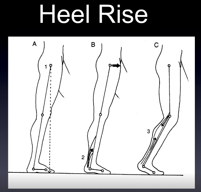
Since the foot is usually planted when it fires, we must look at its closed chain function (how it functions when the foot/insertion is fixed on the ground), which is predominantly maintenance of the medial longitudinal arch, with minor contributions to the transverse metatarsal and lateral longitudinal arches (6) ; flexion and adduction of the tarsal’s and metatarsals and eccentric slowing of anterior translation of the tibia during ankle rocker. It is also an external rotator of the lower leg and is the prime muscle which decelerates internal rotation of the tibia and pronation. As the origin and insertion are concentrically brought towards each other during early passive heel lift it becomes a powerful plantarflexor and inverter of the rearfoot.  There is also a  component of ankle stabilization via posterior compression of the tarsal’s and adduction of the tibia and fibula.

Alas, there is much more than the typical open chain functions of plantar flexion, adduction and inversion. Perhaps it is some of these other, closed chain functions, that cause the “progressive nature of posterior tibial tendon dysfunction"(7)?

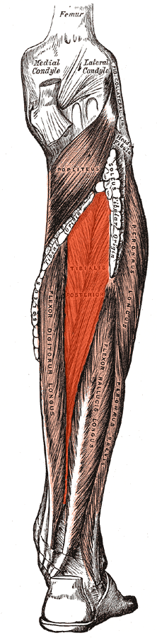
This muscle is easily accessed through the posterior compartment, lying deep in the midline to the gastroc and soleus (8,9). Care should be taken to avoid the sural nerve, often found between the heads of the gastroc and becoming superficial (and palpable) in the midline where it exits from the inferior junction of the 2 heads of the gastroc (10). The posterior tibial artery, vein and nerve lie on top of the tibialis posterior for the upper 2/3 of its route through the calf, becoming superficial (and palpable) in the distal 1/3 of the lower leg (11). I find starting laterally or medially and angling your fingers, instrument or the needle medially or laterally seems to work best. Make sure to count your layers!


1. Ling SK, Lui TH. Posterior Tibial Tendon Dysfunction: An Overview.  Open Orthop J. 2017 Jul 31;11:714-723. doi: 10.2174/1874325001711010714. eCollection 2017. link to free full text: https://www.ncbi.nlm.nih.gov/pmc/articles/PMC5620404/

2. Blake R.L., Anderson K., Ferguson H. Posterior tibial tendinitis. A literature review with case reports. J. Am. Podiatr. Med. Assoc. 1994;84(3):141–149. doi: 10.7547/87507315-84-3-141. [PubMed] [Cross Ref]

3. Supple K.M., Hanft J.R., Murphy B.J., Janecki C.J., Kogler G.F. Posterior tibial tendon dysfunction. Semin. Arthritis Rheum. 1992;22(2):106–113. doi: 10.1016/0049-0172(92)90004-W. [PubMed] [Cross Ref]

4. Semple R, Murley GS, Woodburn J, Turner DE. Tibialis posterior in health and disease: a review of structure and function with specific reference to electromyographic studies. Journal of Foot and Ankle Research. 2009;2:24. doi:10.1186/1757-1146-2-24.

link to free full text: https://www.ncbi.nlm.nih.gov/pmc/articles/PMC2739849

5. Murley GS1, Menz HB2, Landorf KB2.  Electromyographic patterns of tibialis posterior and related muscles when walking at different speeds. Gait Posture 2014 Apr;39(4):1080-5. doi: 10.1016/j.gaitpost.2014.01.018. Epub 2014 Feb

6. Kaye RA1, Jahss MH.  Tibialis posterior: a review of anatomy and biomechanics in relation to support of the medial longitudinal arch. Foot Ankle. 1991 Feb;11(4):244-7.

7. Rabbito M, Pohl MB, Humble N, Ferber R. Biomechanical and Clinical Factors Related to Stage I Posterior Tibial Tendon Dysfunction.  J Orthop Sports Phys Ther, Epub 12 July  2011.doi:10.2519/jospt.2011.3545.

 8. Howitt S, Jung S, Hammonds N. Conservative treatment of a tibias posterior strain in a novice triathlete: a case report. J Can Chiropr Assoc. 2009 Mar;53(1):23-31.   link to free full text: https://www.ncbi.nlm.nih.gov/pmc/articles/PMC2652628/

9.   Lou XF, Yang XD, Jiang SH, Sun CY, Zhang RF.[Study on angle and depth of needle insertion in acupuncture at Zusanli (ST 36)]. Zhongguo Zhen Jiu. 2006 Jul;26(7):483-6

10. https://functionalanatomyblog.com/2009/11/26/the-sural-nerve-anatomy-and-entrapment/

11. Enrique Ginzburg, ... Norman M. Rich VASCULAR ANATOMY OF THE EXTREMITIES in Current Therapy of Trauma and Surgical Critical Care, Mosby; Pages 467–472 2008

A little more on the tibialis posterior (or any tendon for that matter)....

We tend to think of the etiology of tendinopathies as being overuse or biomecanically stressful situations, which are often true, but have you thought about vascularization? My partner Dr Allen wrote a great post on vascularization in a yo yo professional here. It has to make you think...

I wrote about posterior tibial tendinopathies in a post a few days ago. A recent paper shows that vascularization can be a major player in posterior tibial tendinopathies (1). Tendon blood flow tends to decrease with age and compression, which often results from increased mechanical load. This decreased vascularization, at least theoretically, can contribuute to the probability of tissue damage as tissue compliance and flexibilty will be compromised (LER). Oxygen consumption of ligaments and tendons is 7.5% lower than skeletal muscle, which may contribute to longer healing times (2,3).

So, how can we increase vascularization?
 

  • Aerobic conditioning increases mitochondrial content, myoglobin content and capillary number, among other things, due to increased demand (4).
  • Manipulation, mobilization and massage, locally as well as segmentally, can increase blood flow, at least temporarily (5,6). The L4-S1 segments of the posterior tibial nerve are the segmental levels for the tibialis posterior .
  • How about some moist heat to cause local peripheral vasodilation?
  • I wrote a commentary on how needling can effect local vasodilation here (7), based on this article (8).
  • Tissue engineering with various techniques is an upcoming and promising field (9)
  • the early research on laser therapy (photobiomodulation) has shown some positive outcomes (10)

Finally, there is some great information about medications and current research from The Angiogenesis Foundation (11)

Adding a modality like needling or acupuncture, combined with exercise and some moist heat and perhaps some laser may help to improve your outcomes. There appears only upside potential and the methods are easy to apply and implement.


1. Geideman WM, Johnson JE. Posterior tibial tendon dysfunction. J Orthop Sports Phys Ther 2000;30(2):68‐

2. Patla CE, Abbott JH. Tibialis posterior myofascial tightness as a source of heel pain: diagnosis and treatment. J Orthop Sports Phys Ther 2000;30(10):624‐

3. Vailas AC, Tipton CM, Laughlin HL, et al. Physical activity and hypophysectomy on the aerobic capacity of ligaments and tendons. J Appl Physiol 1978;44(4):542-546.

4. https://www.cdc.gov/nccdphp/.../chap3.pdf

5.Pickar JG Neurophysiological effects of spinal manipulation. Spine J. 2002 Sep-Oct;2(5):357-71.

6. http://emedicine.medscape.com/article/324694-overview

7. http://www.rehabchalktalk.com/dry-needling-and-vascular-changes

8. BMC Complement Altern Med. 2015; 15: 72. Published online 2015 Mar 20. doi:  10.1186/s12906-015-0587-6PMCID: PMC4426539 Intensive vasodilatation in the sciatic pain area after dry needling

Lets take another look at the tibialis posterior

As cinincians , we often needle and treat the tibialis posterior for posterior tibial tendon dysfunction, platar fasicits, patellofemoral joint pain, and a host of other conditions. Lets take a look at some of the anatomy and see why it is a big player in these conditions. 

The tibialis posterior takes its origin from the proximal posterior tibia, fibula and interosseous membrane. It is deep to the tricep surae and more superior than the flexors hallucis and didgitorum longus. The tendon descends medially, travels around the medial malleolus and divides into 3 portions: plantar, main and recurrent components. It inserts into all the tarsals and metatarsals 2-4.(1) Note that it DOES NOT insert into the 1st metatarsal. There must be a good reason for this, no?

The tibialis posterior acts to plantar flex and invert the foot as well as help eccentrically control eversion of the foot. It fires from initial conact to almost terminal swing. This assists in plantart flexion of the foot from initial contact to loading response, eccentric slowing of the foot during pronation from loading response to misdstance and concentric contraction to assist in and speed up supination from midstance to terminal stance.  When you look at the EMG studies for walking (2,3) , you will see that it starts ramping down activity just after midstance as the peroneus longus starts to ramp up more (firing from just after loading response to pre swing, with a bust of activity from midstance on). 

So, with all this talk, there has to be a reason, right? Think about this. In order to move forward in the gravitational plane and have high gear push off (ie, pushing off the base of the hallux), the 1st ray needs to descend to gain purchase on the ground (2,4, 5) . This is largely through the actions of the peroneus longus, extensor hallucis brevis and flexor digitorum brevis (6,7). The function of the peroneus longus should be obvious with its attachment to the base of the 1st metatarsal. The extensor hallucis brevis moves the axis of the 1st MTP downward when it contracts, as discussed here and here (8, 9). The flexor digitorum brevis moves the axis of metatarsalphalangeal joints 2-5 dorsally and posterior which effectively moves the axis of the head of the 1st metatarsal phalangeal joint ventral and anteriorly. This is requisite for you to have adequate hallux dorsiflexion of about 60 degrees to toe off normally. 

OK, so what about the tibialis posterior? 

Remember that the tib posterior attaches to most of the proximal bottom of the foot with the exception of the 1st metatarsal base? In the area of the 1st ray, the tib posterior attaches to the navicular. When it contracts, it will pull the navicular posterior and inferior, effectively rasing the base of (and lowering the head of) the 1st metatrsal. If it attached to the 1st metatarsal, its base would be pulled posterior and inferior which would raise the head of the 1st ray, exactly what we are trying NOT TO DO

Armed with this clinical tidbit, can you see how posterior tibial tendon dysfunction can be involved with so many foot and therefore lower kinetic chain problems? If you can’t descend the 1st ray, the foot will need to toe off its lateral aspect, with less effectiveness of the calcaneocuboid locking mechanism (more on that here (10) and here (11)), so problems with propulsion off of an “unlocked” foot. Can you see how the forefoot may be somewhat more everted in this situation? Can you see how this would contribute to more calcaneal eversion and sustained midfoot pronation from midstance through the rest of the gait cycle?  What muscle is sitauted to help maintain the arch as well as decelerate pronation? Tibialis posterior. What muscle will be called into play to assist the gastroc/soleus to help propel you forward? Tibialis posterior. You get the picture.

The tibialis posterior. An important player in the gait game. A great muscle to needle thatpays clinical dividends in more ways than you can imagine. 


1. Bubra PS, Keighley G, Rateesh S, Carmody D. Posterior Tibial Tendon Dysfunction: An Overlooked Cause of Foot Deformity. Journal of Family Medicine and Primary Care. 2015;4(1):26-29. doi:10.4103/2249-4863.152245.

 2. Michaud T. Foot Orthoses and Other Forms of Conservative Foot Care. Thomas Michaud Newton, MA 1993

3. ValmasseyR. Clinical Biomechanics of the lower extremities. Mosby, St Louis, Philadelphia. 101-107: 1996

4. Inman VT, Ralston HJ, Todd F. Human Walking. Baltimore, Williams and Wilkins, 1981

5. Scranton PE, et al. Support phase kinematics of the foot.  In Bateman JE, Trott AW (eds). The Foot and Ankle. New York, Thieme-Stratton, 1980

6. Perry J. Gait Analysis: Normal and Pathological Function. Thorofare, NJ, Slack 1992

7. The Pathokinesiology Service and the Physical Therapy Department. Observational Gait Analysis. Rancho Los Amigos National Rehabilitation Center Downey, CA 2001

8. https://tmblr.co/ZrRYjxFOn2hk

9. https://tmblr.co/ZrRYjxFSJ4Yz

10. https://tmblr.co/ZrRYjx1MjeIVN

11. https://tmblr.co/ZrRYjxToM8SI

tumblr_nwjdvaVDhF1qhko2so1_1280.jpg
tumblr_nwjdvaVDhF1qhko2so2_r1_1280.jpg
tumblr_nwjdvaVDhF1qhko2so3_r1_1280.jpg

L sided medial knee pain in a TKR patient

So, why does this gals L knee hurt, posterior and medial on the left?

  • L total knee replacement 6 years ago; she thinks they used too large a size, we would have to agree.
  • occassional peripatellar discomfort
  • current pain descending hills and stairs, posterior and medial on the left

Physical findings

  • tenderness at posterior, medial aspect of knee at the top of the tibial plateau
  • positive anterior and posterior drawer +2
  • McMurrays for clicking with valgus and varus stresses
  • negative valgus/varus stress
  • all muscles test strong except for one, which one is it?

Read on…

Here is our theory:

This particular muscle fires at heel strike and again from loading response until toe off (you can look at the diagram above if needed). It also acts as an acessory posterior cruciate ligament (PCL).

Think about the forces on the knee while descending hills or stairs. The momentum will carry the femur forward (or anteriorly). There needs to be something to reststrain this; enter the PCL.

Because of the laxity (and instability), the poplitues needs to fire to take up the slack. Palpation confirmed it being tender throughout its course, with most at the tibial attachment. The attachment is largest here, so that makes sense. The muscle also tested weak.

We gave her popliteus and 1 leg balancing exercises in addition to doing acupuncture (origin/insertion work) as pictured. 5 days later she was 60% improved. She may need to return to her ortho, depending on her response to additional care.

Think about the popliteus the next time someone has posterior medial knee pain, especially when descending.