Two things we hope you see right away when viewing this video.

.

We talked about this woman in yesterday‘s post and, when viewing from the front, we wanted to bring out a few salient points

Notice how her foot progression angle is diminished bilaterally. Normally the foot will “toe out“ somewhere between 12 and 20° when the foot hits the ground and hers are nearly straightahead. This can be due to numerous things such as femoral retro torsion, internal tibial torsion, or subtalar version. All of these things will often have the foot point medially when examining a patient on the table and placing the knee in the sagittal plane. In this particular case, she has internal tibial torsion.

The next thing we would like you to look at is noting how her knee falls “outside“ the sagittal plane. In other words, instead of the knees pointing straight ahead, the point slightly out laterally. This is a cardinal sign of internal tibial torsion, especially in a gait analysis.

So what’s a clinician to do?

In this particular case, there’s nothing really to “fix“ as these are hard deformities that are often congenital. Our job is to help the patient to compensate and the best way possible so that they can remain pain and as injury free as possible. We discussed remediation in the post yesterday, So please refer back to that for what we did


Dr Ivo Waerlop, one of The Gait Guys

#invertedforefoot #forefootsupinatus #forefootvarus #pronation #forefoot #gaitanalysis #decreasedprogressionangle #toeingin

Metatarsalgia happens...

So a patient presents with forefoot pain, worse in the am upon awakening, with 1st weight bearing that would improve somewhat during the day, but would again get worse toward the end of the day and with increased activity. It began insidiously a few months ago (like so many problems do) and is getting progressively worse. Rest, ice and ibuprofen can offer some relief. You may see a dropped metatarsal head and puffiness and prominence in that area on the plantar surface of the foot, maybe not. Maybe you do a diagnostic ultrasound and see a lesion of the plantar plate as well? How did it get there? 

image courtesy of Tom Michaud: with permission

image courtesy of Tom Michaud: with permission

Lets look at the anatomy of the short flexors of the foot, as well as some biomechanics of the foot, ankle and hip. 

The flexor digitorum brevis (FDB) is innervated by the medial plantar nerve and arises from the medial aspect of the calcaneal tuberosity, the plantar aponeurosis (ie: plantar fascia) and the areas bewteen the plantar muscles. It travels distally, splitting at the metatarsal phalangeal articulation (this allows the long flexors to travel forward and insert on the distal phalanges); the ends come together to divide yet another time and each of the 2 portions of that tendon insert onto the middle of the middle phalanyx (1) 

As a result, in conjunction with the lumbricals, the FDB is a flexor of the metatarsophalangeal and proximal interphalangeal joints. In addition, it moves the axis of rotation of the metatasophalangeal joints dorsally, to counter act the function of the long flexors, which, when tight or overactive, have a tendency to drive this articulation anteriorly .Do you see any subtle extension of the metatarsophalangeal joint and flexion of the proximal interphalangeal joints on your exam?

We know that the FDB contracts faster than the other intrinsic muscles (2), playing a role in postural stability (3) and that the flexors temporally should contract earlier than the extensors (4), assumedly to move this joint axis posteriorly and allow proper joint centration. When this DOES NOT occur, the metatarsal heads are driven into the ground, causing irritation and pain.

If there is also a loss of ankle rocker this problem is made (much) worse. Why? Because, with the loss of one rocker, another must make up for the loss: ankle rocker decreases, forefoot rocker has to increase; this equals increased metatarsal head pressure. 

If you have been with us for any length of time, you know that ankle rocker and hip extension are intimately related, as one should equal the other, something we call “The “Z” angle”, that you have probably (hopefully?) read about here before. 

So what is the fix? Getting the FDB back on line for one. 

  • How about the toe waving exercise? 

  • How about the lift spread reach exercise? 

  • How about retraining ankle rocker and improving hip extension?

  • How about an orthotic with a metatarsal pad in the short term? 

  • How about some inflammation reducing modalities, like acupuncture, ice laser and pulsed ultrasound. 

  • Maybe some herbal or enzymatic anti inflammatories?


Dr Ivo Waerlop, one of The Gait Guys.

#gait #footpain #metatarsalgia #metatarsalpain #anklerocker #hipextension #thegaitguys


1. http://en.wikipedia.org/wiki/Flexor_digitorum_brevis_muscle

2. Tosovic D1, Ghebremedhin E, Glen C, Gorelick M, Mark Brown J.The architecture and contraction time of intrinsic foot muscles.J Electromyogr Kinesiol. 2012 Dec;22(6):930-8. doi: 10.1016/j.jelekin.2012.05.002. Epub 2012 Jun 27

3.Okai LA1, Kohn AF. Quantifying the Contributions of a Flexor Digitorum Brevis Muscle on Postural Stability.Motor Control. 2014 Jul 15. [Epub ahead of print]

4. Zelik KE1, La Scaleia V, Ivanenko YP, Lacquaniti F.Coordination of intrinsic and extrinsic foot muscles during walking.Eur J Appl Physiol. 2014 Nov 25. [Epub ahead of print]


Subtle clues to an LLD?

Leg length discrepancies, whether their functional anatomical, have biomechanical consequences north of the foot. This low back pain patient exhibited 2 signs. Can you tell what they are?

can you see the difference ?

can you see the difference ?

how about now?

how about now?

compare right to left

compare right to left

compare right to left

compare right to left

can you see the difference in the Q angles?

can you see the difference in the Q angles?

Look at the first picture and noticed how the left knee is hyper extended compared to the right. Sometimes we see flexion of this extremity. This is to "functionally shorten" that extremity.

Now look at the Q angles. Can you see how the left QL angle is greater than the right? This usually results from a long-term leg length discrepancy where the body is attempting to compensate by increasing the valgus angle of that knee, effectively shortening the extremity.

Dr Ivo Waerlop, one of The Gait Guys

#subtle #clues #LLD #leglengthdiscrepancy #leglengthinequality #thegaitguys #gaitabnormality

NO hip internal rotation? Forget the glutes, have you looked at the femur?

Screen Shot 2019-02-14 at 3.13.23 PM.png

Some developmental versions involve the femur. The degree of version is the angle between an imaginary line drawn through the condyles of the femur and an imaginary line drawn through the head and neck of the femur. This is often referred to as the femoral neck angle or FNA.

Beginning about the 3rd month of embryological development (Lanz and Mayet 1953) and reaches about 40 degrees (with an average of 30-60 degrees) at birth. It then decreases 25-30 degrees by adulthood to 8-20 degrees with males being at the lower and females at the upper end of the range.

The FNA angle, therefore, diminishes about 1.5 degrees a year until about 15 years of age. Femoral neck anteversion angle is typically symmetrical from the left side to the right side.

What causes torsion in the first place? By the sixth month in utero, the lumbar spine and hips of the fetus are fully flexed, so perhaps it is positional. Other sources say it coincides with the degree of osteogenesis. There is a growing consensus that muscular forces are responsible, particularly the iliopsoas  or possibly the medial and lateral hip rotators.

Additional changes can occur after birth, particularly with sitting postures. “W” sitting or “cross legged” sitting have been associated with altering the available range of motion and thus the FNA, with the range increased in the direction the hip was held in; W sitting causing increased internal rotation and antetorsion and cross legged causing external rotation and retro torsion.

There are at least 3 reasons we need to understand torsions and versions, They can alter the progression angle of gait, they usually affect the available ranges of motion of the limb and they can alter the coronal plane orientation of the limb.

1. fermoral torsions often alter the progression angle of gait.  In femoral antetorsion torsion, the knees often face inward, resulting in an intoed gait and a decreased progression angle of the foot. This can be differentiated from internal tibial torsion (ITT) by looking at the tibia and studying the position of the tibial tuberosity with respect to the foot, particularly the 2nd metatarsal. In ITT, the foot points inward while the tibial tuberosity points straight ahead. In an individual with no torsion, the tibial tuberosity lines up with the 2nd metatarsal. If the tibial tuerosity and 2nd met are lined up,  and the knees still point inward, the individual probably has femoral ante torsion. Remember that a decreased progression angle is often associated with a decreased step width whereas an increased angle is often associated with an increased step width. See the person with external tibial torsion in the above picture?

2. Femoral torsions affect available ranges of motion of the limb. We remember that the thigh leg needs to internally rotate the requisite 4-6 degrees from initial contact to midstance (most folks have 40 degrees) If it is already fully internally rotated (as it may be with femoral retro torsion), that range of motion must be created or compensated for elsewhere. This, much like internal tibial torsion, can result in external rotation of the affected lower limb to create the range of motion needed.

Femoral retro torsion results in less internal rotation of the limb, and increased external rotation.

Femoral ante torsion results in less external rotation of the limb, and increased internal rotation.

3. femoral torsions usually do not effect the coronal plane orientation of the lower limb, since the “spin” is in the transverse or horizontal plane.

The take home message here about femoral torsions is that no matter what the cause:

  •  FNA values that exist one to two standard deviations outside the range are considered “torsions”

  • Decreased values (ie, less than 8 degrees) are called “retro torsion” and increased values (greater than 20 degrees) are called “ante torsion”

  • Retro torsion causes a limitation of available internal rotation of the hip and an increase in external rotation

  • Ante torsion causes an increase in available internal rotation  of the hip and decrease in external rotation

  • Femoral ante torsion will be perpetuated by “W” sitting (sitting on knees with the feet outside the thighs, promoting internal rotation of the femur)

  • Femoral antetorsion will be perpetuated by sitting cross legged, which forces the thigh into external rotation.

Dr Ivo Waerlop, one of The Gait Guys

#gait, gait analysis, #thegaitguys, #femoraltorsion, #antetorsion, #retrotorsion

image from: http://boneandspine.com/what-is-anteversion-and-retroversion/

image from: http://boneandspine.com/what-is-anteversion-and-retroversion/

Femoral versions and torsions?

While searching for something else, we ran across this post. A pretty good lay discussion and explanation about femoral torsions. Technically, versions are NORMAL variations or limb rotations that are within accepted limits and TORSIONS are pathological, when it measures 2 or greater standard deviations from the mean and is considered pathological. Femoral versions are the angular difference between the transcondylar and transcervical axes. The femur is normally anteverted (1). 

We liked the last section talking about how to compensate for them and "acceptable" work arounds and biomechanics. 

https://b-reddy.org/2013/05/09/talking-about-hip-retroversion/

1. Staehli L in: Fundamentals of Pediatric Orthopedics Lippincott Williams & Wilkins, Jun 15, 2015 p 144

tumblr_nhdevsLYyN1qhko2so3_400.jpg
tumblr_nhdevsLYyN1qhko2so4_r1_250.jpg
tumblr_nhdevsLYyN1qhko2so1_1280.jpg
tumblr_nhdevsLYyN1qhko2so2_250.jpg

Keeping it Objective.

For clinicians and some die hard foot geeks, we often like to keep things objective. What could be more objective than an angular measurement? A few important measurements when examining or radiographing feet can give us information about clinical decision making (not that we suggest radiographs for mensuration purposes unless you are a surgeon, but when they are already available, why not put them to good use ?). When things fall outside the accepted range, or appear to be heading that way, these numbers can help guide us when to intervene. 

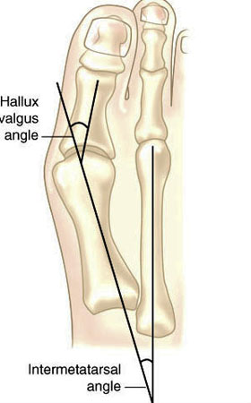
Hallux valgus refers to the big toe headed west (or east, depending on the foot and your GPS). In other words, the proximal and distal phalanyx of the great toe (hallux) have an angle with the 1st metatarsal shaft of typically > 15 degrees. This angle, called the Hallux Valgus Angle (HVA above) is used to judge severity, often for surgical intervention purposes but can guide conservative management as well. 

Metatarsus Primus Varus (literally, varus deformity of the 1st metatarsal) often accompanies Hallux Valgus. It describes medial deviation of the 1st metatarsal shaft, greater than 9 degrees. This angle is called the intermetatarsal angle and is measured by the angle formed by lines drawn parallel along the long axis of the 1st and 2nd metatarsal shafts. 

One other measurement is the Distal Metatarsal Articular Angle, which measures the angle between the metatarsal shaft and the base of the distal articular cap (ie, where the cartilage is) of the 1st metatarsal. This typically should be less than 10 degrees, preferably less than 6 degrees. Remember, these are static angles, things can change with movement, engagement, weight bearing strategies and shoes. What you see statically does not always predict dynamic angles and joint relationship.s

Are you doing surgery? Perhaps, as a last resort. Hallux valgus and metatarsus primus varus can be treated conservatively.

How do you do that?

The answer is both simple and complex.

The simple answer is: anchor the head of the 1st ray and normalize foot function. This could be accomplished by:

  • EHB exercises to descend the head of the 1st metatarsal
  • exercise the peroneus longus, to assist in descending the head of the 1st metatarsal
  • short flexor exercises, such as toe waving, to raise the heads of the lesser metatarsals relative to the 1st
  • work the long extensors, particularly of the lesser metatarsals to create balance between the flexors and extensors
  • consider using a product like “Correct Toes” to normalize the pull of the muscles and physically move the proximal and distal phalanyx of the hallux
  • wear shoes with wide toe boxes, to allow the foot to physically splay
  • consider using an orthotic with a 1st ray cut out, to help descend the head of the 1st metatarsal

This is by no means an exhaustive list and you probably have some ideas of your own. 

The complex answer is that in the above example, we have only included conservative interventions for the foot and have not moved further up the kinetic (or neurological chain). Could improving ankle rocker help create more normal mechanics? Would you accomplish this by working the anterior leg muscles, the hip extensors, or both? Could a weak abdominal external oblique be contributing? How about a faulty activation pattern of the gluteus medius? Could a congenital defect or genetic be playing a role? We have not asked “What caused this to occur in the 1st place?”

Examine your patients and clients. Understand the biomechanics of what is happening. Design a rehab program based on your findings. Try new ideas and therapies. it is only through our failures that we can truly learn.

The Gait Guys

references used:

http://www.bjjprocs.boneandjoint.org.uk/content/90-B/SUPP_II/228.3

http://www.slideshare.net/ANALISIS/hallux-valgus-2008-pp-tshare

http://www.orthobullets.com/foot-and-ankle/7008/hallux-valgus

http://www.slideshare.net/bahetisidharth/hallux-valgus-31768699?related=1

tumblr_mo8dnmvLAB1qhko2so1_1280.jpg
tumblr_mo8dnmvLAB1qhko2so2_1280.jpg

So, what kind of shoes do I put this guy in?


The answer is, well…it depends.

This gentleman has a large Q angle (need to know more about Q angles? click here). The second photo is taken from above looking down at his knee.


If he has medial (inside) knee pain (possibly from shear forces), you would want to unload the medial knee, so a more flexible shoe that would allow more pronation of the foot and INCREASE the amount of valgus would open the medial joint space and probably be more appropriate.

If he had lateral (outside) knee pain (possibly from compressive forces), then a shoe with more support (like a motion control shoe) would help to unload the lateral knee and create more space may be appropriate. And that just covers the local knee issue. What if he has a pes planus and needs more than a “more stable” shoe ? And, what if that pes planus is rigid and won’t accept a more rigid arch supporting device ? What are you gonna do then ?

The caveat?

There are no hard and fast rules AND there is no substitute for examining the person and asking LOTS of questions BEFORE putting them in a shoe. You must approach each case on a case-by-case basis with all factors brought into the fold to make the best clinical decision.  Simply watching them walk, as you have heard it over and over again here on The Gait Guys, will lead you into wrong assumptions much of the time. Sometimes the obvious fix is not possible or won’t be tolerated by the person’s foot, knee, hip or body.  So, sometimes you have to settle with something in-between. 

Need to, or dying to, know more? Take our 3 part National Shoe Fit Program and be a shoe guru!

Email us at thegaitguys@gmail.com for details.