

The Sartorius: insertional tendinitis and medial knee pain?
/We all see folks with medial knee pain, many times women, with the pain located just below the medial tibial plateau. It often results from running, but sometimes with jumping sports like basketball as well. It has been our experience that these people are often diagnosed with an MCL type injury, but when you examine them further, they do not really fit the bill. All the ligaments are stable and there is no tenderness at the joint line. The is often tenderness at the pes anserine, but who is driving the bus here?
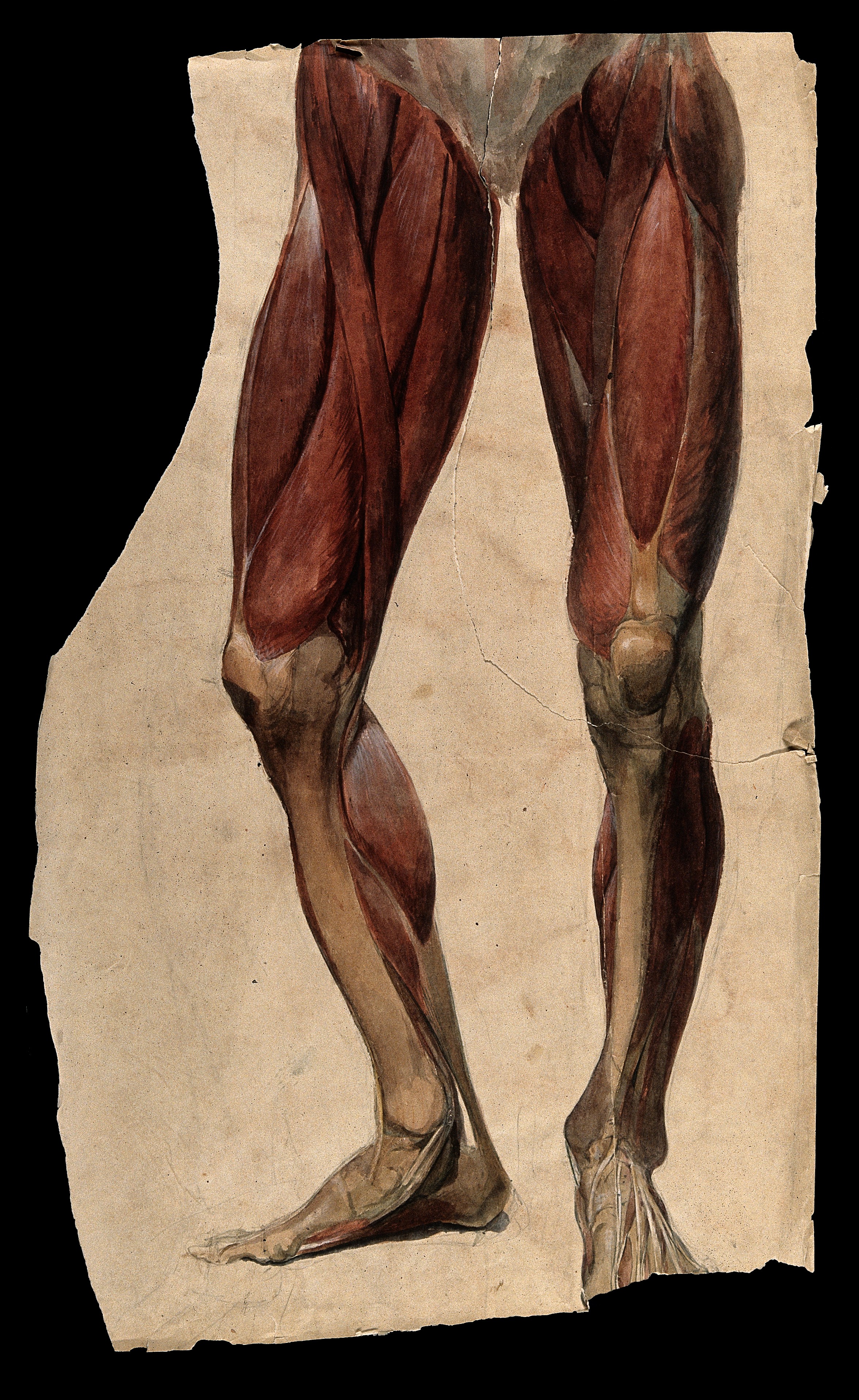
image source: https://commons.wikimedia.org/wiki/File:Muscles_and_tendons_of_the_legs_and_feet;_écorch_́figur_Wellcome_V0008276.jpg
The sartorius originates from the anterior compartment of the thigh. During an ideal gait cycle, the sartorius fires from toe off through nearly terminal swing (1)
We remember that the abdominals should initiate thigh flexion with the iliopsoas, rectus femoris, tensor fascia lata and sartorius perpetuating the motion. Sometimes, when the abdominals are insufficient, we will substitute other thigh flexors, often the psoas and/or rectus femoris, but sometimes sartorius, especially in people with excessive midfoot pronation. Think about all of the medial rotation occurring at the knee during excessive midfoot pronation and when overpronation occurs, the extra compensatory external rotation that must occur to try and bring the knee back into the sagittal plane. The sartorius is positioned perfectly for this function, along with the semitendinosus which assists and external rotation in closed chain. This is why it is often implicated as the culprit in many cases of pes anserine bursitis (or as we like to say “sartorius insertional tendinitis” (2-3)
Some other things you may find interesting is that it is utilized more in crossing or cutting maneuvers while changing directions while running (4). This makes sense, given its anatomical course and origin/insertion. It can often be overlooked in adductor strains. It can also be avulsed during sprints, particularly in adolescents (5) and because of the course of the lateral femoral cutaneus nerve beneath it, can be the cause of meralgia paresthetica (6). It is proprotionally smaller in females (along with the gracilis and short head of the biceps femoris) (7). And during vertical jumping, is considered an internal rotator, along with the semimembranosis, semitendinosis, gracilis, and popliteus (8).
The sartorius is superficial in the anterior thigh, just under the skin, running from the ASIS, coursing lateral to medial and inserting at the pes anserine at its most superior aspect, just overlying the gracilis. Since it is an external rotator, knee flexor and assists in thigh abduction, you can easliy locate it by placing the patient in a "figure 4" position and having them resist as you pull downward on the leg. Be careful if you are needling this muscle because of the subsartorial canal (ie Hunters canal) lying just beneath it in the middle 1/3 of the thigh, from the apex of the femoral triangle to the adductor hiatus in the adductor magnus. It houses the femoral artery and vein, as well as the saphenous nerve and nerve to the vastus medialis.
Michaud T: in Human Locomotion: The Conservative Management of Gait-Related Disorders 2011
Imani F, Rahimzadeh P, Abolhasan Gharehdag F, Faiz SH. Sonoanatomic variation of pes anserine bursa. Korean J Pain. 2013;26(3):249-54.
Gupta, Aman & Saraf, Abhinesh & Yadav, Chandrajeet. (2013). ISSN 2347-954X (Print) High-Resolution Ultrasonography in PesAnserinus Bursitis: Case Report and Literature Review. 1. 753-757.
Rand MK, Ohtsuki T. EMG analysis of lower limb muscles in humans during quick change in running directions. Gait Posture. 2000 Oct;12(2):169-83.
Manning CJ, Singhai S, Marshall P. Synchronised sartorius avulsions in adolescent sprinter. BMJ Case Rep. 2016 Jul 13;2016.
Hsu CY, Wu CM, Lin SW, Cheng KL. Anterior superior iliac spine avulsion fracture presenting as meralgia paraesthetica in an adolescent sprinter. J Rehabil Med. 2014 Feb;46(2):188-90. doi: 10.2340/16501977-1247.
Behan FP, Maden-Wilkinson TM, Pain MTG, Folland JP. Sex differences in muscle morphology of the knee flexors and knee extensors. PLoS One. 2018 Jan 23;13(1):e0190903.
Cleather DJ. An important role of the biarticular hamstrings is to exert internal/external rotation moments on the tibia during vertical jumping. J Theor Biol. 2018 Oct 14;455:101-108


