Stretching part 2

So, how can we utilize this reflex? 

How about to activate a weak or lengthened muscle?

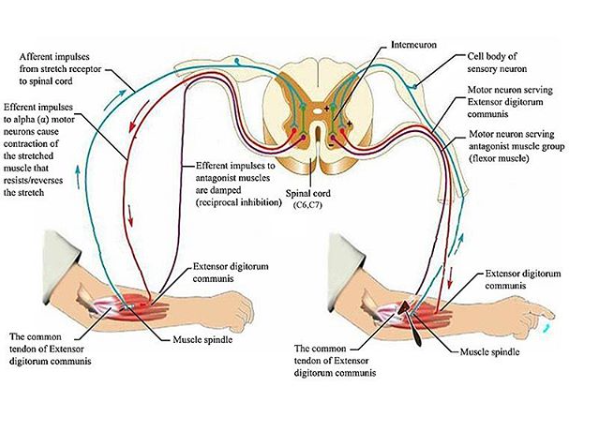
Did you notice the other neuron in the picture? There is an axon collateral coming off the Ia afferent that goes to an inhibitory interneuron, which, in turn, inhibits the antagonist of what you just stretched or activated. So if you acitvate one muscle, you inhibit its antagonist, provided there are not too many other things acting on that inhibitory interneuron that may be inhibiting its activity. Yes, you can inhibit something that inhibits, which means you would essentially be exciting it. This is probably one of the many mechanisms that explain spasticity/hypertonicity

Screen Shot 2020-07-30 at 10.18.23 AM.png


How can we use this? How about to inhibit a hypertonic muscle?
Lets take a common example: You have hypertonic hip flexors. You are reciprocally inhibiting your glute max. You stretch the hypertonic hip flexors, they become more hypertonic (but it feels so good, doesn’t it?) and subsequently inhibit the glute max more. Hmm. Not the clinical result you were hoping for?
How about this: you apply slow stretch to the glutes (ie “reverse stretch”) and apply pressure to the perimeter, both of which activate the spindle and make the glutes contract more. This causes the reciprocal inhibition of the hip flexors. Cool, eh? Now lightly contract the glutes while you are applying a slow stretch to them; even MORE slow stretch; even MORE activation. Double cool, eh?

Don’t believe me, try this on yourself, your clients, patients, willing family members and pets.

Image credit: https://commons.wikimedia.org/wiki/File:The_extensor_digitorum_reflex.jpg

Stretching part 1

Stretching secrets you need to know

OK, maybe they aren’t secrets, but these are some ‘shortcuts” you should know. 

We know from studies out there that static stretching can lengthen muscle (actually add sarcomeres), but you need to do it 30 minutes per day per muscle group. How about something a little faster doc?

How about taking advantage of the stretch reflex and reciprocal inhibition; or the “reverse stretch”

Screen Shot 2020-07-30 at 10.17.35 AM.png


Reciprocal inhibition is a topic we have spoken about before on our blog, social media and our PODcasts. 1st described in1923 by Sherrington, this diagram sums it up nicely. Note the direct connection from the spindle to the alpha motor neuron, which is via a Ia afferent fiber. When the spindle is stretched, and the pathway is intact, the muscle will contract. This its a straight forward stretch or inverse myotatic reflex. 

Remember, ANY kind of stretch or anything that changes the length of the spindle will effect it. So what happens when you do a nice, long, slow stretch? You ACTIVATE the spindle, which activates the alpha motor neuron. If you stretch long enough, you may fatigue the reflex. So why do we give folks long, slow stretches to perform? Good question! Certainly not to “relax” the muscle!

So, how can we utilize this reflex? Read our next post tomorrow!


Image credit: https://commons.wikimedia.org/wiki/File:The_extensor_digitorum_reflex.jpg

Gaining Anterior Length, Through Posterior Strength. A Lesson in Reciprocal Inhibition

tumblr_oco89rcuI61qhko2so2_1280.png
tumblr_oco89rcuI61qhko2so1_1280.png

Gaining Anterior Length, Through Posterior Strength and vice versa….A Lesson in Reciprocal Inhibition

I found a really cool article, quite by accident. I was leafing through an older copy of one of, if not my favorite Journals “Lower Extremity Review” and there it was. An article entitled “Athletes with hip flexor tightness have reduced gluteus maximus activation”. Wow, I thought! Now there is a great article on reciprocal inhibition! This reminded me of a piece we wrote some time ago

What is reciprocal inhibition, also called “reciprocal innervation” you ask? The concept, was 1st observed as early as 1626 by Rene Descartes though observed in the 19th century, was not fully understood and accepted until it earned a Nobel prize for its creditor, Sir Charles Sherrington, in 1932.

Simply put, when a muscle contracts, its antagonist is neurologically inhibited (see the diagram above) When your hip flexors contract, your hip extensors are inhibited. This holds true whether you actively contract the muscle or if the muscle is irritated in some manner, causing contraction. The reflex has to do with muscle spindles and Type I and Type II afferents which I have covered in an article I wrote some time ago.

We can (and often do) take advantage of this concept with treating the bellies of hip flexors (iliopsoas, tensor fascia lata, rectus femoris, iliacus, iliocapsularis) and extensors (gluteus maximus, posterior fibers of gluteus medius). This is especially important in folks with low back pain, as they often have increased psoas activity and cross sectional area, especially in the presence of degenerative changes.

There also appears to be a correlation between decreased hip extension and low back pain, with a difference of as little as 10 degrees being significant. Take the time to do a thorough history and exam and pay attention to hip extension and ankle dorsiflexion as they should be the same, with at least 10 degrees seeming to be the “clinical” minimum. Since the psoas should only fire at the end of terminal stance/preswing and into early swing, problems begin to arise when it fires for longer periods.

Can you see now how taking advantage of reciprocal inhibition can improve your outcomes? Even something as simple as taping the gluteus can have a positive effect! Try this today or this week in the clinic, not only with your patients hip flexors, but with all muscle groups, always thinking about agonist/antagonist relationships.


In the moment: Sports medicine  Jordana Bieze Foster: Athletes with hip flexor tightness have reduced gluteus maximus activation  Lower Extremity review Vol 6, Number 7 2014

https://tmblr.co/ZrRYjx1VG3KYy

Mills M, Frank B, Blackburn T, et al. Effect of limited hip flexor length on gluteal activation during an overhead squat in female soccer players. J Athl Train 2014;49(3 Suppl):S-83.

Ciuffreda KJ, Stark L.  Descartes’ law of reciprocal innervation. Am J Optom Physiol Opt. 1975 Oct;52(10):663-73.
Jacobson M Foundations of Neuroscience Springer Science and Business Media, Plenum Press, NY 1993 p 277

http://www.nobelprize.org/nobel_prizes/medicine/laureates/1932/sherrington-bio.html

https://thegaitguys.tumblr.com/post/9708399904/ah-yes-the-ia-and-type-ii-afferents-one-of-our

Arbanas J, Pavlovic I, Marijancic V, et al MRI features of the psoas major muscle in patients with low back pain. Eur Spine J. 2013 Sep;22(9):1965-71. doi: 10.1007/s00586-013-2749-x. Epub 2013 Mar 31.

Roach SM, San Juan JG, Suprak DN, Lyda M, Bies AJ, Boydston CR. Passive hip range of motion is reduced in active subjects with chronic low back pain compared to controls. Int J Sports Phys Ther. 2015 Feb;10(1):13-20. Erratum in: Int J Sports Phys Ther. 2015 Aug;10(4):572.

Paatelma M Karvonen E Heiskanen J Clinical perspective: how do clinical test results differentiate chronic and subacute low back pain patients from “non‐patients”? J Man Manip Ther. 2009;17(1):11‐19.[PMC free article] [PubMed]

Evans K Refshauge KM Adams R Aliprandi L Predictors of low back pain in young adult golfers: a preliminary study. Phys Ther Sports. 2005;6:122‐130.

Mellin G Correlations of hip mobility with degree of back pain and lumbar spinal mobility in chronic low‐back pain patients. Spine. June 1988;13(6):668‐670. [PubMed]

Lewis CL, Ferris DP. Walking with Increased Ankle Pushoff Decreases Hip Muscle Moments. Journal of biomechanics. 2008;41(10):2082-2089. doi:10.1016/j.jbiomech.2008.05.013.

Nodehi-Moghadam A, Taghipour M, Goghatin Alibazi R, Baharlouei H. The comparison of spinal curves and hip and ankle range of motions between old and young persons. Medical Journal of the Islamic Republic of Iran. 2014;28:74.

Daniel Moon , MD, MS; Alberto Esquenazi , MD Instrumented Gait Analysis: A Tool in the Treatment of Spastic Gait Dysfunction JBJS Reviews, 2016 Jun; 4 (6): e1. http://dx.doi.org/10.2106/JBJS.RVW.15.00076

Kilbreath SL, Perkins S, Crosbie J, McConnell J. Gluteal taping improves hip extension during stance phase of walking following stroke. Aust J Physiother. 2006;52(1):53-6.

Taking advantage of the stretch reflex and reciprocal inhibition; or the “reverse stretch”Reciprocal inhibition is a topic we have spoken about before on the blog (see here). The diagram above sums it up nicely. Note the direct connection from the s…

Taking advantage of the stretch reflex and reciprocal inhibition; or the “reverse stretch”

Reciprocal inhibition is a topic we have spoken about before on the blog (see here). The diagram above sums it up nicely. Note the direct connection from the spindle to the alpha motor neuron, which is via a Ia afferent fiber.  When the spindle is stretched, and the pathway is intact, the uscle will contract. What kind of stimulus affects the spindle? A simple “stretch” is all it takes. Remember spindles respond to changes in length. So what happens when you do a nice, slow stretch? You activate the spindle, which activates the alpha motor neuron. If you stretch long enough, you may fatigue the reflex. So why do we give folks long, slow stretches to perform? Certainly not to “relax” the muscle!

How can we “use” this reflex? How about to activate a weak or lengthened muscle? Good call.

Did you notice the other neuron in the picture? There is an axon collateral coming off the Ia afferent that goes to an inhibitory interneuron, which, in turn, inhibits the antagonist of what you just stretched or activated. So if you acitvate one muscle, you inhibit its antagonist, provided there are not too many other things acting on that inhibitory interneuron that may be inhibiting its activity. Yes, you can inhibit something that inhibits, which means you would essentially be exciting it. This is probably one of the many mechanisms that explain spasticity/hypertonicity

How can we use this? How about to inhibit a hypertonic muscle?

Lets take a common example: You have hypertonic hip flexors. You are reciprocally inhibiting your glute max. You stretch the hypertonic hip flexors, they become more hypertonic (but it feels so good, doesn’t it?) and subsequently inhibit the glute max more. Hmm. Not the clinical result you were hoping for?

How about this: you apply slow stretch to the glutes (ie “reverse stretch”) and apply pressure to the perimeter, both of which activate the spindle and make the glutes contract more. This causes the reciprocal inhibition of the hip flexors. Cool, eh? Now lightly contract the glutes while you are applying a slow stretch to them; even MORE slow stretch; even MORE activation. Double cool, eh?

Try this on yourself. Now go try it on your clients and patients. Teach others. Spread the word.

Reciprocal Inhibition anyone? Thanks to The Manual Therapist (Erson Religioso) for this great post.

What they are doing here is taking advantage of what Sherrington know many years ago. Activating a muscle (agonist for a movement) will inhibit the muscle with the opposite action (antagonist for a movement), through a disynaptic, post synaptic pathway. It is a great way to gain additional movement and remove or reduce muscular inhibition. Try it!

Making your stretching more effective. 
While I was making linguine and clam sauce for my family, one of my favorite foods that I haven’t had in quite some time( and listening to Dream Theater of course) I was thinking about this post.  Then I remem…

Making your stretching more effective. 

While I was making linguine and clam sauce for my family, one of my favorite foods that I haven’t had in quite some time( and listening to Dream Theater of course) I was thinking about this post.  Then I remembered about voice recognition on my iMac.  Talk about multitasking!

What do you agree that stretching is good or not, you or your client still may decide to do so possibly because of the “feel good” component. Make sure to see this post here on “feel good”  part from a few weeks ago. 

If you do decide to stretch, make sure you take advantage of you or your clients neurology.  There are many ways to do this. One way we will discuss today is taking advantage of what we call myotatic reflex.

The myotatic reflex is a simple reflex arc. The reflex begins at the receptor in the muscle (blue neuron above) : the muscle spindles (nuclear bag or nuclear chain fibers). This sensory (afferent) information then travels up the peripheral nerve to the dorsal horn of the spinal cord where it enters and synapses in the ventral horn on an alpha motor neuron.  The motor neuron (efferent) leaves the ventral horn and travels back down the peripheral nerve to the contractile portion of the myfibrils (muscle fiber) from which the the sensory (afferent) signal came (red neuron above).  This causes the muscle to contract. Think of a simple reflex when somebody taps a reflex hammer on your tendon. This causes the muscle to contract and your limb moves.

Nuclear bag and nuclear chain fibers detect length or stretch in a the muscle whereas Golgi Tendon organs tension. We have discussed this in other posts here.   With this in mind, slow stretch of a muscle causes it to contract more, through the muscle spindle mechanism.

Another reflex that we should be familiar with is called reciprocal inhibition. It states simply that when one muscle (the agonist) contracts it’s antagonist is inhibited (green neuron above).  You can find more on reciprocal inhibition here.

Take advantage of both of these reflexes?   Try this:

  • do a calf stretch like this: put your foot in dorsiflexion, foot resting on the side of the doorframe.
  • Keep your leg straight.
  • Grab the the door frame with your arms and slowly draw your stomach toward the door frame. 
  • Feel the stretch in your calf; this is a slow stretch. Can you feel the increased tension in your calf? You could fatigue this reflex if you stretched long enough. If you did, then the muscle would be difficult to activate. This is one of the reasons stretching seems to inhibit performance. 
  • Now for an added stretch, dorsiflex your toes and try to bring your foot upward.  Did you notice how you can get more stretch your calf and increased length? This is reciprocal inhibition at work!

There you have it, one neurological tool of many to give you increased length.The next time you are statically stretching, take  advantage of these reflexes to make it more effective.

 The Gait Guys. Teaching you more  about anatomy, physiology, and neurology with each and every post. 

image from :www.positivehealth.com

tumblr_m2j60zuBR31qhko2so1_1280.jpg
tumblr_m2j60zuBR31qhko2so2_r2_1280.jpg

Neuromechanics Weekly: PART 2:

Stretching increases strength in contralateral muscles?

Lots of cool links in this post. please try and find time to check them out.

Figure it out?  Ever wonder about some of the magic behind some of those manual therapy techniques that are out there ? Sometimes it is not magic at all !

There are 2 related reasons we can think of to cause this seemingly odd length-strength phenomenon (OK, there are more, but this is what we are going to cover today):

  • Reciprocal Inhibition
  • Crossed extensor reflexes/responses

We remember reciprocal inhibition (as demonstrated in LEFT picture above) is when we activate or stimulate a muscle, the Ia afferent from that muscle stimulates that same muscle to contract (this is how a simple reflex arc works) and, through an inhibitory interneuron, inhibits the antagonist muscle on the opposite side of the joint.

Remember, that Ia afferents go to muscle spindles (don’t remember? look here); they respond to LENGTH changes. Wouldn’t you say stretching affects length? If we were talking about the R tricep surae group, we would be inhibiting the R anterior compartment.

But wait, the article said it affects the opposite side….Of course, there is more…

The picture on the right shows the crossed extensor response or reflex (don’t remember? look here). In a nutshell, when you FIRE the flexors on one side, you INHIBIT the extensors on the same side (sound like reciprocal inhibition? It should… it is : ) You also FIRE the extensors on the opposite side while INHIBITING the flexors on the opposite side. (Yes, the opposite side extensors will inhibit the opposite side flexors as well. Yes, this is also reciprocal inhibition).

But wait, that means the opposite calf would be weaker, not stronger, right?

It would be weaker if being called upon to be used at that moment in time, BUT in the study, stretching increased ROM of the stretched calf 8%, with a 1% loss of ROM of the opposite calf (study summary).

Hmm… sounds like shortening to me. That would mean that those spindles (ie the opposite calf)  would be MORE RESPONSIVE to stretch (ie a change in length; and coincidentally, the Golgi’s more responsive to the tension change) . And what happens when we preload a neuronal pool? The likelihood of firing is increased (like doing a Jendrassik maneuver to increase a reflex). The rest is neural adaptation (strength gains initially are due to increased efficiency of the nervous system. For a review to see our video on this, click here)

Interesting that one of the comments on the article was “I don’t have the full text of the paper but a summary prepared by Chris Beardsley and Bret Contreras states that one of the mechanisms for crossover in the case of unilateral strength training is thought to be modulation at the spinal cord level.”   Could they be talking about reciprocal inhibition and crossed extensor responses?

Wow! Very cool! And to think, you knew the answer. We are proud of you!

Ivo and Shawn…Neuro Geeks too!  And applying it to gait, running and motor patterns of all types !