Curly toes?

-We have all seen them. We like them, we hate them, we despise them, we scratch our heads.

-The question becomes why?

IMG_8467 (1).jpg

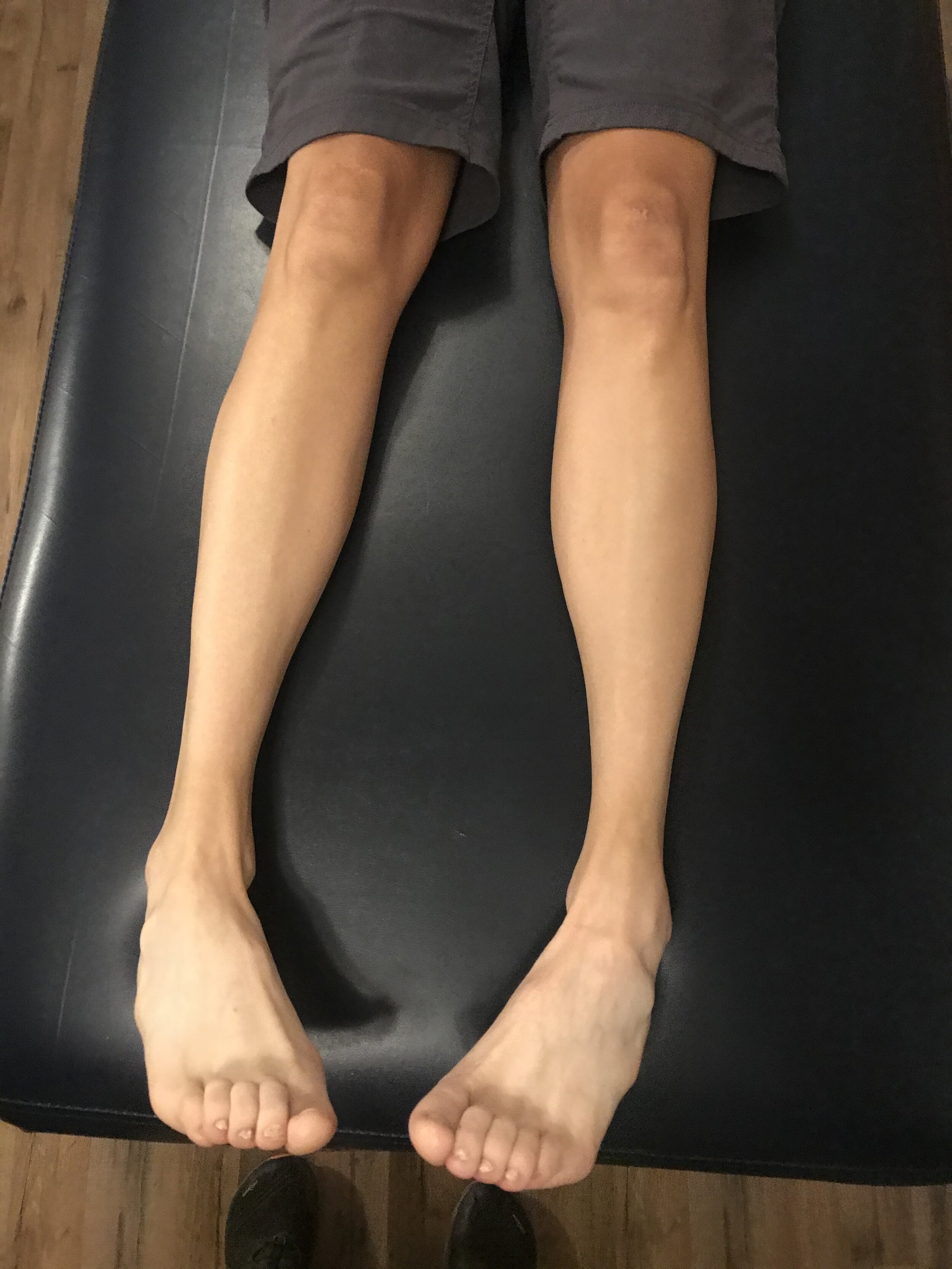
-it’s pretty obvious from the picture that there is dominance of the flexor muscles and not enough intrinsic strength in the extensor muscles. Look at the prominence of the extensor tendon‘s and posturing of the toes.

– Flexor dominance occurs essentially because of too much activity in the central nervous system, particularly the lower brainstem, over activating the flexors and shutting down (reciprocally inhibiting) the extensors.

-This doesn’t necessarily mean that it is a neurological problem however the nervous system is what’s driving the bus here. Extensor tone is largely regulated by the cerebellum and vestibular system with the flexor tone being regulated by the cortex as well as lower, sub cortical systems.

– The cerebellum and vestibular system get the majority of their input from joint and muscle And joint mechanoreceptors as well as the vestibular apparatus. Their output is predominantly to axial extensor muscles as well as muscles which would be directly affected, from a gravitational standpoint, from those systems as well.

– When we don’t have enough afferent information traveling in from these systems, the flexor systems have a tendency to predominate. Think about protective posture’s and DNS work.

Driving the extensors and working on posture/balance/coordination and perhaps long, sustained stretching of the flexor musculature can help to end the bane of curly toes. 

–so let’s go ahead and make those feet, lower extremity, lower kinetic chain muscles and joints and core more competent and help these folks out.

#curlytoes #flexordominance #toeproblem #toeproblems #footproblem #footproblems 

Short leg and Pronation

Dr Allen was ON FIRE on tonites onlinece.com lecture Biomechanics 322). Hope you will join us again (or next time if you missed us). We talked about many of the aspects of a static exam and how it effects weight bearing in the foot. The word "short leg" came up more than once, and yes, from Dr Allen : )

Remember, as the foot pronates more on one side, the center of gravity will move medially. You will often see more toe clenching (and resultant quadratus plantae weakness) on the more pronatory side and more toe elongation on the more supinatory side. You will often also see more splay and elongation on the pronatory side, and less elongation and less splay on the supinatory side. Remember, these are guidelines and not rules, and there are ALWAYS exceptions.

Screen Shot 2018-01-27 at 8.00.13 AM.png

Ankle Rocker Revisited....

How many times have we talked about ankle rocker and its importance? So how are YOU measuring ankle rocker? Are you looking at it on the table? On the ground? Weight bearing? Knee flexed or extended (or both?). The knee is extended at initial contact, flexes through midstance, extends at terminal stance and pre swing and flexes again during swing phase until extending at the end of terminal swing for initial contact again.

What you see on the table may not (and many times doesn't)  translate to real life. Someone with limited ankle dorsiflexion non weight bearing may have normal amounts during gait and vice versa. With gravity in place and a functioning (or malfunctioning) vestibular system, things can change rapidly. Remember that the vestibular system drives the extensors and if inhibited, you will often have flexor dominance. Talk about a tight gastroc/soleus group!


"These findings indicate that nonweightbearing and weightbearing measurements of ankle DF PROM with knee extension should not be used interchangeably and that weightbearing ankle DF PROM with the knee extended is more appropriate for estimating ankle DF during gait."

Kang MH, Oh JS. Relationship Between Weightbearing Ankle Dorsiflexion Passive Range of Motion and Ankle Kinematics During Gait. J Am Podiatr Med Assoc. 2017 Jan;107(1):39-45. doi: 10.7547/14-112.


"There is no relationship between a static diagnosis of ankle dorsiflexion at 0° with dorsiflexion during gait. On the other hand, those subjects with less than -5° of dorsiflexion during static examination did exhibit reduced ankle range of motion during gait."


Gatt A, De Giorgio S, Chockalingam N, Formosa C. A pilot investigation into the relationship between static diagnosis of ankle equinus and dynamic ankle and foot dorsiflexion during stance phase of gait: Time to revisit theory? Foot (Edinb). 2017 Mar;30:47-52. doi: 10.1016/j.foot.2017.01.002. Epub 2017 Feb 6.


Subtle Clues to Ankle Rocker Pathology: How good are your powers of observation ?

There are clues showing you there is motor pathology to ankle dorsiflexion, if you are paying close enough attention.

When we see motor pathology in ankle dorsiflexion we immediately begin to think about impairment to hip extension range of motion, gluteal strength, motor coordination and many other issues.

Here is a simple case. Observation skills are your greatest superpower when it comes to figuring out many gait and movement problems. But, you have to know what to look for and know what they mean before you can even hope to know how to fix things.
This is a simple video. It shows active ankle dorsiflexion in supination. We asked the client (a runner with right heel and persistent sesamoid pain following a healed sesamoid fracture) to perform simple ankle dorsiflexion. This is what we saw.

It should be clear to the observer that the end of the video shows attempted right dorsiflexion pulls the 2-5 toe extensors into the pattern quite aggressively and as a dominating faction. One can see toe abduction and extension with surprisingly little help from the long hallux toe extensor (EHL).  Dorsiflexion also fatigued early on the right. There is only one reason that the lesser toe extensors (EDL & EDB) are being over recruited, it’s because the EHL and tibialis anterior are weak and/or inhibited or have been pattern corrupted for one reason or another. Depending on this smallest of anterior compartment muscles over the EHL and tib anterior will mean that ankle rocker (dorsiflexion) is impaired. It also means that abnormal forefoot valgus posturing is expected (we could make a case for valgus or varus depending on other variables present). Passive ROM assessment confirmed the impaired ankle rocker with barely greater than 90 degrees ankle dorsiflexion ROM. This impairment will possibly do many things including:

  • premature heel rise
  • premature gastrocsoleus engagement
  • accentuated rear foot eversion (Rearfoot pronation)
  • midfoot pronation
  • strain of plantar fascia
  • premature forefoot loading response (strong clue for clients sesamoid fracture and persistent pain)
  • anterior/ posterior shin splints
  • hallux VALgus /bunion formation
  • long toe flexor dominance and many other things.

This clinical find plays nicely into the clients multiple symptoms (plantar pain and sesamoid problems) and functional gait pathology.
Restoring proper motor hierarchy and synchrony to the ankle dorsiflexion team (tib anterior, peroneus tertius, EDL, EHL) will reduce the need for solitary group overuse and impart forces where they should be when they need to be present. Impair the synchrony and problems ensue.

Help your client achieve the motion at the ankle mortise and they do not have to pass the buck into the foot.  Always test for skill, endurance and strength. Endurance is the most often forgotten assessment.  If endurance is lost early, the brain will begin to block out that end range of motion because it cannot be trusted, and thus posterior compartment tightness will be detected. This is an often common source of regional achilles and para-achilles tendonopathy. If your clients symptoms take time during activity to develop looking at the endurance of motor patterns may give the clue to your solution. 

Simple case, but you have to know your normal gait parameters, know functional anatomy and know how impaired mechanics factor into injury. 

Shawn and Ivo

The gait guys

tumblr_n06qsgxeEf1qhko2so1_1280.jpg
tumblr_n06qsgxeEf1qhko2so2_1280.jpg
tumblr_n06qsgxeEf1qhko2so3_1280.jpg

What do you do with these Dogs?

Take a good look at these feet. Hard to not cringe, we know. In this photo, the gentleman’s feet are relaxed! Imagine what it they will look like with some additional long flexor tone!

So, keeping in mind his tibial varum (bend in the tibia) and uncompensated forefoot varus (inability to get the head of his 1st ray down to the ground), what can we do?

  • how about we increase extensor strength? He could do the lift, spread, reach exercise while tripod standing. He could do the toe waving exercise.   He could do shuffle walks.
  • teach him to stretch his long toe flexors. Frequently. 20-30 mins minimum; daily
  • you could manipulate his feet to ensure better biomechnics
  • you could massage his feet to improve mobility and circulation
  • you could facilitate his long toe extensor muscles
  • you could inhibit his long toe flexor muscles
  • you could improve ankle dorsiflexion by showing him how to stretch the calves, 20-30 mins daily
  • you could improve ankle dorsiflexion by making sure he has adequate hip extension
  • he could wear correct toes, to improve the biomechanical advantage of the long toe extensors
  • he could wear shoes with a wider toe box
  • he could wear shoes with less ramp delta (or drop)
  • he could wear shoes with less torsional rigidity

and the list goes on. There are many simple things you teach a person with feet like this. many of them we have introduced you to here on the blog. Spend some time. Learn some cool stuff. Read the blog. Follow us on Facebook. Attend a Biomechanics class we teach the 3rd Wednesday of each month on onlinece.com . Check out our Youtube Channel. Consider furthering your education and taking the National Shoe Fit Program.

The resources are there. All you need to do is dig a little deeper.

We are The Gait Guys and we are all things gait.

The Power of Observation: Part 2

Let’s take a closer look at yesterdays post and the findings. If you are just picking up here, the post will be more meaningful if you go back and read it. 


The following are some explanations for what you were seeing:

torso lean to left during stance phase on L?

if he has a L short leg, he will need to clear right leg on swing phase. We have spoken of strategies around a short leg in another post. This gentleman employs 2 of the 5 strategies; torso lean is one of them

increased progression angle of both feet?

Remember he has femoral retroversion. You may have read about retrotorsion here. He has limited internal rotation o both thighs and must create the requisite 4-6 degrees necessary to walk. He does this by spinning his foot out (rotating externally).

decreased arm swing on L?

This is most likely cortical, as he seems to have decreased proprioception on both legs during 1 leg standing. Proprioception feeds to the cerebellum, which in turn fires axial extensors through connections with the vestibular system. Diminished input can lead to flexor dominance (and extensors not firing). Note the longer stride forward on the right leg compared to the left with less hip extension (yes, we know, a side view would be helpful here).

circumduction of right leg?

This is the 2nd strategy for getting around that L short leg.

clenched fist on L?(esp when standing on either leg)

see the decreased arm swing section. This is a subtle sign of flexor dominance, which appears to be greater on the right.

body lean to R during L leg standing?

This is again to compensate for the L short leg. He has very mild weakness of the left hip abductors as well, more when moving or using them in a synergistic fashion (ie functional weakness) than to manual testing.

Well, what do you think? Now you can see how important the subtle is and that gait analysis may complex than many think.

We are and we remain, the Geeky Guru’s of Gait: The Gait Guys

tumblr_mo22vpR5zv1qhko2so1_1280.jpg
tumblr_mo22vpR5zv1qhko2so2_1280.jpg
tumblr_mo22vpR5zv1qhko2so3_1280.jpg

So, what’s in a test? The standing tripod test

Many of you probably recognize this as the standing tripod test (see here for video of standing tripod exercise). You have the individual stand on both foot tripods (center of calcaneus, head of 1st metatarsal and head of 5th metatarsal). Then have the person lift one leg and remain on the other tripod. This individual was featured in last thurdays post.

watch for:
collapse of arch
body lean
hip sway
falling to either side
spontaneous combustion (OK, this is a RARE complication).

What do we see here?

top picture, L leg

  • collapse of arch
  • forefoot eversion
  • valgus angulation of knee
  • pelvic shift to L
  • arm moves to compensate on right

middle picture, R leg

  • mild collapse of arch
  • pronounced pelvic shift to left
  • body lean to R
  • compensatory arm movement on L

Bottom picture

  • note the pronounced appearance of the head of the 1st met on the L foot
  • bilateral hallux abducto valgus most likely means bilateral uncompensated forefoot varus
  • more hammering (flexion) of digits on the R foot
  • note the prominence of the tail or tubercle of the 5th metatarsal on the L foot

Some questions for you:

Q: why does he have a pelvic shift to the left in both r and L leg standing?

A: look at the feet. He is able t make a better tripod on the L foot, probably because of the prominence of the head of the 1st metatarsal. also note the valgus angulation of the knee, which helps to shift the center of mass to the midline. this is most likely a long term compensation

Q: Why does he have more body lean to the R during r leg standing?

A: see previous question AND he probably has weaker hip abductor muscles on the right

Q: did you notice that the hand and forearm were more supinated in the top (L standing) picture than the middle (r standing) picture (where he is more pronated)? What gives?

A: Wow, this is some subtle stuff, eh? Look to the brain. remember coordinate arm swing? (if not, look here and here) Supination accesses more of the extensors of the arm and pronation more of the flexors. When we have less extensor activity (remember flexor dominance? if not, click here) you have a tendency to use your flexors more to compensate (you use what you have available to you). It appears that he has a much tougher time standing on his r leg (judging from the increased compensation)

Q: Wow,  nice floors! Are they hardwood?

A: No, laminate

The Gait Guys. Helping you help others each and every post. Keep your eyes and your mind open : )