tumblr_ni72j0wHZ11qhko2so2_1280.jpg
tumblr_ni72j0wHZ11qhko2so1_1280.jpg
tumblr_ni72j0wHZ11qhko2so3_1280.jpg
tumblr_ni72j0wHZ11qhko2so4_1280.jpg
tumblr_ni72j0wHZ11qhko2so5_1280.jpg

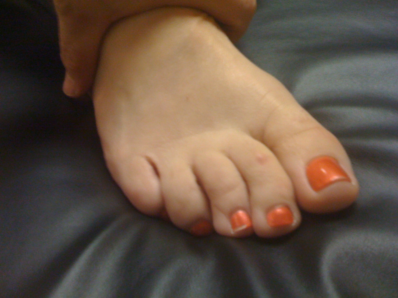
A clear cut case of Form follows Function.  Leave a deforming force long enough and the body will accommodate. 

When the lateral quadratus plantae (QP) is weak and the flexor digitorum longus pulls unopposed (relying on the QP to properly orient the long flexor pull) for too long the 4th and 5th toes and drift medially and spin inwards toward the midline of the foot (as seen in the photo). Then, as the 4th toe presses down on the fleshy pad of the 5th toe, over time the fleshy pad is pancaked and triangulated. Then, with repeated pressure a corn like hardness becomes of the tip of that triangluted tissue, it resembles a hard callus. A corn is a coalescing of the skin cells into a tighter formation, a reaction to fend off repeated pressure and friction.  Form follows prolonged function.  Shave these things down and they will come back, unless you get to the root source of the problem, which could be all the way up the chain. 

-Dr. Allen

tumblr_nozmmaOxAI1qhko2so1_500.png
tumblr_nozmmaOxAI1qhko2so2_540.png

Ahh yes, the lumbricals. 

One of our favorite muscles. And here it is in a recent paper! This one is for all you fellow foot geeks : )

Perhaps the FDL (which fires slightly earlier than the FHL) and FHL (which fires slightly later and longer) at loading response, slowing pronation and setting the stage for lumbrical function from midstance to terminal stance/preswing (flexion at the metatarsal phalangeal joint (it would have to be eccentric, if you think about this from a closed chain perspective) and extension (actually compression) of the proximal interphalangeal joints.

“The first lumbrical arose as two muscle bellies from both the tendon of the FDL and the tendinous slip of the FHL in 83.3 %, and as one muscle belly from the tendon of the FDL or the tendinous slip of the FHL in 16.7 %. These two muscle bellies subsequently merged to form the muscle belly of the first lumbrical. The second lumbrical arose from the tendinous slips of the FHL for the second and third toes as well as the tendon of the FDL in all specimens. The third lumbrical arose from the tendinous slips of the FHL for the third and fourth toes in 69.7 %, and the fourth lumbrical arose from the tendinous slip of the FHL for the fourth toe in 18.2 %. Some deep muscle fibers of the fourth lumbrical arose from the tendinous slip of the FHL for the second toe in 4.5 %, for the third toe in 28.8 %, and for the fourth toe in 15.2 %.”

Hur MS1, Kim JH, Gil YC, Kim HJ, Lee KS. New insights into the origin of the lumbrical muscles of the foot: tendinous slip of the flexor hallucis longus muscle. Surg Radiol Anat. 2015 May 12. [Epub ahead of print]

tumblr_mwzu9gTBzS1qhko2so1_1280.jpg
tumblr_mwzu9gTBzS1qhko2so2_400.gif
tumblr_mwzu9gTBzS1qhko2so3_r1_1280.jpg
tumblr_mwzu9gTBzS1qhko2so4_r1_1280.jpg

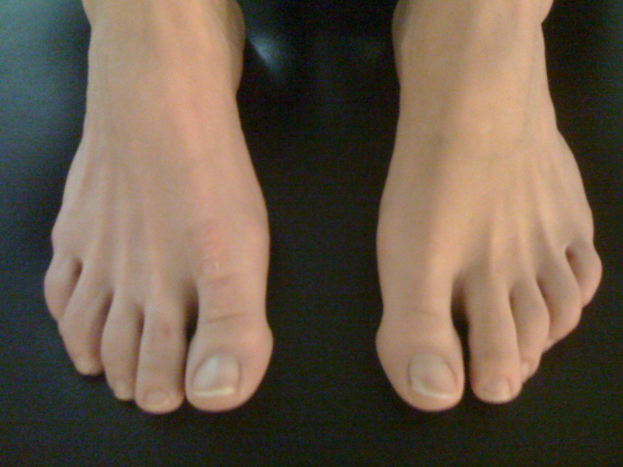
Is your 5th toe curled under ? What do you do when “this little piggy” can’t go wee wee wee all the way home.

Have a look at the 4 photos above.  You will see this curling of the lesser toes quite often in your practice, and when you know what it means it can help to guide your thinking, both from a diagnostic and treatment perspective.  

You should have noticed in the photos that the 4th and 5th toes curl under and are hyper-flexed, and this is at rest.  So, what does this mean ?

It means that the long flexors are overactive, the extensors are underactive, and the adduction pull of the long flexors is unopposed by the under appreciated quadratus plantae muscle.

Look at the clinical drawing. The quadratus plantae has 2 heads, a medial head and a lateral head.  Being able to clinically test these two heads will give you much insight into the function of the foot and when you see these outer two toes curling under, as you see in the photo, you will always see weakness of the lateral head of the quadratus plantae.  

The quadratus plantae arises from two heads separated from each other by the long plantar ligament. The medial head is larger and more muscular, attached to the medial calcaneus;  the lateral head is smaller and more tendinous, attaching to the lateral border of the inferior surface of the calcaneus and the long plantar ligament.  The two portions join and end in a flattened band which inserts into the lateral, upper and under surfaces of the tendons of the flexor digitorum longus, usually the second, third, and fourth toes.

But this time, if you have studied the drawing, you should notice the oblique line of pull of the long flexors.  This should in fact create this undesirable curling effect of the lateral two toes since they are so far out on the oblique line of pull. However, if you look at the insertion of the lateral head of the quadratus plantae you should be able to conclude that this head is designed to offset this oblique pull of the outer two long flexor tendons.  The quadratus creates a posterior pull on the outer long flexor tendons ensuring that the curling effect (as seen in the photo) is nullified. Thus, we have a clinical presentation of a weak lateral head of the quadratus plantae (and probably a few others which we will not discuss here so as to not dilute the purpose of today’s post). Now you just have to figure out why it is weak or if there is a biomechanical reason for its insufficiency

  • is there a foot type presenting itself that makes it difficult for this muscle to create sufficient posterior pull to offset the tremendous leverage of the long flexors? Maybe a forefoot varus, which gives the flexor tendons a mechanical advantage or a forefoot valgus which puts the quadratus plantae at a mechanical disadvantage? (Taking our National Shoe Fit Certification Program will help you get closer to understanding many of these issues.)
  • Are their other anatomical variants like an increased forefoot width or bunions (medial or tailor’s)
  • is there excessive rear or midfoot pronation?
  • Shoe choice problem ?

Some folks do have adequate function of the quadratus plantae. Note the lovely feet in the last picture … .  they must have strong lateral quadratus plantae and abductors of the lateral foot and toes ! And, they have great toe separation, thus great intrinsic interossei muscles, and nice flat toes (great balance between flexors and extensors).

So, what do you do?

  • you could do a surgery, amputate or fuse some of the joints to make them look better. Extreme for a problem like this
  • you could ignore the issue and hope it goes away. (in all likelihood it will worsen)
  • you could give them long flexor, toe scrunching Towel-curling, marble-grasping exercises , like you see all over the internet…and give the flexor digitorum longus even more of a mechanical advantage, and make the problem worse
  • you could give them exercises to increase the function of the long extensors, which would increase the mechanical advantage of the quadratus plantae. like the shuffle walk; lift, spread and reach and tripod standing exercises (hmm…sounding better)
  • be a real clinician and in addition to looking at the foot, look north of the foot to see what might be causing the problem (loss of ankle rocker, insufficient gluteal activity, loss of internal rotation of the hip, etc) Hmmm; sounding like a good idea too…

The Gait Guys. Hammering it home, day after day, about the importance of gait and giving you clues to be a better _________ (insert athlete, coach, trainer, clinician, shoe fitter, rehab specialist…).

tumblr_lkowdng74B1qhko2so2_1280.jpg
tumblr_lkowdng74B1qhko2so1_540.png

The Quadratus plantae (Flexor accessorius) muscle. Do you have foot pain ?

(*There are two pictures here on the blog. Move your cursor over to the side of the photo and you will see that you can toggle between the photo and anatomy pic).

This is a great, but highly overlooked, muscle.  The QP acts to assist in flexing the 2nd to 5th toes.  Equally important is its effect of offsetting the oblique pull of the long toe flexor group (flexor digitorum longus). It has two heads, medial and lateral.  The medial head is attached to the calcaneus, while the lateral head originates from the lateral border of the calcaneus, in front of the lateral process of the calcaneal tuberosity and the long plantar ligament.

The fact that we just love, and one that we believe is often overlooked is the acute angle at which the muscle heads attach into the tendons of the flexor digitorum longus (see picture) and has a rather dramatic alignment effect on the lateral 3 digits (since the line of pull on the long flexor tendons to these 3 digits is most dramatically changed by the purely posterior pull of the Quadratus Plantae.  As you can see in this stripped down anatomy picture, without the QP pulling on the tendons of the FDL to these 3 lateral toes, those toes will have to curl medially and gently flex (*see the photo, a classic presentation!)  By having a competent and active QP that oblique line of pull of the FDL /long flexors is rearranged to be more of a pure posterior pull and you will not see this classic lateral 3 digit curl and medial drift. This action is accentuated in a cavus foot type, where the pull of the FDL will be accentuated, due to the mechanical advantage afforded it and relative adduction of the forefoot with respect to the rear foot.

In the photo you can see a classic representation of a deficient Quadratus Plantae, in this case the patients lateral head was dramatically weaker than the medial, but both were weak.  So, summary time….if you know your anatomy, know your biomechanics, and if you can test the muscle bundles specifically……..then you can see why form follows function (and in this case, why form has followed dysfunction).  As we always say, “ya gotta know your stuff”, and you have to test what you suspect……there are other things that could also do this……so, let your eyes gain info, let your brain process and prove or disprove the information.

we are…….the gait guys !