Which foot exercises activate the intrinsics?

So, your goal is to strengthen the intrinsics. What exercise is best? Probably the most specific one, right? Well....maybe. These 4 exercises seem to all hit them.

This study looked at the muscle activation of the abductor hallucis, flexor digitorum brevis, abductor digiti minimi, quadratus plantae, flexor digiti minimi, adductor hallucis oblique, flexor hallucis brevis, and interossei and lumbricals with the short foot, toe spreading, big toe extension and lesser toes extension exercises with T2 weighted MRI post exercises (perhaps not the best way to look at it) and shows they all work to varying degrees.

"All muscles showed increased activation after all exercises. The mean percentage increase in activation ranged from 16.7% to 34.9% for the short-foot exercise, 17.3% to 35.2% for toes spread out, 13.1% to 18.1% for first-toe extension, and 8.9% to 22.5% for second- to fifth-toes extension."

Gooding TM, Feger MA, Hart JM, Hertel J. Intrinsic Foot Muscle Activation During Specific Exercises: A T2 Time Magnetic Resonance Imaging Study. Journal of Athletic Training. 2016;51(8):644-650. doi:10.4085/1062-6050-51.10.07.

link to full text: https://www.ncbi.nlm.nih.gov/pmc/articles/PMC5094843/

Toe sardines. What have we done to our feet ?

Note that form follows function. If you are observant, you will see the deformation of the 5 digit, just like in this case as the quadratus weakens and the long flexors dominate. The toe begins to spin laterally, and thus the plantar toe pad begins to deform medially, look closely, you can see that here in the video.

Does this look like your foot ? There are a few subtle issues here. 

In the foot, the toe that delineates abduction and adduction of the toes is the 2nd toe. The 2nd toe is considered the anatomic middle of the digits and forefoot. Any toe or movement that moves away from the 2nd toe is abduction and any movement towards the 2nd toe is adduction. This is obviously different than in the hand where the 3rd digit, the one you use during road rage, is the reference digit. Next time you are questioned, tell them you threw them your reference finger, not “the bird”, it is a more accurate descriptor.

In this foot, note how neatly and tightly packed the cute little toes are, all snuggled up to their brothers and sisters. Remember, form follows function. Obviously function has been low on these fellas, at least in abduction.  This often comes from snug toe box footwear and lack of abduction (toe spread) use.  But make no mistake, this is a weak foot.

Today we wish to really focus your attention to an old topic, just a revisit. We can see the 4th and 5th toes curl under from the probably weak lateral head of the quadratus plantae thus encouraging unopposed oblique pull of the long flexors of the digits (FDL). See this post here for an explanation of this phenomenon.  There is also obvious imbalance between the long and short flexors and extensors in these toes, the long flexors are expressing more tone, and that means the long extensors are deprived. 

Note that form follows function. If you are observant, you will see the deformation of the 5 digit, just like in this case as the quadratus weakens and the long flexors dominate. The toe begins to spin laterally, and thus the plantar toe pad begins to deform medially, look closely, you can see that here in the video. This spin can carry the toe nail so far laterally sometimes that the nail can begin to touch the ground during gait and cause painful nail lifting with even some losing the nail. 

There is plenty of life left in this foot, but you have to get to it quickly and get them in lower heeled shoes if tolerable and ones with a wider toe box.  The client needs to be retaught how to access the toe extensors and abductors. Lumbrical retraining, which is a recurrent topic here on our blog, should also be instituted. 

Shawn Allen, one of the gait guys

tumblr_ntnuoif8Tj1qhko2so1_r1_500.jpg
tumblr_ntnuoif8Tj1qhko2so2_r1_1280.jpg

Pincher nails.  Who knew !?

Written by Dr. Shawn Allen

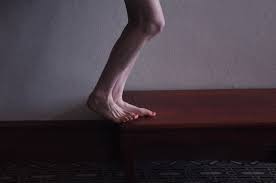
*note: there are two photos here in today’s blog post, look for the side scroll arrows and click on the small box in the upper left corner if you cannot see the photo

 We have seen this one for years in our clinics but we never got around to researching it and pondering the condition more deeply.  Here is our mantra for today, Form follows Function.

Studies seem to be undecided on the cause of this entity. Some suggest that pincer nails are caused by lack of upward mechanical forces on the toe pad where others remark that they can be observed amongst the healthy mechanical walkers. Hitomi’s study suggests that the affected toes fail to receive adequate physical stimulation from proper toe and forefoot loading. Please read on, this gets interesting.

According to Hitomi’s study, in both the barefoot and shod state,

“the pincer nail group had significantly lower pressure on the first toe than the control group. In both the barefoot and shod state, the peak pressure area was mostly the metatarsal head area in the pincer nail group, whereas it was mostly the first toe area in the control group. Binomial logistic regression analysis revealed that peak pressure area was a significant risk factor for pincer nail development.”

This seems to suggest that there is insufficient or aberrant use of downward pressure on the toes and into the toe pads. Hitomi speaks of the locale of the peak pressure, seemingly proposing from this study that it should not be under the metatarsal heads. This, in our experience and thinking, could suggest that more long flexor dominance is present. This long flexor activity seems to create some disfunction not only in the activity of the lumbrical muscles but also altered pressures in the metatarsal (MET) heads.  It certainly alters distal toe pressures which can alter skin and nail responses (see our blog post on subungal hematomas for more on this topic where we discuss principles of counter pressure and shear forces). We try to teach a “spread and reach with long flat toes” approach to our clients in correcting bad habits such as toe hammering and gripping (which are often a result of flawed biomechanics elsewhere).

The nail bed is very rich in vasculature (hence the cause of the dreaded hematoma, the black toenail) and nerve endings.  The nail bed is a derivative of the epidermis containing keratin which gives it its hard nature. The nail consists of the nail plate, the nail fold, the nail matrix, the sterile matrix and the hyponychium. There are many factors that go into the formation of a normal nail, including blood flow, nutrition, local neurogenic factors and not to forget, mechanical loading issues. Failure of any of these issues can lead to softening, brittle, thinning, diseased or malformed nails. The nail grows from a nail root in front of the cuticle and grows distally at a slow but (usually) steady rate.  It is interesting to note that the long extensor tendon (EDL) attachment is close to the proximal nail bed root area thus it brings forward thinking of possible imbalances between long and short flexors and extensor tendons/muscles and their patterns of imbalance in toe gripping and hammering that could cause a change in function which could drive a change in form.  We have all heard it, form follows function, why should this area be any different ?

Hitomi also mentioned something interesting in his study, the observation that bed ridden clients seem to have a predilection to pincer toes.  This at least seems to fit the aberrant loading patterns, in this case an absence of. The study also started some interesting thinking in us when it mentioned a hypothesis,

“that human nails are constitutively equipped with an automatic shrinkage function that allows them to adapt to daily upward mechanical forces.”

This was a fascinating hypothesis to us. It seems to make sense. If constant downward pressure on the toe pads were present, the toe nails would always be undergoing a flattening and spreading response so it could make sense that the nails have a built in curve and shrinkage function offsetting and adapting to the constant distorting pressures (the flattening and spreading forces).  Hence, some possible clarity in Hitomi’s hypothesis that pincer nails are caused by lack of (and in our thinking, distorted) upward mechanical forces on the toe pad.  And, when those distorting pressures are placed elsewhere (ie. the MET heads or tips of the toes as in our subungal hematoma hypothesis) or faulting gripping or hammering loading the automatic shrinkage function is left to dominate.

We think Hitomi’s hypothesis is correct. Here is why (this is paraphrased from our blog post on subungal hematomas and our revolutionary thinking on why they occur and it seems to fit well with pincer nail formation as well).

…  when the skin is pulled at a differential rate over the distal phalange (from gripping of the toes rather than downward pressing through the toe pad) there will be a net lifting response of the nail from its bed as the skin is drawn forward of the backward drawn phalange  (there is a NET movement of skin forward thus lifting the nail from its bedding).  For an at-home example of this, put your hand AND fingers flat on a table top. Now activate JUST your distal long finger flexors so that only the tip of the fingers are in contact with the table top (there will be a small lifting of the fingers). There should be minimal flexion of the distal fingers at this point. Note the spreading and flattening of the nail.  Now, without letting the finger tip-skin contact point move at all from the table, go ahead and increase your long flexor tone/pull fairly aggressively. You are in essence trying to pull the finger backward into flexion while leaving the skin pad in the same place on the table. Feel the pressure building under the distal tip of the finger nail as the skin is RELATIVELY drawn forward.]   This is fat pad and skin being drawn forward (relative to the phalange bone being drawn backward) into the apex of the nail. Could this be magnifying the curvature of the nail and not offsetting the “automatic curving and shrinkage” function of the nail ? We think it is quite possible.

So, there you have it. We will dive deeper on this topic another time, but after reading Hitomi’s study our brain’s started buzzing because we had discussed this process similarly a few years back in our Subungal Hematoma blog post.

And, if you are thinking about chronic repeated ingrown toe nails with this clinical entity, your thoughts are clearly on a logical path.  There is a correlation it seems.

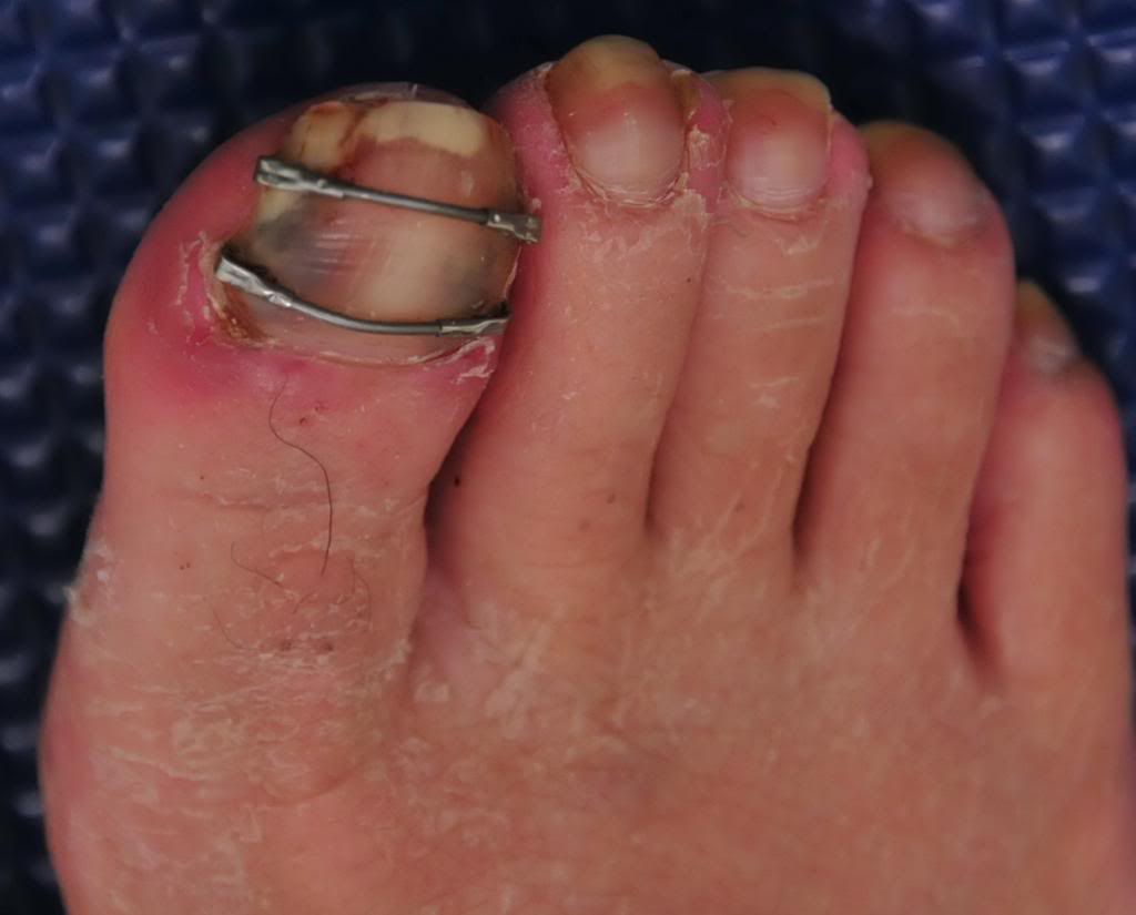
And, as for the horrific metal bar correction you see in the other photo above, this too is new to our eyes.  It seems rather medieval, something one might see in the gallows of yesteryear.  And if that doesn’t curl your hair and make you nauseated, try looking at what this one guy did, a DIY remedy (caution, not for the feint of heart). https://www.mja.com.au/journal/2005/182/4/diy-pincer-nail-repair-brace-yourself

ShawnAllen, one of the gait guys

References:

Foot loading is different in people with and without pincer nails: a case control study  Hitomi Sano1*, Kaori Shionoya2 and Rei Ogawa1  Journal of Foot and Ankle Research 2015, 8:43

tumblr_nozmmaOxAI1qhko2so1_500.png
tumblr_nozmmaOxAI1qhko2so2_540.png

Ahh yes, the lumbricals. 

One of our favorite muscles. And here it is in a recent paper! This one is for all you fellow foot geeks : )

Perhaps the FDL (which fires slightly earlier than the FHL) and FHL (which fires slightly later and longer) at loading response, slowing pronation and setting the stage for lumbrical function from midstance to terminal stance/preswing (flexion at the metatarsal phalangeal joint (it would have to be eccentric, if you think about this from a closed chain perspective) and extension (actually compression) of the proximal interphalangeal joints.

“The first lumbrical arose as two muscle bellies from both the tendon of the FDL and the tendinous slip of the FHL in 83.3 %, and as one muscle belly from the tendon of the FDL or the tendinous slip of the FHL in 16.7 %. These two muscle bellies subsequently merged to form the muscle belly of the first lumbrical. The second lumbrical arose from the tendinous slips of the FHL for the second and third toes as well as the tendon of the FDL in all specimens. The third lumbrical arose from the tendinous slips of the FHL for the third and fourth toes in 69.7 %, and the fourth lumbrical arose from the tendinous slip of the FHL for the fourth toe in 18.2 %. Some deep muscle fibers of the fourth lumbrical arose from the tendinous slip of the FHL for the second toe in 4.5 %, for the third toe in 28.8 %, and for the fourth toe in 15.2 %.”

Hur MS1, Kim JH, Gil YC, Kim HJ, Lee KS. New insights into the origin of the lumbrical muscles of the foot: tendinous slip of the flexor hallucis longus muscle. Surg Radiol Anat. 2015 May 12. [Epub ahead of print]

tumblr_nd32fvOzjx1qhko2so3_r1_1280.png
tumblr_nd32fvOzjx1qhko2so2_1280.png
tumblr_nd32fvOzjx1qhko2so1_1280.png
tumblr_nd32fvOzjx1qhko2so4_r1_500.png

Part 2: “Standing on Glass” Static Foot/Pedograph Assessment

* note (see warning at bottom): This is a static assessment dialogue. One cannot, and must not, make clinical decisions from a static assessment. The right and left sides are indicated by the R and L circled in pink. There are 4 photos here today.

Blue lines: Last time we evaluated possible ideas on the ORANGE lines here, it would be to your advantage to start there. 

We can see a few noteworthy things here in these photos. We have contrast-adjusted the photo so the pressure areas (BLUE) are more clearly noted. There appears to be more forefoot pressure on the right foot (the right foot is on the readers left), and more rearfoot pressure on the left (not only compare the whiteness factor but look at the displacement of the calcaneal fat pad (pink brackets). There is also noticeably more lateral forefoot pressure on the left. There is also more 3-5 hammering/flexion dominance pressure on the left.  The metatarsal fat pad positioning (LIME DOTS represent the distal boundary) is intimately tied in with the proper lumbrical muscle function  (link) and migrates forward toward the toes when the flexors/extensors and lumbricals are imbalanced. We can see this fat pad shift here (LIME DOTS). The 3-5 toes are clearly hammering via flexor dominance (LIME ARROWS), this is easily noted by visual absence of the toe shafts, we only see the toe pads. Now if you remember your anatomy, the long flexors of the toes (FDL) come across the foot at an angle (see photo). It is a major function of the lateral head of the Quadratus plantae (LQP) to reorient the pull of those lesser toe flexors to pull more towards the heel rather than on an angle. One can see that in the pressure photos that this muscle may be suspicious of weakness because the toes are crammed together and moving towards the big toe because of the change in FDL pull vector (YELLOW LINES). They are especially crowding out the 2nd toe as one can see, but this can also be from weakness in the big toe, a topic for another time. One can easily see that these component weaknesses have allowed the metatarsal fat pad to migrate forward. All of this, plus the lateral shift weight bearing has widened the forefoot on the left, go ahead, measure it. So, is this person merely weight bearing laterally because they are supinating ? Well, if you read yesterday’s blog post we postulated thoughts on this foot possibly being the pronated one because of its increased heel-toe and heel-ball length. So which is it ? A pronated yet lateral weight bearing foot  or a normal foot with more lateral weight bearing because of the local foot weaknesses we just discussed ? Or is it something else ? Is the problem higher up, meaning, are they left lateral weight bearing shift because of a left drifted pelvis from weak glute medius/abdominal obliques ?  Only a competent clinical examination will enlighten us.

Is the compensation top-down or bottom up, or both in a feedback cycle trying to find sufficient stability and mobility ? These are all viable possibilities and you must have these things flowing freely through your head during the clinical examination as you rule in/rule out your hands-on findings.  Remember, just going by a screen to drive prescription exercises from what you see on the movement screen is not going to necessarily fix the problem, it could in fact lead one to drive a deeper compensation pattern. 

Remember this critical fact.  After an injury or a long standing problem, muscles and motor patterns jobs are to stabilize and manage loads (stability and mobility) for adequate and necessary movement. Injuries leave a mark on the system as a whole because adaptation was necessary during the initial healing phase. This usually spills over during the early movement re-introduction phase, particularly if movement is reintroduced too early or too aggressively.  Plasticity is the culprit. Just because the injury has come and gone does not mean that new patterns of skill, endurance, strength (S.E.S -our favorite mnemonic), stability and mobility were not subsequently built onto the apparently trivial remnants of the injury.  There is nothing trivial if it is abnormal. The forces must, and will, play out somewhere in the body and this is often where pain or injury occurs but it is rarely where the underlying problem lives.

Come back tomorrow.  We will try to bring this whole thing together, but remember, it will just be a theory for without an exam one cannot prove which issues are true culprits and which are compensations. Remember, what you see is often the compensatory illusion, it is the person moving with the parts that are working and compensating not the parts that are on vacation.  See you tomorrow friends !

Shawn and ivo, the gait guys

* note: This is a static assessment dialogue. One cannot, and must not, make clinical decisions from a static assessment. As in all assessments, information is taken in, digested and then MUST be confirmed, denied and/or at the very least, folded into a functional and clinically relevant assessment of the client before the findings are accepted, dismissed and acted upon. As we always say, a gait analysis or static pedograph-type assessment (standing force plate) is never enough to make decisions on treatment to resolve problems and injuries. What is seen and represented on either are the client’s strategies around clinical problems or compensations.  Today’s photo and blog post are an exercise in critical clinical thinking to get the juices flowing and to get the observer thinking about the client’s presentation and to help open up the field to questions the observer should be entertaining.  The big questions should be, “why do i see this, what could be causing these observances ?”right foot supinated ? or more rear and lateral foot……avoiding pronation ?

Unilateral heightened toe extensor tone.
What do we have here ? Well, it is obvious. The left foot is showing increased short extensor tone (EDB: extensor digitorum brevis) and heightened long flexor tone (FDL: flexor digitorum longus). This is the …

Unilateral heightened toe extensor tone.

What do we have here ? Well, it is obvious. The left foot is showing increased short extensor tone (EDB: extensor digitorum brevis) and heightened long flexor tone (FDL: flexor digitorum longus). This is the classic pairing for hammer toe development.  We also know from this post (link) and from this post (link) that this presentation is closely related with lumbrical weakness and distal fat pad migration.

So, at an assessment took we like to play games. Mental games to be precise. When we see something like this we immediately begin the mental gyrations of “what could have caused this, and what could this in turn be causing”. Remember, what you see is often not the problem, rather your clients compensation around the problem.  In this case, what goes through your mind ?  Without deep thought, our knee jerk thoughts are:

  • possible loss of ankle rocker dorsiflexion (the increased EDB tone can be recruited to help drive more ankle dorsiflexion indirectly)
  • plantar intrinsic weakness ?
  • flip flops or slip on shoes where the heel is riding up and down inside the shoe/sloppy fit ?  (initiating a gripping response from the FDL)
  • weak tib anterior (recruiting EDB to help)
  • weak peroneus tertius (recruiting EDB again)
  • Ankle /foot instability (more FDL gripping will help gain ground purchase)
  • lateral ankle instablity (same thing, more gripping)
  • Weak gastrosoleus (since the FDL is a posterior compartment neighbor it can kick into high gear and help with posterior comparment function, we have a whole video case based around this issue, check this out ! )
  • premature departure off of the good side leg, and thus an abrupt loading response onto this affected side can challenge the frontal plane of the body and thus require more grip response at the foot level.
  • how about simple weakness of the lumbricals or FDB , the short flexors. The long flexors will have to make up for it and present like this.  
  • the list goes on and on … .

These are just some quick cursory thoughts, and by NO means a complete exhaustive list.  Just some quick thoughts.

But what about hip function ?  if ankle rocker is blocked in terminal stance and the FDL fire like this what will that do to hip extension ? Well, heel rise will be premature because of the limitation and thus hip extension will be abbreviated. Thus glute function will be impaired to a degree.  This can become a viscous cycle, each feeding off of each other.

This diagnostic stuff is a tricky and difficult game. If you think you can diagnose or fix a problem from just changing what you see you are mistaken, unless you like driving compensation patterns and future injuries into your clients.   There must be a hands on examination and assessment with an intact educated brain attached to the process.

Just some mental gymnastics for you today.  

Shawn and Ivo

the gait guys

tumblr_n4swdbdmHS1qhko2so2_1280.jpg
tumblr_n4swdbdmHS1qhko2so1_1280.jpg

The case of the missing toes.

OK, a bit dramatic but as you can see in the plantar view above, all you can see is the toe pads, the rest of the digit shafts are hidden.  

This is a classic example of a foot imbalance. We have talked about this many times before but the attached video link here  ( http://youtu.be/IIyg7ejYNOg ) shows it very well.  Read on.

There is shortness and increased resting tone in the short toe extensors (EDB, extensor digitorum brevis) and long toe flexors (FDL=flexor dig. longus) with insufficiency in the short flexors and long extensors. This pairing creates a hammer toe effect.  In the video, you can see that these toes are showing early hammering characteristics, but not yet rigid ones. The key word there is, “yet” so this is still a correctable phenomenon at this point.  You can also clearly see the distal migration of the metatarsal fat pad. The fat pad has migrated forward of the MET heads and is being pulled forward by the excess tension in the long toe flexors. As this imbalance in the toe flexors and extensors develops, the forefoot mechanics get impaired and the lumbricals (which anchor off off the FDL) become challenged. Their contributory biomechanics, amongst other things, help to keep the fat pad in place under the metatarsal heads. You can see in this video link above that by proximally migrating (towards the heel) just the fat pad back under the MET heads the resting mechanics of the toes changes, for the better.  

Remember the other functions of the lumbricals ?  their other major functions, namely: thinking from a distal to proximal orientation (a closed chain mode of thinking), they actually plantarflex the metatarsal on the fixed phalynx, assist in dorsiflexion of the ankle, and help to keep the toes from clawing from over recruitment of the flexor digitorum longus.

Here is another blog post we did on a similar presentation.http://thegaitguys.tumblr.com/post/14766494068/a-case-of-plantar-foot-pain-during-gait-this

Proper balance of the toe flexors and extensors, and their harmony with lumbricals and fat pad amongst other things will give healthy long flat toes that can help the proximal biomechanics of the foot.  If you have neuromas, metatarsalgia, hammer toes, claw toes, migrating toes, bunions or hallux valgus amongst many other things, this might be a good place to start.   

There are exercises that can help this presentation, but understanding “the why” is the first step.

Shawn and Ivo

The Gait Guys

Does this foot look like your foot ? 
There are a few subtle issues here. At first glance this foot looks half-way decent but upon further observation you should note the subtle drift of all of the toes.  In the foot, the toe that delineates abducti…

Does this foot look like your foot ? 

There are a few subtle issues here. At first glance this foot looks half-way decent but upon further observation you should note the subtle drift of all of the toes.  In the foot, the toe that delineates abduction and adduction of the toes is the 2nd toe. The 2nd toe is considered the anatomic middle of the digits and forefoot. Any toe or movement that moves away from the 2nd toe is abduction and any movement towards the 2nd toe is adduction. This is obviously different than in the hand where the 3rd digit is the reference digit.  

In this foot, look at the shape of the 2nd and even the 3rd digit, they have a curve to them. Remember, form follows function and the dead give away here is that the hallux (the big toe) is drifting into adduction towards the 2nd digit. This is referred to as early hallux valgus and it is accompanied by early evidence of a bunion at the medial foot at the metatarsophalangeal joint.  When the shaft of the hallux is not in line with the shaft of the metatarsal long bone we get the angulation between the two causing the hallux valgus.  This is often from excessive pronation (either rearfoot, midfoot and/or forefoot) that collapses the tripod, splays the distal MET head via its dorsiflexion, and the development of complicated long and short hallux flexor muscle dysfunction as well as abductor hallucis (transverse and oblique head) disfunction further driving the hallux pull medially.  When the distal toes are engaged on the ground and there is still forefoot pronation occurring through the medial tripod support, the toes will be forced into a twist or spin, and in time you will get toes that appear drifted or windswept like these toes appear.  A similar phenomenon occurs at the lateral foot and a Tailor’s bunion begins to occur there as the forefoot begins to widen as the MET heads separate and the toes funnel medially (often provoked to do so by pointed footwear).  

We can also see the 4th and 5th toes curl under from the probably weak lateral head of the quadratus plantae thus encouraging unopposed oblique pull of the long flexors of the digits (FDL). See this post here for an explanation of this phenomenon.  

This is a fairly typical foot that we see in our practices.  This is not a far-gone foot but one has to catch this foot at this stage or it is rather difficult to resuscitate back to a healthy foot. Like a spinal scoliosis, once a bunion and  hallux valgus gets too far, it becomes an issue of symptom management rather than repair.  Hallux abduction must be retaught, tripod skills must be retaught, intrinsic foot muscle strength must be regained as well as strength and endurance of the tibialis anterior and toe extensors to help raise the arch again and control pronation. Sometimes a temporary orthotic can help the person to passively regain some degree of competent tripod while homework earns the changes. In some cases, an orthotic needs to be a permanent intervention if tripod stability cannot be adequately achieved.  But, we never give up and neither should you or your client, amazing things can happen over long periods of time when correction is forced.

There is plenty of life left in this foot, but you have to get to it quickly and get them in lower heeled shoes if tolerable and ones with a wider toe box.  Support the midfoot with an orthotic or built up foot bed, if necessary, but don’t leave it there. It is a crutch, and even crutches are intended to be put aside at some point. 

Shawn and Ivo, The gait guys

tumblr_m9sytjXhaA1qhko2so1_540.png
tumblr_m9sytjXhaA1qhko2so2_540.png

The Toe Waving Exercise: Part 3  The Lumbricals

Welcome to Friday, Folks. Part 3 of this series, just in time for the weekend.

In part 1 of this series, we looked at the flexor digitorum longus muscle, and in part 2 the  extensor digitorum longus. In part 3, we will look at some often overlooked muscles, the plantar lumbricals.

Watch the video from the 1st post again. Note the flexion at the metatarsal phalangeal joint.

The lumbricals of the foot attach proximally to the sides of adjacent  tendons of the flexor digitorum longus (with the exception of the 1st, which only attaches to the medial side) and attach distally to the medial aspect of the head of the proximal phalynx; they then continue on to the extensor hoods in toes 2-5. Their open chain function (ie. the foot is off the ground) is described as flexion of the metatarsal phalangeal joint and extension of the proximal and distal interphalangeal joints. They also compress the metatarsal-phalangeal and inerpahlangeal joints, providing stability to the foot. There is also a small adductory moment to counteract abductory shear, due to the tendon passing medial to the metatarsal-phalangeal joints.

The lumbricals are most active from midstance to preswing. That means they act predominantly in closed chain (or, when the foot is on the ground). Thinking along these lines, the lumbricals (along with the other intrinsic muscles of the foot) play a role in maintaining the medial longitudinal arch of the foot and stabilization of the forefoot during stance phase and rearfoot during preswing.

Thinking now, as we are sure you are, in a closed chain fashion, from a distal to proximal orientation, they actually flex the metatarsal on the phalynx, assist in dorsiflexion of the ankle, and help to keep the toes from clawing from over recruitment of the flexor digitorum longus (which we say happen in last weeks post here).

Now think about the changes which can occur with in the gait cycle due to dysfinction of this muscle. A shortened step length, diminished ankle rocker, increased forefoot rocker and premature heel rise. Now we will need increased extension at the metatarsophalangeal joints (particularly the 1st), shifting the tendon of the lumbricals upward and behind the transverse metatarsal joint axis, causing even more extension now at this joint. Chronically over time, this causes displacement of the fat pads anteriorly from under the metatarsal heads.How many patients have you seen with metatarsal head pain?

What about the changes up the kinetic chain and the musculoskeletal implications of muscle inhibition, overfacilitation and joint dysfunction, often with neurological sequalae. Because of the lack of ankle dorsiflexion, you have less hip extension, so you borrow some from the lumbar spine, with increased compressive forces there and an increase in the lordosis, which causes an increase in the thoracic kyphosis and cervical lordosis. We need to get this leg up and forward to continue our progression ahead, so now we fire our hip flexors instead of the obliques. This further fuels inhibition of the glutes, compounding the loss of hip extension. How about a little increased shoulder flexion on the contralateral side to assist getting that leg forward? Don’t forget that we have altered the thoracic kyphosis and thus changed scapulo humeral mechanics. Neck/ shoulder pain from bad feet? Maybe.

Look to the base; it is where many problems begin. Consider function in the context of where it occurs. Have and promote an adequate foot tripod. The Toe Wave is one step in the right direction.

The Gait Guys….Foot Geeks….Gait Geeks…..Shoe Nerds……Yup. If you are reading this, then you are one of us now. Help us to spread the word….


all material copyright 2012 The Homunculus Group/The Gait Guys. All rights reserved. Don’t rip off our stuff! If you ask us nicely we will probably let you use it.

The Toe Waving Exercise, Part 2

In part one of this series, we talked about the importance of the short flexors of the toes (FDB or flexor digitorum brevis) in forming and maintaining the foot tripod. In this installment, we discuss another important muscle used in this exercise,  the extensor digitorum longus (EDL).

We have shown you time and time again, dominance of the long flexors in gait, which cause biomechanical imbalances. We remember that through reciprocal inhibition, the log flexors will reciprocally inhibit the long extensors, so increased activity in the former, means decreased activity and activation in the latter.

The balanced activity of the long flexors and long extensors helps to create harmony during gait. Working the long extensors with this exercise (along with others, like tripod standing, toes up walking and the shuffle gait exercises) helps one to achieve this balance.

The Gait Guys; promoting foot and gait competency every day here, on Youtube, Facebook, Twitter, and in our offices and yours.


all material copyright 2012 The Homunculus Group/The Gait Guys. All rights reserved. If you rip off our stuff, you will never have an adequate foot tripod and will have gait problems for the remainder of your days.

In our series of questions we get…

Dear Gait Guys
I was reading an article the other day about the foot and intrisic muscles to gain more insight into the function and how to re-train these muscles. I am having a difficult time trying to g…
In our series of questions we get…

Dear Gait Guys

I was reading an article the other day about the foot and intrisic muscles to gain more insight into the function and how to re-train these muscles. I am having a difficult time trying to give patients exercises for intrisic muscles when everyone seems to say something different. The most recent I have read is that the best way to retrain the lumbricals is stand on your toes and walk up steps. I can see some logic in this but also seems a very generic exercise and would encompass alot more flexor driven muscles that are likely already strong. I was wondering if you have a more specific exercise that would be simple and easy for patients to do?

Dear Lumbricals

We would have to agree with you that the exercise is very generic and would cause overuse of the flexors, though it would stimulate lumbrical function.

As you are aware, the lumbricals attach proximally to the sides of adjacent tendons of the flexor digitorum longus (with the exception of the 1st, which only attaches to the medial side) and attach distally to the medial aspect of the head of the proximal phalynx and continue on to the extensor hoods in toes 2-5 .

Their typical function is described as flexion of the proximal phalynx and extension of the proximal and distal interphalangeal joints. They have the unique ability to compress the metatarsal-phalangeal and inerpahlangeal joints. There is also a small adductory moment to counteract abductory shear, due to the tendon passing medial to the metatarsal-phalangeal joints (michaud). These are open chain functions. Unless you are in the habit of waving to people with your toes, they often are used quite differently. But this brings upa good point and excellent exercise we call “waving the toes”.

They are performed by holding the great toe in dorsiflexion (hopefully, without assistance) and flexing the other toes at the MTP joint, while keeping the PIP and DIP in extension. This requires and intact and functional EDL (with good motor control!)

Another exercise is sitting with the foot relaxed and concentrating on flexing the toes (2-5) without clawing (similar to above, without the Hallux extended.

Remember they work from mid to terminal stance, but you need to develop skill before endurance or strength.

We hope this helps, 

The Gait Guys

A case of plantar foot pain during gait.
This client came to see us after a surgical proceedure to remove a dead (osteonecrosis) medial sesamoid under the 1st metatarsal head and a later surgery to fix a progressing hammer toe of the 2nd digit. What…

A case of plantar foot pain during gait.

This client came to see us after a surgical proceedure to remove a dead (osteonecrosis) medial sesamoid under the 1st metatarsal head and a later surgery to fix a progressing hammer toe of the 2nd digit. What we really want you to see is the huge divot/depression under the 2-3 metatarsal heads. Also note the accumulation and relocation of the normal MET head fat pad now located distal to the MET heads.  It is as if the fat pad is trying to hitch a ride on the toes now ! This is a case of Metatarsalgia secondary to fat pad displacement (displaced from the divot area to the flexor crease) secondary to surgical sequelae. 

What is additionally cool in this case is the fact that this client has an almost complete webbing of the 2-3 toes so many of the normal independent muscular functions are no longer independent. After the surgeries this person presents with tremendous loss of flexor and extensor function of the 2-3 toes.  Lumbrical testing was most obviously impaired, completely absent in fact, in these 2-3 toes. On the ground the patient was also unable to achieve any flexion-press of the toes into the ground, he was able to flexion/hammer curl which will obviously put them at risk for hammer toes in the future.  But what is important here is that without the ability to PRESS the toes into the ground particularly while in stance phase the lumbricals will not help to hold the fat pad in its normal location under the MET heads. Nor will they be able to to perform their other major functions, namely: thinking from a distal to proximal orientation (a closed chain mode of thinking), they actually plantarflex the metatarsal on the fixed phalynx, assist in dorsiflexion of the ankle, and help to keep the toes from clawing from over recruitment of the flexor digitorum longus.

This client’s MET head pain is obviously caused by lack of cushioning of the head since the fat pad is displaced. There are plenty of other biomechanical abberancies now, the Windlass mechanism will never be the same becuase it is without one of the sesamoids, the hallux short flexor (FHB) is impaired on the medial head without the sesamoid so hallux flexion will become a problem.  Do we really want to see such compromise of the medial tripod ? Heck no, we need sesamoid implants ! There is a novel idea ! When a sesamoid is taken out we need to replace it ! Think about it !

There is so much more to this case, but we will stop here. It’s Christmas after all ! This poor lady was told to wish from Santa for a medial sesamoid implant under the tree and a sudden spontaneous activation of the lumbricals to retract the fat pad back under the MET head so as to reduce her pain.  Hey, wishing can’t hurt !

Merry Christmas and Happy Holidays to you all gang, whatever your faith we wish you well,

from Shawn and Ivo…… The Gait Guys

(PS: we included below more from the body of the article we wrote long ago called “The Lost Lumbricals”.  So for those of you who wish to geek out more on Christmas, read on …

______________

EXCERPTS FROM “THE LOST LUMBRICALS”

The lumbricals of the foot attach proximally to the sides of adjacent  tendons of the flexor digitorum longus (with the exception of the 1st, which only attaches to the medial side) and attach distally to the medial aspect of the head of the proximal phalynx and continue on to the extensor hoods in toes 2 through 5. Their typical function is described as flexion of the proximal phalynx and extension of the proximal and distal interphalangeal joints. They have the unique ability to compress the metatarsal-phalangeal and interphalangeal joints. These are “open chain” functions as described, unless you are in the habit of waving to people with your toes, they often are used quite differently in the gait cycle with the foot affixed to the ground.

The lumbricals are most active from midstance to preswing. That means they act predominantly in the closed chain. The lumbricals, along with the other intrinsic muscles of the foot, play a role in maintaining the medial longitudinal arch of the foot.  Along with the interossei, they play a role in stabilization of the forefoot during stance phase and rearfoot during preswing. One author has proposed that overpronation is due to a lack of neuromuscular control of the intrinsic foot muscles to stabilize the tarsal and metatarsal bones and therefore modulate the speed of pronation.

Thinking from a distal to proximal orientation (a closed chain mode of thinking), they actually plantarflex the metatarsal on the fixed phalynx, assist in dorsiflexion of the ankle, and help to keep the toes from clawing from over recruitment of the flexor digitorum longus.

Clawing toes during gait, which are considered abnormal, are defined as extension of the metatarsophalangeal articulation, and flexion of the proximal and distal interphalangeal joints result from a foot attempting to stabilize itself during the terminal stance and preswing phases of gait.  This is an attempt to help propel the body forward, often accompanied by overactivity of the flexor digitorum longus, tibialis posterior, flexor pollicus longus, and gastroc soleus groups. Overactivity of these groups causes reciprocal inhibition of the long toe extensors and ankle dorsiflexors (tibialis anterior for example), causing the toes to buckle further and a loss of ankle dorsiflexion; in short, diminished ankle rocker.

Now think about the changes in the gait cycle in the above scenario. There will be a resultant shortened step length, diminished ankle rocker, increased forefoot rocker and premature heel rise. This will necessitate an increased extension at the metatarsophalangeal joints, shifting the tendon of the lumbricals upward and behind the transverse metatarsal joint axis, causing even more extension now at this joint. Chronically over time, this causes displacement of the fat pads anteriorly from under the metatarsal heads and is one of the main reasons metatarsal head pain (metatarsalgia). In the past have you made the apparent simple diagnoses of metatarsalgia, shin splints, stress fractures or Morton’s neuroma without knowing a more plausible cause ?  Do you now feel you have better answers to these clinical phenomena ?

Now think about changes up the kinetic chain and the potential musculoskeletal implications of muscle inhibition, overfacilitation and joint dysfunction, often with neurological sequelae. With lumbrical dysfunction (weakness) and the resultant lack of ankle dorsiflexion, you have less hip extension.  So, you borrow some from the lumbar spine, with increased compressive forces there and an increase in the lordosis, which causes an increase in the thoracic kyphosis and cervical lordosis. We still need to get this leg up and forward to continue our progression ahead, so now we fire our hip flexors instead of the abdominal obliques. And because there needs to be cooperation of the abdominals and hamstrings to maintain pelvis neutrality, this further fuels inhibition of the gluteals thus further compounding the loss of hip extension. Now how about a little increased shoulder flexion on the contralateral side to assist getting that leg forward? Don’t forget that we have altered the thoracic kyphosis and thus changed scapulo humeral mechanics. Now neck/shoulder pain all from bad feet?  Maybe. These muscles developed and exist for a good reason, do your best not to dismiss them and their function the next time you see a tortured foot.

When patients have continued dysfunction, consider the base and where it all begins. Consider function in the context of where it occurs. Proper evaluation of the feet and gait can provide valuable clues as to the etiology or manifestation of continued problems. Important? You decide.

When the Short Toe Extensors Try to Rule the World ! 
A case of a runner with forefoot pain. 
This is a runner of ours, one of the fastest young men in the state  of illinois, top 10 in the country in mid-distance, top 20 in the USA in  cross countr…

When the Short Toe Extensors Try to Rule the World !

A case of a runner with forefoot pain.

This is a runner of ours, one of the fastest young men in the state of illinois, top 10 in the country in mid-distance, top 20 in the USA in cross country.

He came in with left forefoot plantar pain.  He explained (in a matter of words) that he was having pain at full forefoot loading at heel rise /push off.

We watched him walk, saw this visual problem present itself in dynamic motion (yup, no stop frame video on this one, not when you see it about 10 times a month !) and noted a subtle left lateral hip/pelvis shift past what would be considered normal for frontal plane mechanics.

On the table this is a photo of his feet.  What do you see ?

We see a suspected (which you will try to confirm on examination) increase in short extensor (EDB, extensor digitorum brevis) muscle tone.  Increased long extensor (EDL, extensor dig. longus muscle) tone would have represented itself with the distal toes also extended but here we see a relative dominance of the long flexors (FDL, Flexor dig. longus) with the heightened short flexor increase.

We also see more confirmation of heightened long flexor tone (FDL) by the degree of heavy callus formation on the very tip of the 2nd toe (it was on all 4 lateral toes but the photo is not clear enough to demonstrate).  You can also see supporting evidence of heightened long flexor dominance by the subungual hematoma (bleeding under the 2nd toe nail). (How does this correlate ? Well, in most runners with excessive long flexor tone/use not only do they flex and claw so much in the shoes that the callus is on the tip of the toes but the nail also begins to lift as the  nail is caught on the sock liner of the shoe as the toe flexes, slowly, mile by mile pulling the toe nail from the nail bed thus bleeding underneath it).  Yes, it is NOT from the toes hitting the front end of the shoe !

Our examination confirmed weakness of all lumbrical muscles and of the flexor digitorum brevis and lateral quadratus plantae.  The patient could feel the strength/engagement difference as compared to testing on the right foot of the same muscle groups (we always compare side to side, for us and for the patient’s awareness).  The extensor digitorum brevis muscle mass on the lateral dorsum of the foot was tender as were the tendons along their course.  There was also weakness higher up in the kinetic chain at the lower division of the transversus abdominus and internal abdominal oblique, and frontal plane hip stabilizers (gluteus medius; anterior-middle-and posterior divisions).The 2nd and 3rd metatarsal heads were remarkably tender to palpation and it was obvious that the metatarsal fat pads had migrated distally from the lumbrical muscle weakness.

Sometimes a grasp response by the long flexors can represent a propioceptive /balance deficit during single leg stance phase so be sure to test those centers as well (cerebellar, vision, joint position sense, inner ear-vestibular apparatus). 

So, what is the take away for the non-medical person, the runner next door if you will ?  Lets just say, symmetry wins and when asymmetry is apparent, bring it up to the people that do your body work.  Hopefully, what you and they see will be assessed in a clinical light, and as a team you can get to the bottom of what is not working…….and in this case…..what was causing not only the plantar foot pain, but the left lateral hip sway outside the frontal plane.

———we are, The Gait Guys……Shawn and Ivo