Whoa!

Whoa! What's wrong with #172?

 Where do we start? Look at all of that tibial and genu varum! Notice how his knee is outside the sagittal plane? This means that he also has internal tibial torsionand he is rotating his foot out to create the requisite 4-6° internal rotation needed to move forward. It also looks like he has limited internal rotation of the thighby the positioning of his body.  This could be due to femoral retroversion as this commonly occurs with internal tibial torsion. Check out the interesting hand posturing bilaterally. Notice the extended thumb and wrist on the left? He may be trying to fire into his extensor pool to help gain more hip extension.  I sure wish we had a Sideview. Thankfully his pelvis is relatively level, isn't it? No, it actually isn't. That's just his shirt. Look closely at the tops of the iliac crests and you will see what I am talking about. Did you catch the slight head tiltto the right? With that much tibial and genu varum his center of gravity is moving to the left and he needs to tilt his head to the right to equalize things out.  What about the posterior rotation of the left shoulder? Again probably this is due to a lack of or failure to use internal rotation of the left hip.

Lots to talk about on this picture and we will do some more next time.

What internal tibial torsion and tibial varum looks like in a world champ.

You can be an effective athlete with internal tibial torsion and tibial varum.

The video of world champ Mirinda Carfrae shows it beautifully on the right side during loading. The question is always, "how durable is your given anatomy ?". 

How durable is your compensation ? And is there a cost when your endurance runs outor when the load gets too high ?  Those are the big questions you have to ask.

In this video, stop the play at 34-37 seconds, keep playing that loop over and over again until you can clearly lock this in your head -- internal tibial torsion and tibial varum.  See how far laterally she appears in initial weight bearing ? See the appearance of her apparent "in-toe"?  You cannot correct this. You would be a fool to tell them to toe out more -- this would take her knee outside the sagittal plane. You leave this one alone. You make your athlete durable, giving them frontal plane and rotational-axial plane work to control those torsional forces during loading and unloading.  The difference been you and her, is she has done this loading a trillion times, safely and built durability on the entire chain from foot to spine so the tissues can tolerate it. The question is, will there be a limit for her ? Will there be a point where the bone and soft tissues say they have had enough ?  This is the golden question. 

Some folks with this can be assisted by more step width separation, getting away from a Cross Over gait but Mirinda has a beautiful running form.  However, in this particular video so does show some cross over gait, very narrow foot separation, and this magnified what you are seeing during her foot strike.  In many other videos she does not cross over if you have studied her running form elsewhere.

As she says in her video, being a world champ is all about the details, details like we pointed out here today.

Know your anatomical variances. Know how they play out, and how they fail.

Shawn and Ivo, the gait guys

Limitations: The powers of observation will help you.
Physical examination, FMS, DNS, gait analysis … . . these are all very important tools for the coach, trainer, therapist, clinician.  They will all offer information and lead the “th…

Limitations: The powers of observation will help you.

Physical examination, FMS, DNS, gait analysis … . . these are all very important tools for the coach, trainer, therapist, clinician.  They will all offer information and lead the “therapy giver” in a direction for intervention.  But when something doesn’t match up with the basic standard protocols, you have to go outside the standard box.  We have all been there and today is just a little reminder not to get caught up in the “proceedures” and merely running through protocol without an engaged brain putting the pieces together.  

Here we see 2 classic examples of deviations from the mean, the client on the left has drifted further outside the frontal plane because of tibial varum and a little genu varus.  The client on the right has imploded deep into the frontal plane via rigid pes planus foot collapse and genu valgum.  These will both affect your physical screenings for these clients. And keep in mind, and this is probably the most important point of today’s blog post, either client may have good or bad strategies around their anatomy.  In other words, some clients will have great compensations to limit further functional pathology, and some will have poor compensation strategies, and thus, both will have different physical exam findings, different screenings and different neuromotor patterns embedded deep into their CPGs (central pattern generators).   Put yet another way, all of the scenarios discussed may/will have varying screening assessment outcomes but for different reasons.  If you know the cause of these faults and the impaired neuro-recruitment patterns that are likely, your assessments will make more sense, and so will your exercise/therapy/rehab prescriptions.  If you do not understand the fundamental differences (ie long bone torsions or various femoral-neck shaft angles, foot types such as an uncompensated forefoot valgus etc) , one could prescribe therapies that will not address the underlying problems, rather they might address the compensations and strategies found with these client’s challenges.

It can get sloppy messy.  Wear a bib.

Dig for the roots, don’t mow the grass…… Shawn and Ivo, The Gait Guys

The Dual Density Foam Running Shoe.
This goes along nicely with yesterdays post. Note the photo attached. This is a great example of something we all see everyday. A laterally tipped foot in a stability shoe.  Clearly a shoe that has been mis-prescr…

The Dual Density Foam Running Shoe.

This goes along nicely with yesterdays post. Note the photo attached. This is a great example of something we all see everyday. A laterally tipped foot in a stability shoe.  Clearly a shoe that has been mis-prescribed for the wrong reason. Or has it ?

This client is clearly tipped laterally in the shoe, forcing supination.  Did this client self fit the shoe themselves in a discount store ? Were they fitted in a retail running store ? Where did things go wrong ? Or did they ?  The initial knee jerk reaction is to say this is the wrong shoe for this client.  Lets go a little deeper and ask some harder questions and see if you are considering some alternatives.

The assumption is frequently one of, “you are a hyperpronator so you need a stability shoe”. In this case is this person a hyperpronator ?  There is no way to know, not in the shoe.  On initial knee jerk observation this looks like a supinator in a stability shoe, a poor match.  But read on …

1. What if this person has significant flat feet, pes planus with severe pronation problems, but they find the stability they need by standing on the outer edge of the foot in the mechanically locked out position (supination).  Perhaps this is a less fatiguing posture, perhaps a less painful posture. This is often a comfort thing for hyperpronators to display.  What you see is not always what you get because there are two types of feet, those that drop or collapse into the weakness and those that fight the collapse and weakness the whole way via an alternative compensation.  You cannot tell by looking, certainly not from this picture of someone in a shoe. There must be a functional assessment and some gait evaluation. 

2. There exists the high arched flexible foot that pronates excessively, quickly and for a long time (this is the flexible cavus foot) and then there is the high arched rigid foot (the equinovarus foot).  The first described foot may need support from a stability shoe even though they have a high arch on presentation/examination and the later described foot can often go right into a neutral non-supportive shoe.  Can you tell either of these from this picture ? No you cannot.

3. Maybe the person in the photo has tibial varum (bowed lower leg) combined with a rearfoot varus and forefoot varus. This could mean they pronate heavily through the midfoot-forefoot and less so through the rearfoot-midfoot. In this case they are still a heavy pronator but not through what is typically noted or detected by significant medial arch collapse.  In this case the dual density shoe is not going to help all that much because the pronation is occurring mostly after the bulk of the shoe’s dual density stability foam has been passed through by the foot. Can this be detected by this photo ? Again the answer is no. The shoe fitter needs to be clinically aware that this type of client needs a forefoot varus posted shoe to help post up that medial tripod (1st metatarsal head).

4. Maybe, just maybe this is a typical rearfoot-midfoot pronating client, excessive mind you, and all they need is some foot and gait retraining to break their old compensation pattern of lateral weight bearing (standing or walking) and with this correct shoe they can then engage a healthier motor pattern. 

Which is it ?

Do you know how to navigate your way through these issues to make the right decision ?  There is no way to know here without seeing the foot naked and moving across the floor, and with a clinical examination to boot.

You can get all these things through our National Shoe Fit Certification program found here.

LINK:  http://store.payloadz.com/results/results.aspx?advsearch=1&m=80204

Email us and we will share the necessary info to get you started.  thegaitguys@gmail.com

Shawn and Ivo, The Gait Guys

A question from a doctor on the topic of limb alignment development.

The following question was forwarded to us from an internist on the USA east coast.

Question:

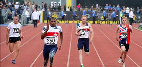
“I have a large number of female patients, many of them elderly.  I have noted that women in our society tend to progress to valgus knee deformity with age, and that TKR (total knee replacement) doesn’t seem to correct that deformity. Men tend more to the varus in our society.  I had formerly chalked that up to inherit gender difference.

3 or 4 years ago, I had occasion to spend a lot of time waiting outside the main Tokyo train station and observed a large number of people coming and going.  I observed the following:
1.  Young women had legs that were either straight or varus.
2.  Young women tended toward toeing in.
3.  They did all this in ridiculous high heels.
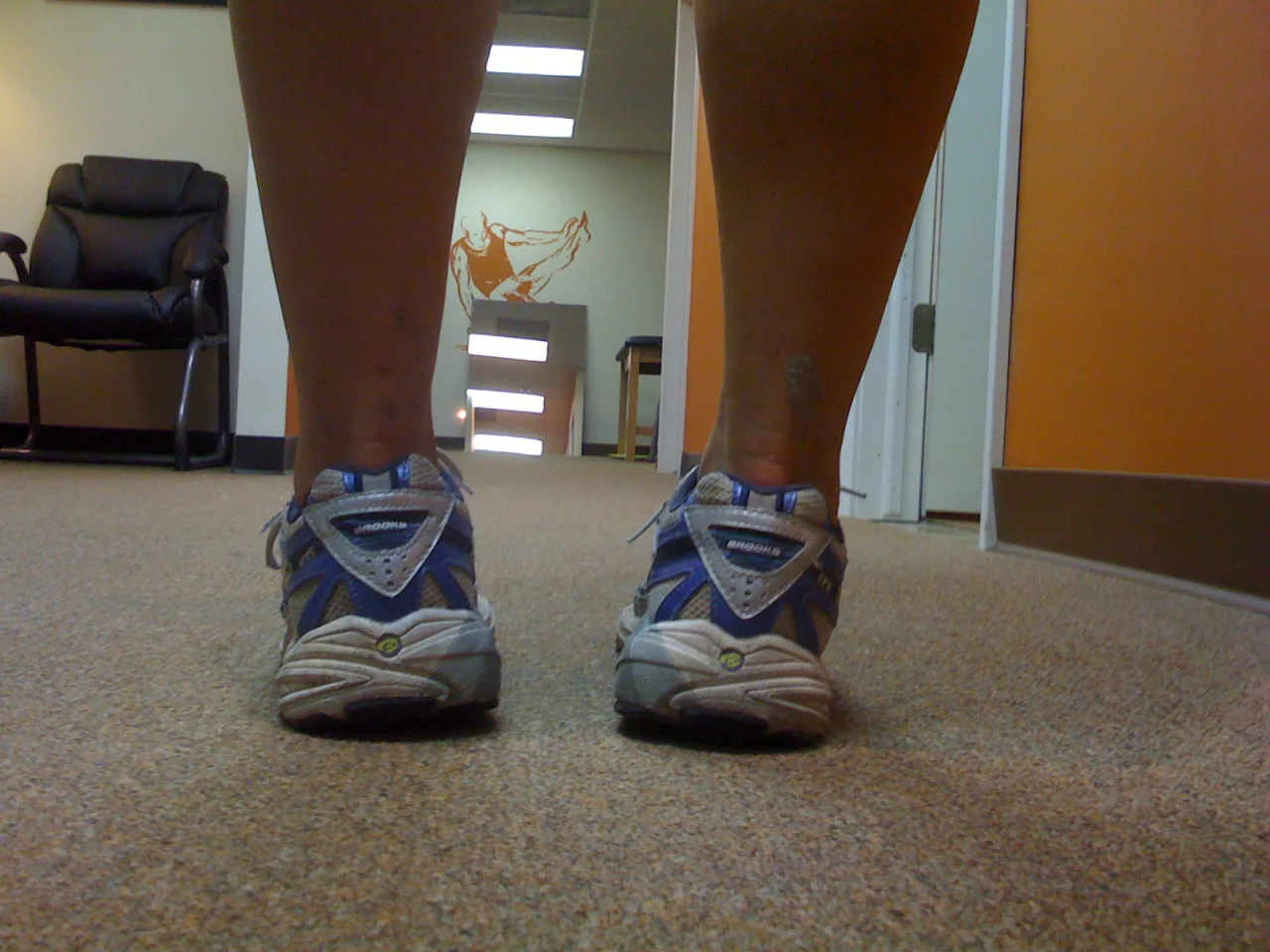
After some thought, I tend to attribute it to prolonged sitting in sesa (knees folded under), though being barefoot or in slippers while inside may also contribute.  Women in our society sit with their legs crossed.  Additionally, extensive shoe wearing leads to foot pronation.
So, could you direct me to someone who might have an interest in this observation and can refer me to any research that might have been done in this area?  I’ve had the dickens of a time trying to find anything on it, or even a specialized area of study that cares about such things.”

The GAIT GUYS RESPONSE:

Thanks for your confidence in us. Here are some thoughts:

Frontal plane deformities (or development) is twofold: genetic (and X linked) and developmental. Children usually go through a varus to straight to valgus to straight development (Ron Valmassey talks about this in his text Clinical Biomechanics of the Lower Extremities). Women generally have larger Q angles (from birth) and this angulation often causes assymetrical epiphyseal development (increased pressure on the lateral malleolus/tibial plateau stunts growth) with overgrowth of the medial femoral condyle. Developmental changes are secondary to weight (obesity causes increase in valgus angle) and posture/muscular devlopment. The increased genu valgus places weight medial to the midline (2nd met) of the foot and the foot accomodates by pronating (often excessively, as noted by both of you). This causes medial rotation of the lower leg and thigh, resulting in lengthening of the glutes (esp G max) resulting in stretch weakness and subsequent over reliance on the gastroc/soleus group for propulsion (remember this group tries to invert the heel in an attempt to cause supination once you go past midstance. Weak intrinsics (as pointed out by Dr Mark) further fuels this cycle. “W” sitting (sometimes a cultural development, as pointed out by Dr Birgit) plays in as well.

As for “toeing in”; may women have the combination of genu valgus with internal tibial torsion (often with femoral retroversion) which makes the condition difficult to treat (the rearfoot needs to be supported, but the forefoot needs to be valgus posted) otherwise the knee is placed outside the saggital plane and the meniscus becomes macerated due to conflicting biomechanics at the knee (Thus the short term fix with orthotics with a return of the pain later).

Yes, high heels and open back shoes are evil as are open backed shoes (we spoke at a convention in Chicago a few years back on this, before some of the research was out).

Thanks for allowing us to participate. below are some references for you.

-The GAIT GUYS…….Ivo and Shawn

______________________________________________________________


J Orthop Sports Phys Ther. 2008 Mar;38(3):137-49.

Differences in lower extremity anatomical and postural characteristics in males and females between maturation groups.

Shultz SJ, Nguyen AD, Schmitz RJ.

Source

Applied Neuromechanics Research Laboratory, Department of Exercise and Sport Science, University of North Carolina at Greensboro, 1408 Walker Ave., Greensboro, NC 27402, USA. sjshultz@uncg.edu

RESULTS:

When comparing maturation groups, limb length, pelvic angle, and tibial torsion increased with maturation, and anterior knee laxity, genu recurvatum, tibiofemoral angle, and foot pronation decreased with maturation. Females had greater general joint laxity, hip anteversion, and tibiofemoral angles, and shorter femur and tibial lengths than males, regardless of maturation group. Maturational changes in knee laxity and quadriceps angles were sex dependent.

CONCLUSIONS:

We observed a general change of posture with maturation that began with greater knee valgus, knee recurvatum, and foot pronation in MatGrp1, then moved toward a relative straightening and external rotation of the knee, and supination of the foot in later maturation groups. While the majority of the measures changed similarly in males and females across maturation groups, decreases in quadriceps angles and anterior knee laxity were greater in males compared to females, and females were observed to have a more inwardly rotated hip and valgus knee posture, compared to males, particularly in later maturation groups.


PMID:
18383647
[PubMed - indexed for MEDLINE]

_______________________________________________________________________

J Bone Joint Surg Br. 1995 Sep;77(5):729-32.

Development of the clinical tibiofemoral angle in normal adolescents. A study of 427 normal subjects from 10 to 16 years of age.

Cahuzac JP, Vardon D, Sales de Gauzy J.

Source

Centre Hospitalier Universitaire de Toulouse-Purpan, France.

Abstract

We measured the clinical tibiofemoral (TF) angle and the intercondylar (IC) or intermalleolar (IM) distance in 427 normal European children (212 male and 215 female) aged from 10 to 16 years. In our study, girls had a constant valgus (5.5 degrees) and displayed an IM distance of < 8 cm or an IC distance of < 4 cm. By contrast, boys had a varus evolution (4.4 degrees) during the last two years of growth and displayed an IM distance of < 4 cm or an IC distance of < 5 cm. Values above these for genu varum or genu valgum may require careful follow-up and evaluation.

Clinical Video Case Study: Tibial Varum with added Post-op ACL complications.

This is a case of ours. This young man had a left total knee reconstruction (Left ACL and posterolateral compartment reconstruction; allograft ligaments for both areas). This video is roughly 3 months post surgery.

Q: What anatomical variants are seen in this individual?

A: Note the genu and tibial varum present. This results in an increased amount of pronation necessary (right greater than left, because of an apparent Left sided short leg length;

* NOTE: post-operatively at this point the client had still some loss of terminal left knee extension. thus the knee was in relative flexion and we know that a slightly flexed knee appears to be a shorter leg. Go ahead, stand and bend your left knee a few degrees, the body will present itself as a shorter leg on that left side with all the body compensations to follow such as right lateral hip shift and left upper torso shift to compensate to that pelvic compensation.)

Normally, in this type of scenario (although we have corrected much of it at this point by giving him more anterior compartment strength and strategy as evidenced by his accentuated toe extension and ankle dorsiflexion strategies, these are conscious strategies at this point for the patient), the functionally shorter left leg has a body mass acceleration down onto it off of the longer right leg stance phase of gait. This sagittal (forward) acceleration is met by a longer stride on the right with an abrupt heel strike (in other words, the client is moving faster than normal across the left stance phase so there is abrupt and delayed heel strike on the right because of a step length increase. (again, this is just commentary, had we videoed this client weeks before this, you would have seen these gait pathologies. This video shows him ~70% through a gait corrective phase with us.)

Again, this client has bilateral tibial varum. You can see this as evidence due to the increased calcaneal valgus (ie. rearfoot pronation; look at the achilles valgus presentation).
He increases his arm swing on the Left to help bring the longer Right lower extremity (relative) through.
if you look closely you can also see early right heel departure which is driven by the increased forward momentum of the body off of the short left limb. In other words, the body mass is moving forward faster than normal onto the right limb (because of the abbreviated time spend on the left “short” leg) and thus the forward propulsed body is pulling the right heel up early and the heel is spinning inwards creating a net external rotation on the right limb (look for the right foot to spin outwards/externally ever so slightly in the second half of the video).

Early heel departure means early mid and forefoot weight bearing challenges and thus reduced time to cope well with pronation challenges. As we see in this case where the right foot is pronating more heavily than the left. You can think of it this way as well, the brain will try to make a shorter leg longer by supinating the foot to raise the arch, and the longer leg will try to shorten by creating more arch collapse/pronation.