The Calcaneo Cuboid Locking Mechanism...Revisited...

Do you know what this is? You should if you treat folks who walk or run!

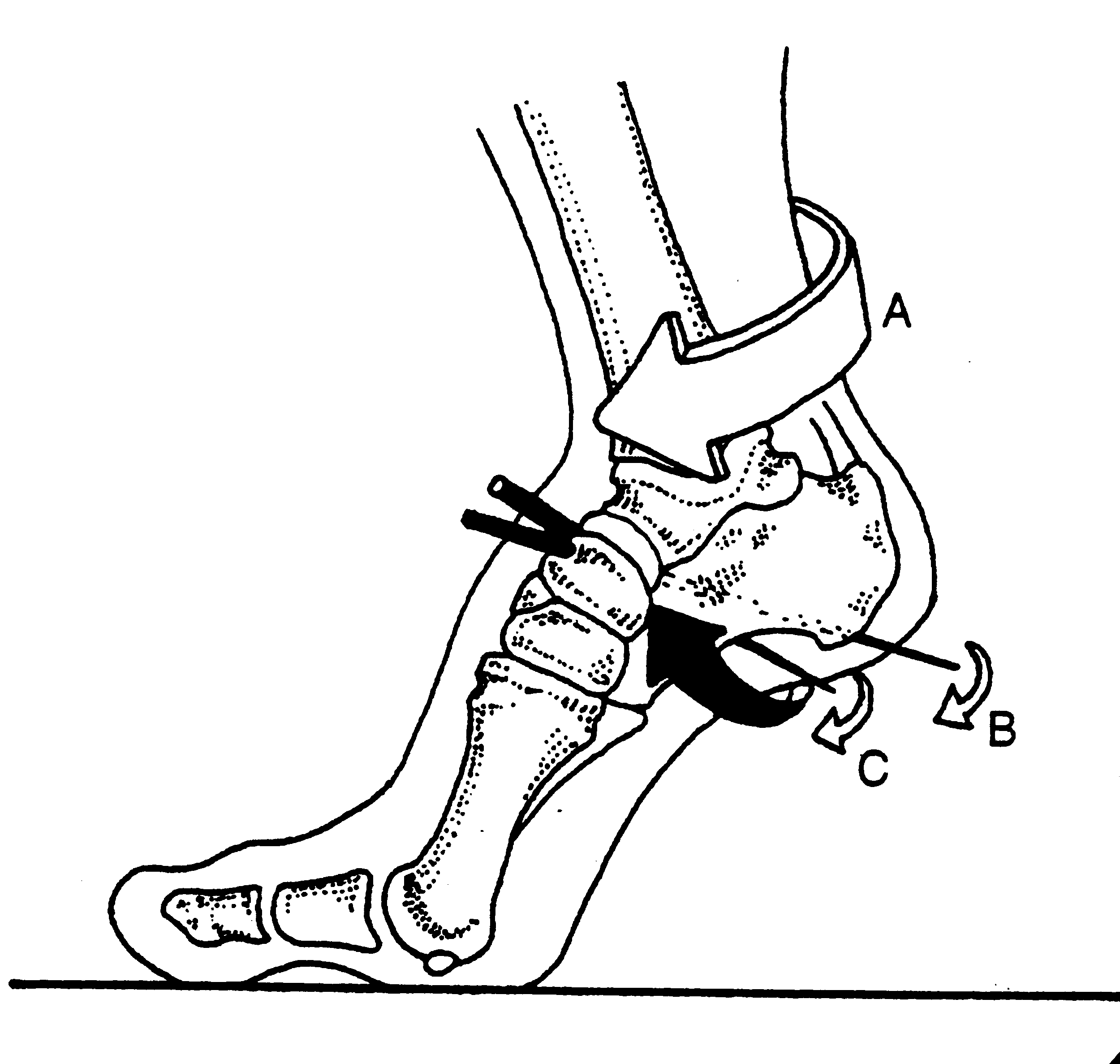
It is the mechanism by which the tendon of the peroneus longus travels behind the lateral malleolus of the ankle, travels underfoot, around the cuboid to insert into the lateral aspect of the base of the 1st metatarsal and adjacent 1st cunieform. Remember the peroneus longus?

The peroneus (or fibularis) longus arises from the head and upper two-thirds of the lateral surface of the fibula, from the deep surface of the fascia, and from the intermuscular septa between it and the muscles on the front and back of the leg; occasionally also by a few fibers from the lateral condyle of the tibia.  You can see from it attachments that it can influence the entire upper lateral leg.

It’s tendon runs down the fibular shaft, wraps around the lateral malleolus, travels obliquely under the foot, crossing the lateral cuboid (which it everts after midstance to help with supination) crosses the sole of the foot obliquely, and inserts into the lateral side of the base of the first metatarsal and lateral aspect of the 1st cunieform.  

It acts from just prior to heel strike to limit excessive rearfoot inversion, through midstance to decelerate subtalar pronation and assists in stabilization of the midfoot articulations, and into terminal stance and pre swing to lock the lateral column of the foot for toe off and plantar flex the 1st ray (creating a good foot tripod), allowing dorsal posterior shift of the 1st metatarsal-phalangeal joint axis (necessary for dorsiflexion of the hallux (big toe)).

When the peroneus longus contracts, in addition to plantar flexing the 1st ray, it everts the cuboid and locks the lateral column of the foot, minimizing muscular strain required to maintain the foot in supination (the locked position for propulsion). Normally, muscle strength alone is insufficient to perform this job and it requires some help from the adjacent articulations.

In addition, the soleus maintains spuination during propulsion by plantar flexing and inverting rear foot via the subtalar joint. This is assisted by the peroneus brevis and tertius which also dorsflex and evert the lateral column, helping keep it locked. Can you see why the peroneii are so important?

signs of a faulty calcaneo cuboid locking mechanism

  • weak peroneus longus, brevis and or tertius
  • excessive rear or midfoot pronation
  • low arch during ambulation
  • poor or low gear “push off”
  • subluxated cuboid


The calcaneo cuboid locking mechanism. Essential for appropriate supination and ambulation. Insufficiency, coming to a foot you will soon examine.

Supination, anyone?

Pronation gets all the press; but what about its counterpart, supination? There could not be one without the other. If anything, supination is at least as, if not more important to create propulsion.

Pronation is dorsiflexion, eversion and abduction of the foot. It provides shock absorption. Supination is plantar flexion, inversion and adduction. It helps the foot become a rigid lever so we can GO (Like in Theo Selig’s “Go Dig Go” ).

external leg rotation supination.gif

Locking of the lateral column of the foot (4th and 5th metatarsal, cuboid and calcaneus) is a necessary prerequisite for normal force transmission through the foot and ultimately placing weight on the head of the 1st metatarsal for proper (high gear) toe off . Locking of the lateral column minimizes muscular strain as the musculature (soleus, peroneus longus and brevis, EHL, EDL, FDL and FHL) is usually not strong enough to perform the job on its own.

external rotary moment.gif

This process is initiated by the opposite leg going into swing phase, which initiates dorsiflexion, inversion and abduction of the talus

The peroneus longus tendon aids this process by wrapping around the cuboid (the brevis attaches to the base of the 1st metatarsal) on its way to insert onto the base of the 1st metatarsal. When the peoneus longus contracts, it dorsiflexes and everts the cuboid, which, along with the soleus (which plantar flexes and inverts the subtalar joint) allows dorsiflexion of 4th and 5th metatarsals and “locks” the lateral column. Without this mechanism, there is no locking. Without locking, there is no supination. Without supination, there is little rigidity and inefficient propulsion.

The calcaneo cuboid locking mechanism. Another cool thing you learned about gait today from The Gait Guys.

tumblr_n04k3m4RxJ1qhko2so1_250.png
tumblr_n04k3m4RxJ1qhko2so5_r1_250.png
tumblr_n04k3m4RxJ1qhko2so4_400.png
tumblr_n04k3m4RxJ1qhko2so2_500.png
tumblr_n04k3m4RxJ1qhko2so3_400.png

Oh, it is just a simple ankle sprain. It will heal fine. (Not always ! Sometimes we do not know what we should fear, often because we do not even know it exists.)

When an ankle sprain is far more than an ankle sprain.

_________

Fracture of the Anterior Process of the Calcaneus

We have seen enough of these over our careers that we know they should be on the differential list when an ankle sprain smells fishy. This may be the most frequently missed fracture in the foot because it is not well known and the classic radiographic series often leaves this teeny tiny area poorly laid out on plain film radiographs.  This focal piece of bone, has a critical attachment to the cuboid and navicular so it is critical for stability of the rear-midfoot complex and obviously for mobility of the forefoot on the mid-foot. This Bifurcate ligament (see diagram above) if left unhealed or reattached to its calcaneal base can lead to anatomic instability and serious performance and loading problems. It can be a career ending injury if it is not caught early. This fracture accounts for ~15% of all calcaneal fractures and as we mentioned, it is misdiagnosed as a more severe ankle sprain. The mechanism is a typical inversion sprain mechanism, the most common of ankle sprains, and it can have all of the other typical presentations but with this fracture as a complicating parting gift of the injury. 

Do not miss this one ! When in doubt, refer it out. Don’t leave your client with an unresolving ankle sprain. If you are anxious, as for the imaging and a competent clinical exam. Tenderness over the calcaneocuboid joint that is localized approximately 1 cm inferior and 3 to 4 cm anterior to the lateral malleolus, just distal to the anterior talofibular ligament insertion is of high suspicion.  

This fracture can be serious and lead to prolonged disability and as we said it can be a career ending injury. So do not take that next ankle sprain too lightly. You or your client may pay for it for a lifetime.  

If you do not know it exists, you can’t make the call.  So after today, after reading this short blog post, you are now officially accountable !

Shawn and Ivo

Gait guys and clinical nerds

Supination, anyone? The importance of the calcaneocuboid locking mechanism. Pronation gets all the press; but what about its yin counterpart, supination? There could not be one without the other. Pronation is dorsiflexion, eversion and abduction of …

Supination, anyone? The importance of the calcaneocuboid locking mechanism.

Pronation gets all the press; but what about its yin counterpart, supination? There could not be one without the other.

Pronation is dorsiflexion, eversion and abduction of the foot. It provides shock absorption. Supination is plantar flexion, inversion and adduction. It make the foot into a rigid lever so we can GO (Like in Theo Selig’s “Go Dig Go”. OK, I have been reading that to my kids alot lately…)

Locking of the lateral column of the foot (4th and 5th metatarsal, cuboid and calcaneus) is a necessary prerequisite for normal force transmission through the foot and ultimately placing weight on the head of the 1st metatarsal for proper toe off. Locking of the lateral column minimizes muscular strain as the musculature (soleus, peroneus longus and brevis, EHL, EDL, FDL and FHL) is usually not strong enough to perform the job on its own.

The peroneus longus tendon wraps around the cuboid (and the brevis attaches to the base of the 1st metatarsal) on its way to insert onto the base of the 1st metatarsal. When it contracts, it dorsiflexes and everts the cuboid, which, along with the soleus (which plantar flexes and inverts the subtalar joint) allows dorsiflexion of 4th and 5th metatarsals and “locks” the lateral column.  Without this mechanism, there is no locking. Without locking, there is no supination. Without supination, there is little rigidity and inefficient propulsion.

The calcaneo cuboid locking mechanism. Another cool thing you learned about gait today from The Gait Guys.


all material copyright 2012 The Homunculus Group/The Gait Guys. All rights reserved. If you use our stuff without asking us, we will find you and put valgus wedges in all your shoes.