

Metatarsus Adductus: The Basics
/Metatarsus Adductus: The Basics
A few points to remember:
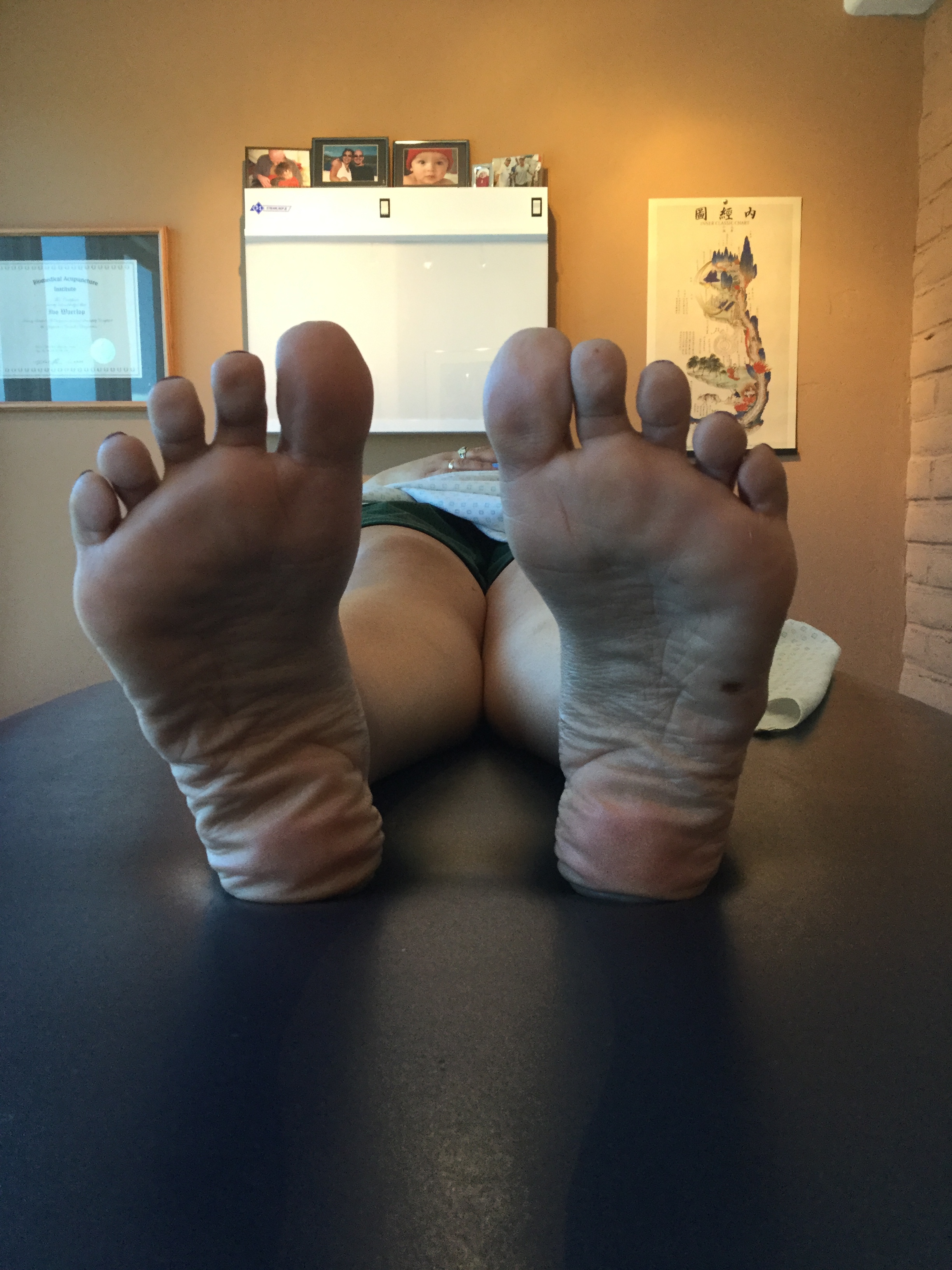
- Metatarsus adductus deformity is a forefoot which is adducted in the transverse plane with the apex of the deformity at LisFranc’s (tarso-metatarsal) joint. The fifth metatarsal base will be prominent and the lateral border of the foot which is convex in shape . The medial foot border is concave with a deep vertical skin crease located at the first metatarso cuneiform joint level. The hallux (great toe) may be widely separated from the second digit and the lesser digits will usually be adducted at their bases (se below). ln some cases the abductor hallucis tendon may be palpably taut just proximal to its insertion into the inferomedial aspect of the proximal phalanx (1)
- To measure the deviation of the metatarsals, the midline of the foot correspondingto bisecting the heel is used as a reference. This is the line that divides the heel pad into equal parts and, when extended, runs through the second toe or the second web space. In mild deformities, the midline of the foot runs through the third toe. In moderate adductus deformities, it falls between the third and fourth toes. In severe deformities the line is lateral to the third web space.(2)
- If detected early, stretching is a common and effective treatment for mild and some moderate cases. The heel is steadied with one hand while the forefoot is abducted in relation to the hind foot. This is done for 5 reps, 5-7 times per day. (2)
- 85% will resolve spontaneously, is caused by intrauterine position, is flexible & resolves spontaneously in more than 90 % of children. (3)
- Though often used interchangeably, the term "metatarsus adductus" is usually reserved for milder cases, where the forefoot is adducted on the hindfoot at the tarso-metatarsal articulation. Metatarsus varus is often reserved for conditions where the matatrsals are actually curved AND the forefoot is adducted on the hindfoot. (4) The term "Metatarsus primus varus" is reserved for feet which have the same neutral or valgus hindfoot and varus forefoot but, in addition, increased divergence of the first and second metatarsals. (5)
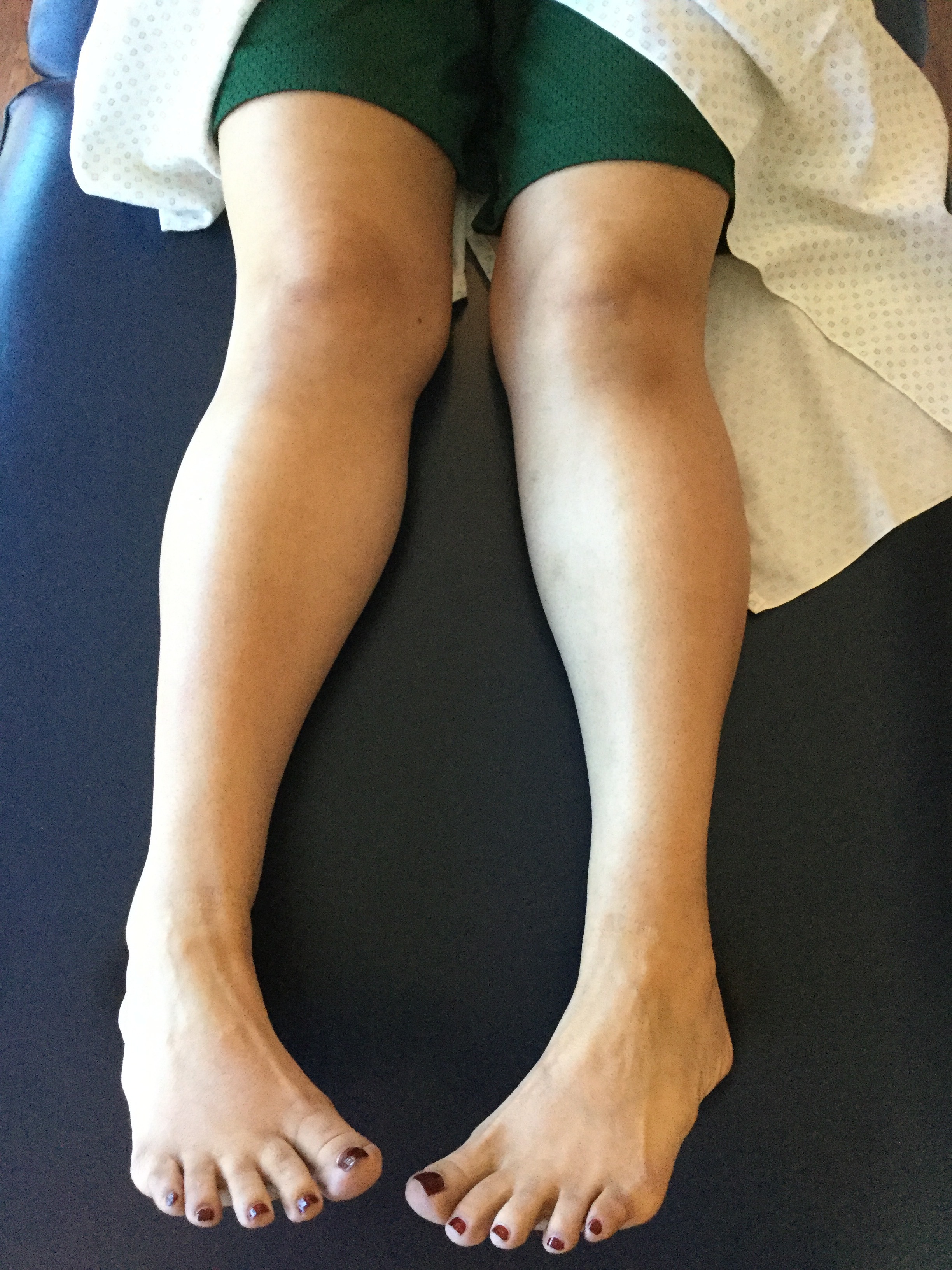
- It is interesting to note that along with forefoot adductus, hip dysplasia and internal tibial torsion are common (6) and this patient has the latter
- Gait abnormalities seen with this deformity include a decreased progression angle, in toed gait, excessive supination of the feet with low gear push off from the lesser metatarsals.
1. Bleck E: Metatarsus adductus: classification and relationship to outcomes of treatment. J Pediatric Orthop 3:2-9,1983.
2. Bohne W. Metatarsus adductus. Bulletin of the New York Academy of Medicine. 1987;63(9):835-838. link to FREE full text: https://www.ncbi.nlm.nih.gov/pmc/articles/PMC1629274/
3. http://www.wheelessonline.com/ortho/metatarsus_adductus
4. Peabody, C.W. and Muro, F.: Congenital metatarsus varus. J. Bone Joint Surg. 15:171-89, 1933.
5. Truslow, W.: Metatarsus primus varus or hallux valgus? J. Bone Joint Surg.23:98-108, 1925.
6. Jacobs J: Metatarsus varus and hip dysplasia. C/inO rth o p 16:203-212, 1960
additional references:
Kane R. Metatarsus varus. Bulletin of the New York Academy of Medicine. 1987;63(9):828-834. link to FREE full text: https://www.ncbi.nlm.nih.gov/pmc/articles/PMC1629282/
Wynne-Davies R, Littlejohn A, Gormley J. Aetiology and interrelationship of some common skeletal deformities. (Talipes equinovarus and calcaneovalgus, metatarsus varus, congenital dislocation of the hip, and infantile idiopathic scoliosis). Journal of Medical Genetics. 1982;19(5):321-328. link to FREE full text: https://www.ncbi.nlm.nih.gov/pmc/articles/PMC1048914/


